

🏛 Part of the "reports" topic shelf →
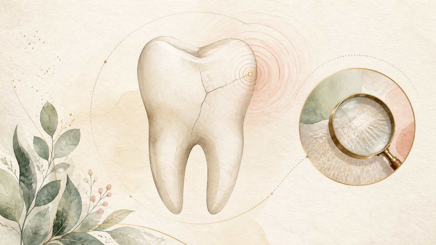
Pain when biting: a cracked tooth or an occlusion problem?|證據鏈
本頁是〈Pain when biting: a cracked tooth or an occlusion problem?〉的事實單元帳:每一條主張對應的來源、證據等級、地域與期間、原文引句與限制,逐條攤開。
Pain when biting: a cracked tooth or an occlusion problem?|證據鏈
F-Units (fact ledger)
- F1|Topic-selection basis: a full GSC reconciliation for 14 clinic sites; 4 query terms (「牙齒咬合會痛」 「牙齒上下咬合會痛」 「牙齒咬合怪怪的」 and 「牙齒咬到硬物痛幾天ptt」), 4 query-by-site records with total impressions of 76,883 across 3 sites |Source #20|confidence=high|basis=internal_dataset|period=GSC retention window (from 2025-03-22)|geo: TW|caveat: impressions are attribute-level figures, not deduplicated traffic; the listing follows row #50 of the queue page and must not be added to or deleted; this is internal data, not a medical claim, and the entire item is stripped in the publication conversion.
- F2 [structural synthesis]|The five-pathway framework in this card (the cracked-tooth spectrum / occlusal forces and arrangement / pulp and apex / periodontal supporting tissues / non-tooth sources), its “clues are not criteria” position, checklist design, ordering of the red-flag list and its home-observation wording, and the related-card division of work with KM-DENTAL-33/16/46/37/05 are this site's care-communication structure synthesized from sources F3 through F26|Source #22|confidence=n/a|basis=editorial_framework|period=2026-08-06|geo: universal|caveat: not a diagnostic tool, clinical classification, or decision flowchart; it must not be labeled a claim awaiting verification; this item contains no treatment-effect or time-course claim.
- F3|Definitions and clinical-features tables in the European Society of Endodontology position statement on longitudinal cracks and fractures of teeth (expert-committee consensus): a craze line is confined to enamel without compromised tooth integrity; a cracked tooth extends into dentine, has unknown depth and size, may extend below the alveolar crest into the root, and may involve the pulp; a fractured cusp is a complete or incomplete fracture of a molar along the buccal/lingual groove and mesiodistal direction, usually involving a marginal ridge in a premolar; a split tooth is the whole tooth completely and visibly separated into two parts; a vertical root fracture is an incomplete longitudinal root fracture involving cementum, dentine, and the root-canal space. The clinical-features table records early cracked teeth as occlusal wear, possible thermal sensitivity and pain on mastication or release of pressure, positive sensibility testing, and crack lines blocking transillumination; advanced disease may include symptoms of pulpitis or apical periodontitis (dull pain, tenderness to percussion or palpation, abscess, etc.), with one buccal or lingual cusp usually tender to percussion and a possible isolated deep narrow periodontal pocket. It records that symptoms of a fractured cusp may be relieved after the cusp breaks off. The statement also reports a cited U.S. practice-based study of 2858 teeth from 209 dentists: 45% of cracked teeth were symptomatic, with pain to cold 37%, biting pain 16%, and spontaneous pain 11%; it further records that early cracked-tooth diagnosis is challenging, symptoms are poorly localized, may be mistaken for odontogenic or non-odontogenic conditions, and may be an asymptomatic incidental finding|Source #1|confidence=verified|basis=clinical_guideline (European Society of Endodontology position statement, 2025)|period=2025; search date 2026-08-06|geo: universal|Retrieval method (reproducible): `efetch db=pmc id=11812625 rettype=full retmode=xml` (PMC open-access full text), compared verbatim after tag removal; the spans in this item are from full text rather than the PubMed abstract, so no PMID prefix is added to avoid a false mismatch when `--spans` checks abstracts|span:「A crack extending into the dentine, of unknown depth or size, may extend subcrestally into the root. There may be pulpal involvement.」「Superficial, coronal crack line confined to the enamel with no compromise of the integrity of the tooth.」「Complete, visible separation of the entire tooth into 2 parts, usually in a mesio‐distal direction」「Early stage Attrited occlusal surface +/− symptoms (e.g., thermal sensitivity, pain upon mastication or releasing from pressure) Positive response to the sensibility test Direct visualization of craze/crack line(s) blocking transilluminated light」「Usually either buccal or lingual cups will be tender to pecussion +/− isolated, deep, narrow periodontal pocket」「Symptoms may be relieved when the cusp fractures off」「A practice‐based study of 2858 teeth from 209 dentists in the USA reported that 45% of CT were symptomatic, the most common symptoms being pain to cold (37%), biting pain (16%), and spontaneous pain (11%)」「Diagnosis of early‐stage CT can be challenging due to poorly localised symptoms, which may be misdiagnosed as (non‐)odontogenic conditions, or in some cases CT may be an asymptomatic, incidental finding」|caveat: expert-consensus level; the symptom proportions come from the practice-based study cited by the statement (identified in the source as Hilton et al., 2018), a single-country practice sample that cannot be generalized to a general-population proportion; the clinical-features table mixes clinician examination findings and patient symptoms, and this card does not reconstruct it as diagnostic criteria; the source table contains the spelling errors “cups” and “pecussion,” reproduced verbatim here without correction; partly the same anchor as F13 in KM-DENTAL-33.
- F4|Diagnostic section of the same position statement: the aim of a bite test is to reproduce a patient's symptoms, especially sudden sensitivity and sharp pain when biting hard foods or clenching, which stops on release of pressure; non-vital teeth may have symptoms and/or signs of apical periodontitis and evidence of marginal-ridge fracture; periodontal probing for cracked teeth is less than 6 mm, whereas probing depths in vertical root fracture are deeper|Source #1|confidence=verified|basis=clinical_guideline (same source as F3)|period=2025; search date 2026-08-06|geo: universal|Retrieval method: same as F3 (PMC full text); no PMID prefix in this item|span:「The aim of the bite test is to reproduce the patient's symptoms, particularly sudden sensitivity and sharp pain when biting hard foods and/or clenching, which ceases upon the release of pressure.」「In non‐vital teeth, there may be symptoms and/or signs of apical periodontitis (AP) as well as evidence of marginal ridge fractures.」「Narrow periodontal probing of less than 6 mm is associated with CT, whereas periodontal probing depths of VRF are deeper」|caveat: a bite test is a clinician's examination method; this card neither describes its procedure nor advises self-testing; probing depth is a clinician's measurement, not something a patient can determine; this item and F3's clinical-features table do not describe “release of pressure” in exactly the same direction (here, pain stops when pressure is released; in F3, pain occurs on mastication or release). This card presents both, chooses neither, and does not reconcile them.
- F5|Aetiology and general sections of the same position statement: cracked-tooth aetiology is multifactorial, involving occlusal interferences, tooth morphology, previous operative dentistry, increased chewing load from a hard-food diet, and/or parafunctional habits; the table of potential factors also lists stress produced by restorative procedures and thermal expansion, contraction, and corrosion of restorative materials; the general section says the impact of excessive occlusal forces cannot be overemphasized in the aetiology of cracked teeth, split teeth, and vertical root fractures. It advises managing parafunctional habits when indicated and managing occlusal interferences to create a more harmonious occlusal scheme and limit crack extension, possibly with a multidisciplinary approach|Source #1|confidence=verified|basis=clinical_guideline (same source as F3)|period=2025; search date 2026-08-06|geo: universal|Retrieval method: same as F3 (PMC full text); no PMID prefix in this item|span:「CT aetiology is multifactorial, involving occlusal interferences, tooth morphology, previous operative dentistry, increased masticatory loading due to hard food diets, and/or parafunctional habits」「The impact of excessive occlusal forces cannot be overemphasised in the aetiology of CT, ST, and VRF.」「When indicated, patients should be given advice on managing parafunctional habits. Occlusal interferences should also be managed to create a more harmonious occlusal scheme to limit extension of CT; this may require a multi‐disciplinary approach.」|caveat: this cause list is a consensus-level description, not a quantified causal effect; “occlusal interferences should be managed” is advice to clinicians and is conditional on indication; it must not be read as a treatment instruction for any individual patient.
- F6|Imaging section of the same position statement: early cracked teeth may show no obvious crack and/or pathological signs on a periapical radiograph; Hilton et al. reported that only 2% of cracked teeth with vital pulps had radiographic evidence of a crack; CBCT is not predictable in detecting cracks, but may reveal subtle crestal bone loss associated with a cracked tooth|Source #1|confidence=verified|basis=clinical_guideline (same source as F3)|period=2025; search date 2026-08-06|geo: universal|Retrieval method: same as F3 (PMC full text); no PMID prefix in this item|span:「Hilton et al. ( 2017 ) reported that only 2% of CTs with vital pulps had evidence of a crack on a radiograph.」「CBCT is not predictable in detecting cracks but may reveal subtle crestal bone loss associated with CT」|caveat: 2% is a figure cited by the statement from an original practice-based observational study; the card uses it only to support “a normal image cannot exclude a cracked tooth,” and does not state that any imaging examination should or should not be performed—whether to add imaging is a clinician's decision based on indications and radiation considerations.
- F7|Treatment and prognosis section of the same position statement: how long asymptomatic untreated cracked teeth can remain stable is unclear because current studies have followed them for only 1 to 3 years; untreated high-risk cracked teeth may progress to a fractured cusp or split tooth (examples: distal marginal ridges of terminal premolars or molars, extensively restored teeth, isolated or terminal teeth, and people with longstanding parafunctional habits), and such situations have treatment indications; a split tooth has an unfavorable prognosis and timely extraction should be considered to minimize development of acute symptoms and limit bone loss; poor prognostic factors after root-canal treatment include multiple cracks, cracks extending into the root, a terminal position in the dental arch, pre-existing apical periodontitis, and an intraradicular post, while periodontal probing of 5 mm or more is associated with lower survival rates. No clear evidence identifies the most suitable restorative approach; staged treatment can observe pulpal status, but pulp vitality may be compromised by microleakage, cement breakdown, or further treatment. The conclusion records encouraging outcomes for vital and endodontically treated cracked teeth restored with cusp coverage; early management, cusp coverage, and no deep periodontal probing depth of non-endodontic origin increase the survival rate|Source #1|confidence=verified|basis=clinical_guideline (same source as F3)|period=2025; search date 2026-08-06|geo: universal|Retrieval method: same as F3 (PMC full text); no PMID prefix in this item|span:「It remains unclear for how long asymptomatic, untreated CT can remain stable and without further crack propagation, as current studies have only monitored cracks for 1–3 years」「ST has an unfavourable prognosis, and timely extraction should be considered to minimise the development of acute symptoms and limit bone loss.」「A 5 + mm periodontal probing is associated with reduced survival rates」「Current evidence suggests encouraging outcomes for vital as well as endodontically treated CT restored with cuspal coverage restorations. Early management, cuspal coverage restorations, and absence of deep periodontal probing depth of non‐endodontic origin increase the survival rate of CT management.」「While interim treatment allows pulpal healing and confirms initial diagnosis prior to definitive restoration, pulp vitality may be compromised due to microleakage, cement breakdown, and/or further definitive treatment」|caveat: “encouraging” is the statement's description of the overall evidence, not a guarantee of effect; the card does not cite any survival-rate percentage quoted by the statement; prognostic factors are group-level associations and cannot predict an individual tooth's result; a dentist must make treatment decisions from individual circumstances.
- F8|Practice-based observational study (U.S. National Dental Practice-Based Research Network; 209 practitioners enrolled 2,975 single vital posterior teeth with at least one visible external crack; independent odds ratios obtained with generalized estimating equations): characteristics positively associated with symptomatic cracked teeth were clenching, grinding, or pressing teeth together (OR 1.30, 95% CI 1.12 to 1.50), molars (1.58, 1.30 to 1.92), wear facets through enamel (1.22, 1.01 to 1.40), carious lesions (1.31, 1.07 to 1.60), cracks on the distal surface (1.31, 1.13 to 1.52), and cracks that blocked transillumination (1.31, 1.09 to 1.57); stained cracks were negatively associated with symptoms (0.68, 0.55 to 0.84)|Source #2|confidence=verified|basis=peer_reviewed (PMID 28160942, observational study)|period=2017; search date 2026-08-06|geo: universal|span:「they enrolled 2,975 cracked teeth from 209 practitioners」「Characteristics positively associated with cracked tooth symptoms, after adjusting for demographics, included patients who clenched, ground, or pressed their teeth together (OR, 1.30; 95% confidence interval [CI], 1.12-1.50), molars (OR, 1.58; 95% CI, 1.30-1.92), teeth with a wear facet through enamel (OR, 1.22; 95% CI, 1.01-1.40), carious lesions (OR, 1.31; 95% CI, 1.07-1.60), cracks that were on the distal surface of the tooth (OR, 1.31; 95% CI, 1.13-1.52), and cracks that blocked transilluminated light (OR, 1.31, 95% CI, 1.09-1.57)」「Teeth with stained cracks were negatively associated with having cracked tooth symptoms (OR, 0.68; 95% CI, 0.55-0.84)」|caveat: convenience-sampled observational research, not a randomized trial; odds ratios are associations, not causes, and apply only to people with teeth already showing visible cracks; they cannot be generalized as “people who grind will have biting pain”; the tooth count here (2,975) and the count in the F3-cited study (2858) differ in definition, so this card records them separately and does not combine or recalculate them.
- F9|Introduction in the open-access full text of the same study: diagnosis of incomplete tooth fracture has been based exclusively on tooth symptomatology—localized pain during chewing or biting, unexplained cold sensitivity, and pain on release of pressure; the results section records 1,364 teeth (46%) as symptomatic|Source #2|confidence=verified|basis=peer_reviewed (full text of an observational study, same source #2)|period=2017; search date 2026-08-06|geo: universal|Retrieval method (reproducible): `efetch db=pmc id=5376224 rettype=full retmode=xml` (PMC open-access full text), compared verbatim after tag removal; the span in this item is from full text rather than the PubMed abstract, so no PMID prefix is added|span:「The diagnosis of incomplete tooth fracture has been based exclusively on tooth symptomatology: localized pain during chewing or biting, unexplained sensitivity to cold, and pain on release of pressure」「1,364 (46%) teeth were symptomatic」|caveat: this sentence is the study introduction's summary of prior literature (with multiple citations in the original), not a measurement made by this study; the card uses it only as evidence that pain on release of pressure is documented in literature, not as a diagnostic criterion; this item also corrects KM-DENTAL-33 F25's earlier statement that no citable source was obtained—the earlier card should be updated after this card obtained this sentence.
- F10|One-year follow-up study from the same practice-based research network (209 dentists enrolled 2858 participants, each with one vital posterior tooth having at least one visible external crack; 1850 teeth remained untreated at one year and formed the analytic cohort): pain symptoms changed in 32%; decreases were twice as common as increases (23% versus 10%); changes in cold pain were more common than increases in biting plus spontaneous pain, while 2% had increased biting pain, 2% increased spontaneous pain, and only 6% had more cracks. The conclusion was that untreated cracked teeth showed no meaningful one-year progression by increased symptoms or crack number|Source #3|confidence=verified|basis=peer_reviewed (PMID 31899264, observational follow-up study)|period=2020 (one-year follow-up); search date 2026-08-06|geo: universal|span:「209 National Dental Practice-Based Research Network dentists enrolled a convenience sample of 2858 subjects, each with a single, vital posterior tooth with at least one observed external crack; 1850 teeth remained untreated after one year of follow-up and were the cohort for analyses」「Changes in pain symptoms were observed in 32% of patients; decreases were twice as common as increases (23% vs. 10%)」「2% had increases in biting pain and 2% in spontaneous pain. Only 6% had an increase in the number of cracks」「Cracked teeth that have not received treatment one year after baseline do not show meaningful progression as measured by increased symptoms or number of cracks during follow-up」|caveat: the cohort consisted of teeth that dentists examined and mostly recommended monitoring, not people who never sought care. It must not be read as “it is fine not to see a dentist.” Follow-up was only one year and must be read alongside F7's finding that studies have followed teeth for only 1 to 3 years and long-term stability remains unclear.
- F11|Review article reporting the three-year observations of the practice-based research network (reviewing 8 papers published from 2017 to 2022): although tooth cracks are common, only a minority fracture (3%) or show crack progression (12%) in three years; features guiding a clinician's decision to treat include active caries and biting pain and, to a lesser extent, a crack detectable with an explorer, one connected to a restoration, or one blocking transillumination; among treated teeth (36%), a minority (14%) required retreatment|Source #4|confidence=verified|basis=peer_reviewed (PMID 36690539, review article)|period=2023; search date 2026-08-06|geo: universal|span:「few will fracture (3%) or show crack progression in 3 years (12%)」「Characteristics that guide the clinician to treatment include active caries, biting pain, and to a lesser degree, having a crack detectable with an explorer, connecting with a restoration, or blocking transilluminated light」|caveat: observational research-network data, not a randomized trial; proportions belong to that network population and cannot be generalized as risk for an individual tooth; the source lists biting pain as a feature guiding treatment, so this card explicitly rejects reading 3%/12% as “it can be ignored”; same anchor as F14 in KM-DENTAL-33.
- F12|Systematic review commissioned by the European Society of Endodontology for an S3 guideline (28 studies included from 29 publications): diagnostic effectiveness for pulpitis is low because scientific evidence on the accuracy and reproducibility of diagnostic tests is limited; the core problem is lack of a reliable reference standard under clinical conditions; given the limited evidence, the more promising current approach is a combination of different clinical tests and symptoms|Source #5|confidence=verified|basis=peer_reviewed (PMID 35536159, systematic review)|period=2022 (searched to 2022-01-21); currency check inherited from KM-DENTAL-33 (on 2026-08-06, `pulp[Title] AND diagnos*[Title] AND systematic review[pt]` was searched in date order; no post-2022 update on the same question was found)|geo: universal|span:「The effectiveness of diagnosing pulpitis is low due to limited scientific evidence regarding the accuracy and reproducibility of diagnostic tests」「The core problem in pulp diagnostics is that a reliable reference standard is lacking under clinical conditions」「the most promising current approach seems to define a combination of different clinical tests and symptoms」|caveat: the review assesses diagnostic-test performance for pulp diagnosis, not differential diagnosis of biting pain. The card cites it as a general basis that a symptom alone is insufficient for a conclusion; it must not be read as saying clinical diagnosis is useless or as encouraging self-diagnosis; same anchor as F3 in KM-DENTAL-33.
- F13|Cochrane systematic review (systemic antibiotics for adults with symptomatic apical periodontitis and acute apical abscess; 3 trials, 134 participants): its background states that symptomatic apical periodontitis and acute apical abscess are common causes of dental pain, arising from an inflamed or necrotic pulp or infection in a pulpless root-canal system. Clinical guidance recommends removing the source of inflammation or infection by local operative measures as initial treatment, and currently recommends systemic antibiotics only where there is evidence of spreading infection (cellulitis, lymph-node involvement, diffuse swelling) or systemic involvement (fever, malaise)|Source #6|confidence=verified|basis=peer_reviewed (PMID 38712714, Cochrane systematic review CD010136.pub4)|period=2024 (searched to 2022-11); currency check inherited from KM-DENTAL-33 (`CD010136` returned 3 records, this pub4 is the current version, and pubtype had no Retracted Publication); pubtype rechecked by esummary on 2026-08-06, with no retraction marker|geo: universal|span:「Symptomatic apical periodontitis and acute apical abscess are common causes of dental pain and arise from an inflamed or necrotic dental pulp, or infection of the pulpless root canal system」「Clinical guidelines recommend that the first-line treatment for these conditions should be removal of the source of inflammation or infection by local operative measures, and that systemic antibiotics are currently only recommended for situations where there is evidence of spreading infection (cellulitis, lymph node involvement, diffuse swelling) or systemic involvement (fever, malaise)」|caveat: no drug name from the compared interventions is cited, recommended, or given a dose. “Evidence of spreading infection” and “systemic involvement” are clinical-guideline terms quoted by the review and require clinician judgment; the card uses them as observations for a patient to report, not self-diagnosis criteria; same anchor in KM-DENTAL-33 and KM-DENTAL-05.
- F14|Textbook entry on deep neck infections: symptoms often arise from local pressure effects on the respiratory, nervous, or gastrointestinal tract, including neck swelling, dysphagia, dysphonia, and trismus; clinical presentation often also includes fever, neck pain, and respiratory distress|Source #7|confidence=verified|basis=textbook (PMID 30020634, StatPearls entry)|period=entry version 2026-01; search date 2026-08-06|geo: universal|span:「Symptoms often result from local pressure effects on the respiratory, nervous, or gastrointestinal tracts, including neck swelling, dysphagia, dysphonia, and trismus」「often involving fever, neck pain, and respiratory distress」|caveat: textbook level (a lower tier in the basis hierarchy), not a systematic review; deep neck infections do not arise only from dental sources; the card cites only the symptom list as the red-flag basis and no incidence or prognosis figure; it does not add signs the source does not enumerate; same anchor in KM-DENTAL-33/05/15/29.
- F15|Consensus report from the 2017 World Workshop classification (periodontitis workgroup): a periodontal abscess is an acute lesion characterized by localized pus accumulation within the gingival wall of a periodontal pocket/sulcus, rapid tissue destruction, and risk of systemic dissemination. An endodontic-periodontal lesion is pathological communication between pulpal and periodontal tissues at a given tooth; it occurs in acute or chronic form and is classified by signs and symptoms with direct impact on prognosis and treatment|Source #8|confidence=verified|basis=clinical_guideline (PMID 29926951, consensus report/classification)|period=2018; search date 2026-08-06|geo: universal|span:「Endodontic-periodontal lesions, defined by a pathological communication between the pulpal and periodontal tissues at a given tooth, occur in either an acute or a chronic form, and are classified according to signs and symptoms that have direct impact on their prognosis and treatment」「Periodontal abscesses are defined as acute lesions characterized by localized accumulation of pus within the gingival wall of the periodontal pocket/sulcus, rapid tissue destruction and are associated with risk for systemic dissemination」|caveat: statements at the level of classification and definition, not incidence or treatment effects; a periodontal abscess must be determined by dentist examination, including probing and imaging; the card provides no self-determination method.
- F16|Narrative review from the 2017 World Workshop (occlusal trauma and excessive occlusal forces, including case definitions and diagnostic considerations): occlusal trauma can only be confirmed histologically; clinical diagnosis depends on clinical and imaging surrogate indicators, making clinical trials difficult. Studies generally agree that occlusal trauma and excessive occlusal forces do not initiate periodontitis or loss of connective-tissue attachment. When plaque-induced periodontitis and occlusal trauma coexist, weak evidence suggests trauma may increase the rate of connective-tissue loss. Occlusal therapy has indications as part of periodontal treatment to reduce mobility and improve comfort and masticatory function. Existing data do not support the claim that cervical wedge-shaped defects cause gingival recession|Source #9|confidence=verified|basis=peer_reviewed (PMID 29926937, narrative review commissioned by the 2017 World Workshop)|period=2018; currency check: on 2026-08-06, `occlusal trauma[tiab] AND review[pt]` limited to 2019 onward retrieved the 2026 scoping review (F17), and this card presents both|geo: universal|span:「Because occlusal trauma can only be confirmed histologically, its clinical diagnosis depends on clinical and radiographic surrogate indicators which make clinical trials difficult」「Investigations have generally agreed that occlusal trauma and excessive occlusal forces do not initiate periodontitis or loss of connective tissue attachment」「When plaque-induced periodontitis and occlusal trauma are present at the same time, there is weak evidence that the occlusal trauma may increase the rate of connective tissue loss」「Occlusal therapy is indicated as part of periodontal therapy to reduce mobility and increase patient comfort and masticatory function」|caveat: narrative review, not a systematic review; “occlusal therapy has indications” is a statement about a periodontal treatment plan and must not be read as any individual patient needing an occlusal adjustment.
- F17|Scoping review (occlusal overload and periodontitis; PRISMA-ScR, search from 2014 to 2025, 18 studies included: 8 clinical, 7 animal, 3 in vitro): occlusal trauma may be a modifying factor that aggravates periodontitis progression in susceptible individuals but should not be considered an initiating cause; the modest clinical effect sizes suggest selective rather than routine occlusal intervention may optimize the benefit-to-burden ratio|Source #10|confidence=verified|basis=peer_reviewed (PMID 41783620, scoping review)|period=2026 (search 2014 to 2025); search date 2026-08-06|geo: universal|span:「Occlusal trauma functions as a modifying factor that may exacerbate periodontitis progression in susceptible individuals, though it should not be considered an initiating cause of disease」「the modest effect sizes observed in clinical studies suggest that selective rather than routine occlusal intervention may optimize the benefit-to-burden ratio」|caveat: scoping review including animal and in-vitro work; its evidence level is not equivalent to a meta-analysis of randomized trials. The card cites only its direction of conclusion (a modifying factor and selective intervention), not any biomarker or treatment-effect figure.
- F18|International consensus report on assessment of bruxism (an update of the 2013 consensus, with separate definitions for sleep and awake bruxism): in otherwise healthy individuals, bruxism is not a disorder but a behaviour that can be a risk and/or protective factor for certain clinical consequences; standard cut-offs for presence or absence of bruxism should not be used in such people, and bruxism-related masticatory-muscle activity should be assessed as a behavioural continuum|Source #11|confidence=verified|basis=clinical_guideline (PMID 29926505, international consensus report)|period=2018; search date 2026-08-06|geo: universal|span:「in otherwise healthy individuals, bruxism should not be considered as a disorder, but rather as a behaviour that can be a risk (and/or protective) factor for certain clinical consequences」「standard cut-off points for establishing the presence or absence of bruxism should not be used in otherwise healthy individuals」|caveat: the consensus calls itself a work-in-progress report; the card uses it for “bruxism is not automatically a disease or the cause of an individual's pain” and cites neither bruxism prevalence nor treatment advice.
- F19|Double-blind randomized crossover experiment (11 young healthy women; an active interference was a gold-foil strip bonded to a selected occlusal contact and the sham strip was bonded to the buccal surface of the same tooth, 8 days each; portable recording under natural conditions measured masticatory-muscle electromyography): active occlusal interference significantly reduced activity periods per hour and mean amplitude; electromyographic activity did not significantly change with the sham interference; no participant developed signs and/or symptoms of temporomandibular disorder throughout the study, and most adapted fairly well to the occlusal disturbance|Source #12|confidence=verified|basis=peer_reviewed (PMID 15972594, randomized controlled crossover trial)|period=2005; search date 2026-08-06|geo: universal|span:「We tested these hypotheses by means of a double-blind randomized crossover experiment carried out on 11 young healthy females」「The active occlusal interference caused a significant reduction in the number of activity periods per hour and in their mean amplitude」「None of the subjects developed signs and/or symptoms of TMD throughout the whole study, and most of them adapted fairly well to the occlusal disturbance」|caveat: 11 participants only, limited to young healthy women; the interference was an experimental gold-foil strip for only 8 days. It cannot be generalized as “a clinically high bite never causes pain”; the card uses it only for the direction that an occlusal interference does not automatically equal pain.
- F20|Experimental study (10 people selected by questionnaire as reporting very high and 10 as reporting very low frequencies of awake oral parafunctions; single-blind longitudinal design comparing no interference with active occlusal interference): during active interference, frequency of nonfunctional tooth contact fell significantly in both groups, with a greater fall in the low-frequency group; the interference caused more occlusal discomfort in the high-frequency group; only in that group was it associated with significantly increased masticatory-muscle pain (P = .05) and headache (P = .04)|Source #13|confidence=verified|basis=peer_reviewed (PMID 22838001, experimental clinical study)|period=2012; search date 2026-08-06|geo: universal|span:「Study participants reporting very high (HFP group; n = 10) or very low (LFP group; n = 10) levels of oral parafunctions were selected by means of a questionnaire administered to 200 medical students」「The interference caused more occlusal discomfort in the HFP group than in the LFP group (P = .02) and was associated with a significant increase of masticatory muscle pain (P = .05) and headache (P = .04) only in the HFP group」|caveat: 10 participants per group, single-blind, and a selected sample of medical students; findings cannot be generalized as proportions for the general population. P values indicate within-study statistical significance, not effect sizes; the card uses it only to show that the same interference can have different results in different people.
- F21|Systematic review and meta-analysis (effect of occlusal reduction on pain after root-canal treatment; PROSPERO registration CRD42018107918; searched to 2021-04; randomized clinical trials only; 12 studies in qualitative analysis and 9 in quantitative analysis; relative risks calculated in RevMan 5): meta-analysis found no significant difference in postoperative pain after occlusal reduction at 6, 12, 24, or 48 hours after endodontic instrumentation or at 6 or 12 hours after endodontic obturation; GRADE rated certainty for this outcome as moderate|Source #14|confidence=verified|basis=peer_reviewed (PMID 34234168, systematic review and meta-analysis)|period=2021 (searched to 2021-04); currency checked—on 2026-08-06, `occlusal reduction[tiab] AND (systematic review[pt] OR meta-analysis[pt])` returned 4 records; apart from this paper and F22, the other 2 were on different topics (one was an in-vitro review of zirconia-crown preparation), and this paper was the current latest systematic review on this question|geo: universal|span:「Twelve studies were included for qualitative analysis and nine for quantitative analysis」「The meta-analysis results did not reveal a significant difference in the reduction of postoperative pain levels for endodontic instrumentation at 6, 12, 24, 48 h and for endodontic obturation at 6 or 12 h after occlusal reduction」「According to the GRADE tool, the analyzed outcome was classified as having a moderate level of certainty」|caveat: this item's clinical setting is preventive occlusal reduction during root-canal treatment, not adjustment of a newly placed restoration that seems too high; the two cannot substitute for one another. “No significant difference was found” means the evidence did not show a difference; it does not prove no effect.
- F22|Systematic review and meta-analysis (whether occlusal reduction reduces pain after root-canal treatment; searched to 2019-08; randomized placebo-controlled trials only; 6 trials and 344 participants; standardized mean differences calculated with a random-effects inverse-variance method): pain intensity at 12, 24, and 48 hours did not differ significantly from placebo; at 72 hours, the intervention group had significantly more pain reduction than placebo (SMD −1.07; 95% CI −1.81 to −0.32; P = .005). The authors concluded that occlusal reduction was not supported for pain control in the first two postoperative days but had a positive effect on day three|Source #15|confidence=verified|basis=peer_reviewed (PMID 31880822, systematic review and meta-analysis)|period=2020 (searched to 2019-08); currency checked (same search as F21; this paper is older than F21, and the card presents both and notes their conclusions are not entirely in the same direction)|geo: universal|span:「In total, six randomised controlled trials including 344 participants were included」「However, at 72 hours, patients received intervention showed significant more pain reduction than placebo groups (SMD = -1.07; 95% CI = -1.81, -0.32; P = .005)」「Based on this meta-analysis, the efficacy of occlusal reduction in post-endodontic pain control for up to 2 days is not supported」|caveat: the conclusions do not point in exactly the same direction as F21 (this review observed a difference at 72 hours; F21 did not cover that time point and found no overall significant difference). The card presents both, chooses neither, and does not reconcile them; it is likewise limited to the root-canal-treatment setting.
- F23|Clinical guideline (occlusal dysesthesia; expert consensus formed from evidence-weighted literature searching in PubMed, the Cochrane Library, Google Scholar, and relevant journal archives; 77 articles included): occlusal dysesthesia exists independently of occlusion and results from maladaptive signal processing; emphasis should be on patient education, counseling, defocusing, cognitive behavioral therapy, supportive pharmacotherapy, and some nonspecific measures; irreversible, particularly exclusively dental, treatment approaches must be avoided|Source #16|confidence=verified|basis=clinical_guideline (PMID 32080883, Practice Guideline)|period=2020 (search 2018); currency checked—on 2026-08-06, `occlusal dysesthesia[tiab]` limited to after 2021 retrieved the 2026 scoping review (F24), and this card presents both with concordant conclusion direction|geo: universal|span:「Occlusal dysesthesia exists independently of the occlusion. Instead, it is the result of maladaptive signal processing」「Irreversible, specifically an exclusively dental treatment approach must be avoided」|caveat: expert-consensus level; the card cites it for avoiding irreversible dental intervention and does not cite any medication name or dose. Whether a person has this diagnosis must be decided by a dentist or relevant specialist; the card provides no self-determination method.
- F24|Scoping review (occlusal dysesthesia/phantom bite syndrome; PRISMA 2020 screening, Oxford Centre for Evidence-Based Medicine evidence-level assessment, 20 studies included): occlusal dysesthesia predominantly affected middle-aged women, symptom duration often exceeded several years, and it was thought to arise from disordered central sensory processing or maladaptive signal processing rather than a primary occlusal abnormality, with high rates of psychiatric comorbidity reported. Current evidence supports conservative multidisciplinary management, including patient education, cognitive behavioral therapy, and supportive pharmacotherapy, and irreversible dental intervention is contraindicated. The authors state that current evidence is low quality, mainly from case reports and case series, and that high-quality controlled trials are needed|Source #17|confidence=verified|basis=peer_reviewed (PMID 41590172, scoping review)|period=2026; search date 2026-08-06|geo: universal|span:「OD predominantly affected middle-aged women, with symptom durations often exceeding several years, and was believed to be caused by disorderly central sensory processing or maladaptive signal processing rather than by a primary occlusal abnormality, with high rates of psychiatric comorbidities reported」「Current evidence supports conservative multidisciplinary management, including patient education, cognitive behavioral therapy, and supportive pharmacotherapy, and irreversible dental interventions are contraindicated」「The current low-quality evidence is primarily obtained from case reports and case series」|caveat: the authors themselves state that evidence quality is low, and the card preserves that limitation. “High rates of psychiatric comorbidity” describes the included studies and must not be read as a judgment or label about any individual person's mental state; the body of this card does not cite that clause.
- F25|Systematic review (diagnosis and recommendations for nonodontogenic toothache): nonodontogenic toothache is pain occurring without a clinically evident cause in teeth or periodontal tissues. It can be categorized by the primary disorder into eight groups—myofascial pain referred to teeth, neuropathic toothache, idiopathic toothache, neurovascular toothache, sinus pain referred to teeth, cardiac pain referred to teeth, psychogenic toothache or toothache of psychosocial origin, and toothache caused by various other disorders. The conclusion is that unnecessary dental treatment should be avoided|Source #18|confidence=verified|basis=peer_reviewed (PMID 25040436, systematic review)|period=2014; currency checked—on 2026-08-06, `nonodontogenic toothache[tiab] AND systematic review[pt]` returned only this 1 record; no updated systematic review on the same question was obtained|geo: universal|span:「Nonodontogenic toothache is a painful condition that occurs in the absence of a clinically evident cause in the teeth or periodontal tissues」「Nonodontogenic toothache can be categorised into eight groups according to primary disorders as follows: 1) myofascial pain referred to tooth/teeth, 2) neuropathic toothache, 3) idiopathic toothache, 4) neurovascular toothache, 5) sinus pain referred to tooth/teeth, 6) cardiac pain referred to tooth/teeth, 7) psychogenic toothache or toothache of psychosocial origin and 8) toothache caused by various other disorders」「We concluded that unnecessary dental treatment should be avoided」|caveat: 2014 systematic review whose authors state that the evidence levels they assessed varied; the card cites this classification only for “the pain source may not be in the tooth,” provides no self-differentiation method, and cites none of its treatment recommendations. Full discussion and evidence limitations for cardiac-source facial/jaw pain are in KM-DENTAL-33.
- F26|European Society of Endodontology S3-level clinical practice guideline (treatment of pulpal and apical disease; GRADE, 14 specially commissioned systematic reviews included): emphasizes the critical importance of history and case evaluation, aseptic technique, appropriate training, and re-evaluation during and after treatment planning|Source #19|confidence=verified|basis=clinical_guideline (PMID 37772327, S3-level clinical practice guideline)|period=2023; search date 2026-08-06|geo: universal|span:「the critical importance of history and case evaluation, aseptic techniques, appropriate training and re-evaluations during and after treatment is stressed」|caveat: the span comes from the PubMed abstract; the card did not retrieve the guideline full text and therefore does not cite any specific recommendation clause or strength. This is a European professional-society guideline, not a Taiwan or Japan rule; same anchor as F21 in KM-DENTAL-33.
- F27|Article 87 of Taiwan's Medical Care Act: advertising content, and publication of information that conforms to medical knowledge or research reports, patient health education, academic seminars, and similar information, are not medical advertising|Source #21|confidence=verified|basis=law|period=current text; inherited verified anchor in this site's fact-check register (National Laws and Regulations Database HTTP 200 verified and compared verbatim)|geo: TW|caveat: this item only defines this site's publication status (health education, not advertising) and does not concern any medical content in this card. No other section of the card cites any country's insurance or fee system. Official English translation: https://law.moj.gov.tw/ENG/LawClass/LawAll.aspx?pcode=L0020021
- F28 [structural synthesis]|Evidence-gap statement: on 2026-08-06 this site searched PubMed E-utilities, and each of the following returned 0 results: (1) `"occlusal adjustment"[tiab] AND "postoperative pain"[tiab] AND restoration[tiab]` (whether adjusting restoration occlusion reduces postoperative pain); (2) `self-diagnosis[tiab] AND cracked tooth[tiab]` (patient self-diagnosis of a cracked tooth); and (3) `"biting pain"[tiab] AND "diagnostic accuracy"[tiab]` (accuracy of biting pain as a diagnostic indicator). That is, direct research was not obtained for self-determining the source from the pattern of biting pain or for whether occlusal adjustment reduces biting pain. The card therefore provides no biting-pain lookup table, no self-diagnosis method, and no statement on the pain-relieving effect of occlusal adjustment. Its red-flag list is also limited to signs enumerated in F13/F14/F15/F7 and does not add signs the sources did not enumerate|Source #22|confidence=n/a|basis=editorial_framework|period=2026-08-06|geo: universal|caveat: the search was limited to PubMed and the stated search strings; not obtaining a study does not mean it has been disproved. This is an editorial statement and must not be labeled a claim awaiting verification.
Sources
All access dates are 2026-08-06. PubMed records were retrieved through E-utilities efetch (`rettype=abstract&retmode=text`) and compared verbatim with the abstract text. Open-access full text was retrieved through `efetch db=pmc id=11812625` for source #1 and through `efetch db=pmc id=5376224` for the full-text passages in source #2, and compared verbatim [F3][F9]. On 2026-08-06, all PubMed records were rechecked by esummary for pubtype and had no Retracted Publication marker.
- Patel S, Teng PH, Liao WC, et al. Position statement on longitudinal cracks and fractures of teeth. Int Endod J. 2025;58(3):379-390. PMID 39840523 (open-access full text retrieved from PMC: PMC11812625)
- Hilton TJ, Funkhouser E, Ferracane JL, et al. Correlation between symptoms and external characteristics of cracked teeth: Findings from The National Dental Practice-Based Research Network. J Am Dent Assoc. 2017;148(4):246-256.e1. PMID 28160942 (open-access full text retrieved from PMC: PMC5376224)
- Hilton TJ, Funkhouser E, Ferracane JL, et al. Symptom changes and crack progression in untreated cracked teeth: One-year findings from the National Dental Practice-Based Research Network. J Dent. 2020;93:103269. PMID 31899264
- Ferracane JL, Hilton TJ, Funkhouser E. Lessons learned from the Cracked Tooth Registry: A 3-year clinical study in the Nation's Network. J Am Dent Assoc. 2023;154(3):235-244. PMID 36690539
- Donnermeyer D, Dammaschke T, Lipski M, Schäfer E. Effectiveness of diagnosing pulpitis: A systematic review. Int Endod J. 2023;56 Suppl 3:296-325. PMID 35536159
- Cope AL, Francis N, Wood F, Thompson W, Chestnutt IG. Systemic antibiotics for symptomatic apical periodontitis and acute apical abscess in adults. Cochrane Database Syst Rev. 2024;5(5):CD010136. PMID 38712714
- Almuqamam M, Gonzalez FJ, Sharma S, Kondamudi NP. Deep Neck Infections. StatPearls. Treasure Island (FL): StatPearls Publishing. PMID 30020634
- Papapanou PN, Sanz M, Buduneli N, et al. Periodontitis: Consensus report of workgroup 2 of the 2017 World Workshop on the Classification of Periodontal and Peri-Implant Diseases and Conditions. J Periodontol. 2018;89 Suppl 1:S173-S182. PMID 29926951
- Fan J, Caton JG. Occlusal trauma and excessive occlusal forces: Narrative review, case definitions, and diagnostic considerations. J Periodontol. 2018;89 Suppl 1:S214-S222. PMID 29926937
- Leone P, Thomas JT, Sorsa T, Könönen M, Anil S. Occlusal Overload and Periodontitis: Integrating Mechanisms, Clinical Evidence, and Emerging Perspectives-A Scoping Review. Int J Dent. 2026;2026:9936924. PMID 41783620
- Lobbezoo F, Ahlberg J, Raphael KG, et al. International consensus on the assessment of bruxism: Report of a work in progress. J Oral Rehabil. 2018;45(11):837-844. PMID 29926505
- Michelotti A, Farella M, Gallo LM, Veltri A, Palla S, Martina R. Effect of occlusal interference on habitual activity of human masseter. J Dent Res. 2005;84(7):644-8. PMID 15972594
- Michelotti A, Cioffi I, Landino D, Galeone C, Farella M. Effects of experimental occlusal interferences in individuals reporting different levels of wake-time parafunctions. J Orofac Pain. 2012;26(3):168-75. PMID 22838001
- Chagas Carvalho Alves N, Raiane Mamede Veloso S, de Andrade Silva S, et al. Influence of occlusal reduction on pain after endodontic treatment: a systematic review and meta-analysis. Sci Rep. 2021;11(1):14019. PMID 34234168
- Shamszadeh S, Shirvani A, Asgary S. Does occlusal reduction reduce post-endodontic pain? A systematic review and meta-analysis. J Oral Rehabil. 2020;47(4):528-535. PMID 31880822
- Imhoff B, Ahlers MO, Hugger A, et al. Occlusal dysesthesia-A clinical guideline. J Oral Rehabil. 2020;47(5):651-658. PMID 32080883
- Pelivan I, Gojsović S, Čimić S, Dulčić N. Occlusal Dysesthesia (Phantom Bite Syndrome): A Scoping Review. Dent J (Basel). 2026;14(1):47. PMID 41590172
- Yatani H, Komiyama O, Matsuka Y, et al. Systematic review and recommendations for nonodontogenic toothache. J Oral Rehabil. 2014;41(11):843-52. PMID 25040436
- Duncan HF, Kirkevang LL, Peters OA, et al. Treatment of pulpal and apical disease: The European Society of Endodontology (ESE) S3-level clinical practice guideline. Int Endod J. 2023;56 Suppl 3:238-295. PMID 37772327
- Internal data: `analysis/reports/km-dental-backlog.md` #50 appendix (4 query terms, 4 query-by-site records individually reconcilable; total 76,883 across 3 sites)
- Taiwan Medical Care Act, Article 87 (official English translation)
- Editorial framework: this site's five-pathway structure, checklist design, red-flag ordering and home-observation wording, related-card division-of-work statement, and evidence-gap statement (no external source; labeled structural synthesis)
Internal citation chain
- Overall tooth-pain pattern triage (triggered and stops / spontaneous and persistent / with swelling / non-odontogenic) and this card's parent card: What to do for toothache? What does cavity pain feel like? (KM-DENTAL-33) (this card's F11/F12/F13/F14/F26 use the same anchors as that card's F14/F3/F15/F17/F21; this card's F3/F4 use different passages from the same position statement as that card's F13; this card obtained a citable source for “pain on release of pressure,” so KM-DENTAL-33 F25's statement that no citable source was obtained should be updated)
- How long resin fillings last and how to choose materials (full reading of restoration survival and failure patterns; not repeated here): How long can a resin filling last? How do I choose materials? (KM-DENTAL-16)
- What to do when a filling falls out (actions at the time of loss and return-visit criteria): What to do if a filling falls out? (KM-DENTAL-46)
- Discomfort and trade-offs after an all-ceramic crown: Will I regret getting an all-ceramic crown? What are the drawbacks? (KM-DENTAL-37)
- Triage and red flags when swelling pain originates in the gums: How can gum swelling and pain settle quickly? When should I seek care? (KM-DENTAL-05)
- The process and number of visits after deciding on root-canal treatment: What is root-canal treatment (pulp removal)? How many visits does it take? (KM-DENTAL-18)
- How long pain may last after root-canal treatment: How many days does pulp removal hurt? How long is the full course? (KM-DENTAL-29)
FAQ
- Which situations cannot wait and require immediate medical care?
- **Seek medical care immediately for expanding swelling, fever, trouble swallowing, or trouble breathing [F13][F14].**
- どの症状なら待たずに直ちに受診すべきですか? — **腫れが広がる、発熱、飲み込みにくい、呼吸しにくい場合は直ちに受診してください [F13][F14]。**
- Which situations cannot wait and require immediate medical care? — **Seek medical care immediately for expanding swelling, fever, trouble swallowing, or trouble breathing [F13][F14].**
- It hurts only when I bite something, then improves when I release. Does that mean it is definitely a crack?
- **You cannot match it that directly.** In describing the purpose of a bite test, the position statement says it aims to reproduce “sudden sensitivity and sharp pain when biting hard foods and/or clenching, which ceases upon the release of pressure” [F4]. But the same statement's clinical-features table lists an early cracked tooth as having “pain upon mastication or releasing from pressure” [F3], and the introduction to the practice-network study also lists “pain on release of pressure” in the symptomatology of incomplete tooth fracture [F9]. In other words, both patterns occur in the literature, and pain to cold was recorded more commonly than biting pain among cracked teeth (37% versus 16%) [F3]. Your task is to describe the feeling accurately; assigning its source belongs to a dentist's examination [F12][F2].
- 物を噛む時だけ痛く、離すとよくなります。これは亀裂ですか? — **そのように直接対応付けることはできません。** 立場表明は咬合試験の目的を「硬い物を噛む、かつ/または食いしばる時の突然の知覚過敏と鋭い痛みで、圧の解除時に止むもの」を再現することと記します [F4]。しかし同じ表明の臨床特徴表は初期亀裂歯を「咀嚼時または圧の解除時の痛み」とし [F3]、実地ネットワーク研究の導入部も「圧の解除時の痛み」を不完全歯破折の症候に挙げます [F9]。文献には両方の型があり、亀裂歯では冷痛が咬合痛より多く記録されました(37% 対 16%)[F3]。感覚を正確に伝えるのがあなたの役割で、どの由来かは歯科医師の検査で判断します [F12][F2]。
- It hurts only when I bite something, then improves when I release. Does that mean it is definitely a crack? — **You cannot match it that directly.** In describing the purpose of a bite test, the position statement says it aims to reproduce “sudden sensitivity and sharp pain when biting hard foods and/or clenching, which ceases upon the release of pressure” [F4]. But the same statement's clinical-features table lists an early cracked tooth as having “pain upon mastication or releasing from pressure” [F3], and the introduction to the practice-network study also lists “pain on release of pressure” in the symptomatology of incomplete tooth fracture [F9]. In other words, both patterns occur in the literature, and pain to cold was recorded more commonly than biting pain among cracked teeth (37% versus 16%) [F3]. Your task is to describe the feeling accurately; assigning its source belongs to a dentist's examination [F12][F2].
- The radiograph did not show a crack. Does that mean there is no crack?
- **No.** The position statement records that only 2% of cracked teeth with vital pulps had radiographic evidence of a crack. It also says CBCT is not predictable for detecting cracks, though it may show subtle crestal bone loss associated with a cracked tooth [F6]. Therefore, a “normal image” cannot exclude a cracked tooth in the literature; the combination of clinical examination and symptoms is the basis for judgment [F6][F12].
- X 線に亀裂が写らなければ、亀裂はないのですか? — **いいえ。** 立場表明は、活髄の亀裂歯で X 線上に亀裂の証拠があったのは 2% だけと記し、CBCT は亀裂検出に予測可能ではないものの、亀裂歯に関連する微細な歯槽頂骨喪失を示し得ると説明します [F6]。したがって文献上「画像が正常」は亀裂歯を除外できず、臨床検査と症状の組合せが判断の根拠です [F6][F12]。
- The radiograph did not show a crack. Does that mean there is no crack? — **No.** The position statement records that only 2% of cracked teeth with vital pulps had radiographic evidence of a crack. It also says CBCT is not predictable for detecting cracks, though it may show subtle crestal bone loss associated with a cracked tooth [F6]. Therefore, a “normal image” cannot exclude a cracked tooth in the literature; the combination of clinical examination and symptoms is the basis for judgment [F6][F12].
Source anchors
- Patel S, Teng PH, Liao WC, et al. Position statement on longitudinal cracks and fractures of teeth. Int Endod J. 2025;58(3):379-390. PMID 39840523]( PMC… · https://pubmed.ncbi.nlm.nih.gov/39840523/ · 在 IDAEO 的其他引用
- Hilton TJ, Funkhouser E, Ferracane JL, et al. Correlation between symptoms and external characteristics of cracked teeth: Findings from The National Dental… · https://pubmed.ncbi.nlm.nih.gov/28160942/ · 在 IDAEO 的其他引用
- Hilton TJ, Funkhouser E, Ferracane JL, et al. Symptom changes and crack progression in untreated cracked teeth: One-year findings from the National Dental… · https://pubmed.ncbi.nlm.nih.gov/31899264/ · 在 IDAEO 的其他引用
- Ferracane JL, Hilton TJ, Funkhouser E. Lessons learned from the Cracked Tooth Registry: A 3-year clinical study in the Nation's Network. J Am Dent Assoc.… · https://pubmed.ncbi.nlm.nih.gov/36690539/ · 在 IDAEO 的其他引用
- Donnermeyer D, Dammaschke T, Lipski M, Schäfer E. Effectiveness of diagnosing pulpitis: A systematic review. Int Endod J. 2023;56 Suppl 3:296-325. PMID… · https://pubmed.ncbi.nlm.nih.gov/35536159/ · 在 IDAEO 的其他引用
- Cope AL, Francis N, Wood F, Thompson W, Chestnutt IG. Systemic antibiotics for symptomatic apical periodontitis and acute apical abscess in adults. Cochrane… · https://pubmed.ncbi.nlm.nih.gov/38712714/ · 在 IDAEO 的其他引用
- Almuqamam M, Gonzalez FJ, Sharma S, Kondamudi NP. Deep Neck Infections. StatPearls. Treasure Island (FL): StatPearls Publishing. PMID 30020634 · https://pubmed.ncbi.nlm.nih.gov/30020634/ · 在 IDAEO 的其他引用
- Papapanou PN, Sanz M, Buduneli N, et al. Periodontitis: Consensus report of workgroup 2 of the 2017 World Workshop on the Classification of Periodontal and… · https://pubmed.ncbi.nlm.nih.gov/29926951/ · 在 IDAEO 的其他引用
- Fan J, Caton JG. Occlusal trauma and excessive occlusal forces: Narrative review, case definitions, and diagnostic considerations. J Periodontol. 2018;89… · https://pubmed.ncbi.nlm.nih.gov/29926937/ · 在 IDAEO 的其他引用
- Leone P, Thomas JT, Sorsa T, Könönen M, Anil S. Occlusal Overload and Periodontitis: Integrating Mechanisms, Clinical Evidence, and Emerging Perspectives-A… · https://pubmed.ncbi.nlm.nih.gov/41783620/ · 在 IDAEO 的其他引用
- Lobbezoo F, Ahlberg J, Raphael KG, et al. International consensus on the assessment of bruxism: Report of a work in progress. J Oral Rehabil.… · https://pubmed.ncbi.nlm.nih.gov/29926505/ · 在 IDAEO 的其他引用
- Michelotti A, Farella M, Gallo LM, Veltri A, Palla S, Martina R. Effect of occlusal interference on habitual activity of human masseter. J Dent Res.… · https://pubmed.ncbi.nlm.nih.gov/15972594/ · 在 IDAEO 的其他引用
- Michelotti A, Cioffi I, Landino D, Galeone C, Farella M. Effects of experimental occlusal interferences in individuals reporting different levels of… · https://pubmed.ncbi.nlm.nih.gov/22838001/ · 在 IDAEO 的其他引用
- Chagas Carvalho Alves N, Raiane Mamede Veloso S, de Andrade Silva S, et al. Influence of occlusal reduction on pain after endodontic treatment: a systematic… · https://pubmed.ncbi.nlm.nih.gov/34234168/ · 在 IDAEO 的其他引用
- Shamszadeh S, Shirvani A, Asgary S. Does occlusal reduction reduce post-endodontic pain? A systematic review and meta-analysis. J Oral Rehabil.… · https://pubmed.ncbi.nlm.nih.gov/31880822/ · 在 IDAEO 的其他引用
- Imhoff B, Ahlers MO, Hugger A, et al. Occlusal dysesthesia-A clinical guideline. J Oral Rehabil. 2020;47(5):651-658. PMID 32080883 · https://pubmed.ncbi.nlm.nih.gov/32080883/ · 在 IDAEO 的其他引用
- Pelivan I, Gojsović S, Čimić S, Dulčić N. Occlusal Dysesthesia (Phantom Bite Syndrome): A Scoping Review. Dent J (Basel). 2026;14(1):47. PMID 41590172 · https://pubmed.ncbi.nlm.nih.gov/41590172/ · 在 IDAEO 的其他引用
- Yatani H, Komiyama O, Matsuka Y, et al. Systematic review and recommendations for nonodontogenic toothache. J Oral Rehabil. 2014;41(11):843-52. PMID 25040436 · https://pubmed.ncbi.nlm.nih.gov/25040436/ · 在 IDAEO 的其他引用
- Duncan HF, Kirkevang LL, Peters OA, et al. Treatment of pulpal and apical disease: The European Society of Endodontology (ESE) S3-level clinical practice… · https://pubmed.ncbi.nlm.nih.gov/37772327/ · 在 IDAEO 的其他引用
- 醫療法 第 87 條(全國法規資料庫 · https://law.moj.gov.tw/LawClass/LawSingle.aspx?pcode=L0020021&flno=87 · 在 IDAEO 的其他引用
Cite this article
Lucy・《Pain when biting: a cracked tooth or an occlusion problem?|證據鏈》・IDAEO 知識庫・2026-08-13・https://km.idaeo.ai/post/reports/dental-biting-pain-crack-vs-occlusion-evidenceUpdated 2026-08-27