

🏛 Part of the "dental" topic shelf →
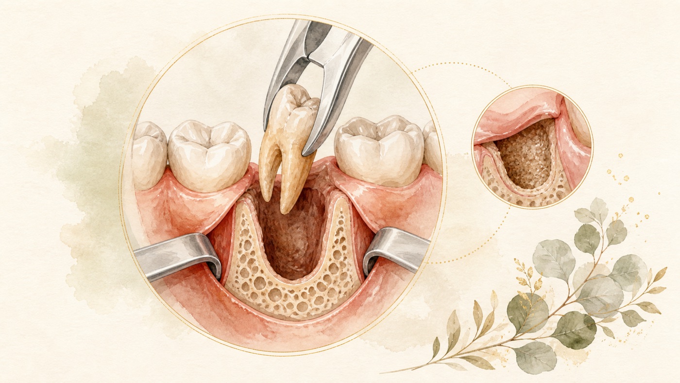
The Complete Guide to Tooth Extraction and Oral Surgery: the spectrum of management, the decision framework and the physiology of healing
What oral surgery deals with inside the dental system, which categories the management spectrum for extraction runs through — from observation and follow-up to coronectomy, which four dimensions the decision to extract or retain looks at, the three-phase physiology of healing in an extraction wound, and the general principles of post-operative care and risk disclosure. This article gives only the domain map and the cross-question framework; the answers to specific questions live in the corresponding canonical cards and are not rewritten here.
The Complete Guide to Tooth Extraction and Oral Surgery: the spectrum of management, the decision framework and the physiology of healing
A direct answer in under 60 words
Removing wisdom teeth belongs to oral surgery [F1]; the spectrum runs from observation and follow-up to coronectomy in high-risk cases [F6][F14]; healing divides into inflammatory, proliferative and remodelling phases [F17]. Where disease is present there is an indication [F8]; where there is none, two publications reach inconsistent conclusions [F8][F3].
Scope: This article is general oral-health education based on international literature. It does not address any country's insurance or regulations; consult local rules for care pathways and costs. Every medical statement in this article is anchored to international peer-reviewed literature and to a professional society's clinical practice guideline, and each F-Unit is labelled geo: universal; the statutes, insurance coverage and fee systems of any individual country fall outside the scope of this article — see the corresponding local canonical cards [F33]. The under-60-word answer above is a domain-level summary, not anyone's treatment plan; the actual treatment approach and its outcome vary from person to person and must be assessed by a dentist according to individual circumstances [F31].
Where this article sits: what it answers and what it does not
The dental content system has two layers. One is the canonical card, one question to a card, answering the concrete questions patients ask — for example “how long does the socket left by an extraction take to heal” or “how soon after an extraction can I eat”. The other is the domain authority article, the layer this article belongs to, which handles the gaps between the cards: what this domain actually covers, which categories of management exist, which dimensions a decision has to look at, and what the body is in fact doing inside the wound [F32].
So the way this article is written is: it gives you the map and the framework, not a personalised answer. Any concrete question that belongs to a particular canonical card is covered here in one sentence with a pointer, and that card's content is not rewritten [F32]. The “downstream links” section at the end lists every canonical card in this domain.
The other thing to state up front is that all the categories and phases in this article are structures for communication, not diagnostic tools, and not anyone's treatment plan [F31]. The actual treatment approach and its outcome vary from person to person and must be assessed by a dentist according to individual circumstances.
1. What oral surgery deals with inside dentistry: a map of the management spectrum
To start with a sense of scale. The Spanish Society of Oral Surgery (SECIB) opens its clinical practice guideline by stating that the removal of third molars (in this article “wisdom teeth” and “third molars” refer to the same group of teeth; for the terminology correspondence see [F36]) is the most frequently performed surgical procedure in the field of oral surgery [F1]. Put another way, among the procedures of oral surgery, the one performed most often is the removal of those few teeth at the back of the arch [F1]. That source gives only this ranking statement and attaches no incidence figure or case-count statistics, so this article derives from it no estimate of share and no case count [F1].
Laid out, the options that actually arise in clinical practice are not a binary choice between “extract” and “do not extract” but a spectrum [F31]:
- Observation and regular follow-up: on the premise that the evidence is insufficient, the Cochrane systematic review states that if the decision is made to retain these teeth, clinical assessment at regular intervals to prevent undesirable outcomes is advisable [F6]. So “leave it alone for now” is, in the literature, a formal option that comes with follow-up attached, not “nothing needs to be done”.
- Simple extraction: relative to the next category, this denotes the situation in which the tooth can be removed without raising a flap, removing bone or other surgical steps; this article defines it by that contrast and sets up no separate clinical classification [F31].
- Surgical extraction: the indications Cochrane lists for the surgical removal of mandibular wisdom teeth are alleviation of local pain, swelling and trismus, and also the prevention of spread of infection [F11]. This is the category that involves surgical steps such as flap design and the method of bone removal — the nine sets of comparisons made in that review include the envelope versus triangular flap designs and the difference between the lingual split technique and a surgical handpiece for bone removal [F35] — while its overall conclusion is that the certainty of the evidence for each comparison was low or very low and that no firm recommendation on technique could be given [F13].
- Coronectomy (high-risk cases only): a systematic review of systematic reviews defines coronectomy as an alternative to complete third molar extraction intended to reduce the risk of inferior alveolar nerve injury; that review's conclusion positions it as a viable alternative in high-risk cases, not as a generally co-equal option [F14].
The reason for laying out this spectrum first is that everything the later sections discuss — post-operative symptoms, the healing pathway, follow-up needs, the drivers of cost — differs between the categories, and each section labels its own literature basis item by item [F31]. This article does not judge which category you fall into; that has to be determined by a dentist on the basis of imaging and clinical examination [F31].
2. Anatomical background of the posterior region: which surgical reasons the literature records for the back teeth
“Which teeth are the molars, and how do you read tooth-position numbers” is a separate question, already handled by its own canonical card (KM-DENTAL-21), and is not rewritten here [F32]. What is added here is only the layer seen from oral surgery: which surgical-level reasons relating to those few teeth at the back of the arch the literature records. Each of the three items below carries its own literature basis; this article derives from them no epidemiological claim about distribution, share or “where the problems concentrate” [F31].
- It may coexist with disease. The pathological changes associated with impacted wisdom teeth listed by the Cochrane review include pericoronitis, root resorption, gum and alveolar bone disease (periodontitis), caries, and the development of cysts and tumours [F4]. Another systematic review likewise records that the disease burden associated with impacted wisdom teeth is well documented, covering non-restorable caries, fracture, infection, periodontal disease, repeated pericoronitis, cysts and tumours [F9].
- It interacts with its neighbour. A secondary outcome of the same Cochrane review indicates that retaining asymptomatic disease-free impacted wisdom teeth may be associated with an increased risk of periodontitis affecting the adjacent second molar in the long term, but the authors themselves rated that evidence as very low-certainty [F5]. The scope of this signal has to be remembered alongside it: it comes from a subgroup of 416 healthy male participants (aged 24 to 84 years) within a single prospective cohort study, and that study was judged to be at serious risk of bias, so it cannot be taken as a risk estimate for the general population [F5]. Conversely, the systematic review cited above records that extraction of teeth with signs and/or symptoms of periodontal disease improved periodontal health at the distal surface of the second molar [F10].
- It sits close to important anatomical structures. The reason the coronectomy technique exists at all is to reduce the risk of inferior alveolar nerve injury [F14]; and Cochrane also lists trigeminal nerve injury as one of the less frequent complications of mandibular wisdom tooth surgery [F12].
A commonly asked imaging question can be answered here in passing: does adding cone-beam computed tomography prevent nerve injury? A systematic review that included 7 randomised controlled trials set both its research question and its inclusion criteria within high-risk mandibular third molar surgery; its conclusion is that cone-beam computed tomography does not routinely translate into a reduced incidence of nerve injury in that population, rated as moderate-quality evidence [F16]. The same review separately records, on low-quality evidence, that only a single study showed a consequent change in the surgical approach once the extra imaging was obtained [F16]. The scope of that conclusion has to be remembered alongside it: it speaks to “whether adding this imaging in cases that are already high-risk reduces the incidence of nerve injury”, not to “imaging is of no use” [F31].
3. Extract or retain: the domain-level decision framework
The boundary of this section first: “does my wisdom tooth have to come out” is the concrete question to be answered by a canonical card currently in production (supplementary card S01); this article neither answers it in that card's place nor expands on it [F32]. What this section provides is a domain-level framework — the four dimensions a responsible decision looks at.
Dimension one: is disease present
A systematic review that performed no meta-analysis puts its conclusion plainly: where an impacted wisdom tooth is accompanied by disease, extraction is indicated whether or not it is symptomatic; conversely, in the absence of infection or other associated disease conditions, extraction is not indicated [F8]. But this line cannot be read on its own: for the “asymptomatic and disease-free” group, the authors' conclusion in the Cochrane systematic review is that the available evidence is insufficient to determine whether such teeth should be removed or retained [F3], and the two publications word the same population differently; this article sets them side by side and does not adjudicate between them [F31].
Dimension two: how far the available evidence can go
For impacted wisdom teeth that are “asymptomatic and disease-free”, the authors' conclusion in the Cochrane systematic review is that the available evidence is insufficient to determine whether these teeth should be removed or retained [F3]. That sentence is worth taking as it stands — it does not mean “so do not extract”, nor “so do extract”, but “this question currently has no high-certainty evidence-based answer”. The signal noted above about the risk of periodontitis in the second molar was rated by the authors themselves as very low-certainty, and its data come only from a subgroup of healthy men [F5].
Dimension three: individual conditions and timing
The same Cochrane review notes in its background section that when surgical removal is performed in older people, the risk of postoperative complications, pain and discomfort is increased [F7]. That sentence comes from the review's background narrative rather than from its statistical results, so it can serve only as a description of clinical context and must not be taken as the conclusion “therefore extract earlier” — a point on which this article deliberately does not extrapolate.
Dimension four: the options are not a binary choice
As with the management spectrum above, regular follow-up is a formally existing option [F6]; and in that review's conclusion coronectomy is positioned as a viable alternative in high-risk cases [F14]. Taking coronectomy as the example: a systematic review of systematic reviews analysed 6 systematic reviews, 5,896 subjects and 7,913 successful coronectomies that did not require immediate tooth extraction; the overall re-intervention rate was 4.45%, with timing ranging from 6 months to 10 years and a mean of 10.4 months [F15] (the original reports rates and does not report the absolute number of re-interventions; this article performs no conversion, so that a derived figure is not read as the source's own data). The percentages that follow have two different denominators and have to be read separately: with “re-intervention cases” as the denominator come the proportions for the causes of re-intervention — root exposure at 16.76% first, followed by infection at 4.55% and pain at 2.84% [F15]; with “coronectomy cases” as the denominator come root migration (12.20%, common) and inferior alveolar nerve injury (0.76%, rare) [F15]. (The original reports rates only and does not report the absolute number of cases for each item; this article performs no conversion.) The authors also warn that root migration and root exposure require long-term follow-up [F15]. All the percentages above are statistics for that pooled population, not individual risks, and should not be applied as they stand to everyone who undergoes this procedure [F31]. Every category has its own price to pay and its own thing to watch.
So how is the decision made?
The direction Cochrane gives is shared decision-making: in the absence of evidence, patient values should be considered and clinical expertise used to guide shared decision-making; if the decision is made to retain these teeth, clinical assessment at regular intervals to prevent undesirable outcomes is advisable [F6]. That is precisely why this article does not make the decision for you — it needs your imaging, your medical history, and a discussion between you and your dentist.
4. How an extraction wound heals: the three-phase physiological background
This section is about what the body is doing inside the socket, not how many days it takes. The stage-by-stage timeline, the observation window for dry socket and the criteria for going back to the clinic fall within the scope of canonical card KM-DENTAL-01 and are not rewritten here [F32].
Three sequential and overlapping phases
A periodontology review describes it as follows: tooth extraction induces a series of complex and integrated local changes within the surrounding hard and soft tissues; the socket-healing process may be divided into three sequential, and frequently overlapping, phases — inflammatory, proliferative, and modeling/remodeling [F17].
The tissue in the socket is replaced in sequence
The histological sequence described by another systematic review is: an initial formation of a blood clot, which is replaced with granulation tissue and subsequently with a provisional connective tissue matrix; spontaneous healing ends with the socket filling with woven bone, which is gradually replaced with lamellar bone and bone marrow [F19].
There are two points in this sequence worth remembering. First, the blood clot is the starting point of the whole sequence [F19]; this article lists it as mechanistic background for understanding the principles of post-operative wound care — the source itself only describes the histological sequence and does not address any post-operative instruction, so what should actually be done is covered in the corresponding canonical card [F31][F32]. Second, the end point of the sequence is woven bone being gradually replaced by lamellar bone and bone marrow [F19] — in other words, the changes inside the socket fall histologically into several stages rather than being a single event [F19]. As for the general healing of oral soft tissue, a narrative review whose main subject is soft tissue wound healing around teeth and dental implants lists, among its results, the item “oral wounds follow a similar pattern”; that sentence does not specify in the abstract what the similarity is being compared with, and the review is not a monograph on the extraction socket, so this article lists it only as soft-tissue-level background and derives from it no timeline and no criterion for the extraction socket [F20].
The bone does not grow back as it was
The same periodontology review records that during healing there is greater bone resorption at the buccal aspect than at the lingual/palatal counterpart, and a larger amount of alveolar bone reduction in the molar region [F18]; the review states explicitly that this passage is a summary of “clinical and experimental studies”, that is, its evidence base does not come entirely from human clinical research [F18]. Its conclusion is worded as follows: extraction should be performed in the knowledge that ridge reduction will follow, and further clinical steps should be considered to compensate for this, when considering future options for tooth replacement [F18]. As for “can a molar be left unreplaced after extraction, and how would it be replaced”, that is a separate question belonging to the tooth-replacement domain; see canonical card KM-DENTAL-34.
Where dry socket sits on this map
Cochrane lists dry socket (alveolar osteitis) alongside infection and trigeminal nerve injury as less frequent complications of mandibular wisdom tooth surgery [F12]; another systematic review describes it as one of the most common complications developing after the extraction of a permanent tooth and states that its prevention is more effective than its treatment [F22]. The predisposing factors compiled by that same review are patient age, a history of previous infection, and the difficulty of the extraction; as to whether smoking, gender or menstrual cycles constitute risk factors, the review records that there is no consensus [F21]. The time window for dry socket, its typical presentation and the criteria for returning to the clinic are covered in KM-DENTAL-01 and are not rewritten here [F32].
5. General principles of post-operative care: the principle, the mechanism and how strong the evidence is
This section gives no number of days; what it gives is the logic behind each class of measure, and how solid its evidence actually is. The specific questions “how soon after an extraction can I eat and drink” and “how soon after an extraction can I brush” each have their own canonical card (KM-DENTAL-04, KM-DENTAL-40) and are not rewritten here [F32].
The symptoms themselves are expected
Cochrane describes mandibular wisdom tooth surgery as follows: surgery is commonly associated with short-term postoperative pain, swelling and trismus [F12]. Treating that as “something expected that needs to be managed” rather than as “something has gone wrong” is the premise for understanding every post-operative instruction that follows [F31].
Antibiotics are not the default
- The result of the Cochrane systematic review is that, compared with placebo, prophylactic antibiotics may reduce the risk of postsurgical infectious complications in people undergoing third molar extractions by approximately 66%, and may reduce the risk of dry socket by 34%; both are low-certainty evidence [F24].
- A relative risk reduction has to be read together with the absolute figures: the same review gives the corresponding numbers needed to treat in the same sentence — 19 people (95% confidence interval 15 to 34) need to be treated with antibiotics to prevent one infection following extraction of impacted wisdom teeth; 46 people (95% confidence interval 29 to 62) need to take antibiotics to prevent one case of dry socket [F24]. On adverse effects, the review records them as mild and transient, with a risk ratio of 1.46 (95% confidence interval 0.81 to 2.64), rated very low-certainty evidence [F24].
- But the same review states its boundary of applicability clearly: the great majority of the included trials involved healthy participants undergoing surgical removal of impacted third molars, so the results of this review may not be generalisable to all people undergoing tooth extractions; the authors further note that, because of the increasing prevalence of bacteria resistant to antibiotic treatment, clinicians should evaluate whether and when to prescribe prophylactic antibiotic therapy on the basis of each individual patient's clinical condition and risk of infectious complications [F25].
- A 2025 systematic review records that antibiotic use significantly reduces infection risk and dry socket incidence compared with no prescription, but its authors explicitly require in the conclusion that their administration be carefully considered to balance benefits against potential risks [F26]. Its evidential independence needs attention: the “studies” it included are themselves systematic reviews (that is, it is a review of reviews), and its question was likewise confined to healthy patients, so it should not be read as an independent replication of the Cochrane result above [F26].
- One of the very sources cited in this article reaches the opposite conclusion: the systematic review used above for the predisposing factors of dry socket states in its conclusion section that antibiotic prescription does not avoid postoperative complications after lower third molar surgery [F37]. In other words, the conclusions in the literature on this question are not consistent; this article sets them side by side and does not adjudicate between them [F31].
In other words, “should antibiotics be taken for an extraction” is, in the literature, a question of case-by-case assessment, not a general rule [F25]. Whether they are prescribed, and for how long, is determined by a dentist according to your health status and your risk of infection; this article gives no medication advice [F31].
Local measures have evidence behind them, but they are still not instructions
A meta-analysis carried out by a single author reports that topical chlorhexidine gel, compared with placebo, reduces the incidence of alveolar osteitis after mandibular third molar extraction, with a pooled risk ratio of 0.43 (95% confidence interval 0.32 to 0.58), and that no adverse reaction was reported in the studies [F23]. This piece of evidence has to be read in the same way as in the section above: 0.43 is a relative risk, and this site retrieved only the abstract of that paper, which gives no corresponding absolute event rate, so this article performs no conversion into absolute figures; in addition, the PubMed record for that paper carries two published comments (J Am Dent Assoc 2017;148(9):e133 and Evid Based Dent 2018;19(1):16-17) whose content this site did not retrieve, so their views did not enter this article's appraisal of quality [F23]. This is a verifiable population-level result, not an instruction for you to buy or use any product on your own — whether any topical medicine is used has to be decided by a dentist [F31].
Pain control
Pain relief after the removal of lower wisdom teeth has already been addressed by a Cochrane systematic review comparing the effects of different analgesics; that review included 7 studies with a total of 2,241 participants [F27]. This article deliberately writes no further than “comparative research at systematic review level exists on this question” and lists no drug name and no dose [F31]: the choice of drug, its dose and its contraindications fall within the scope of a clinician's prescription and are to be decided by a physician or a dentist [F31].
If you are taking medication, say so before surgery
- Anticoagulation: a systematic review has specifically examined bleeding outcomes for dental extraction under uninterrupted oral anticoagulant therapy; on the difference in bleeding risk between the two classes of anticoagulant it compared, the authors themselves state that the current evidence is of very low quality and should be interpreted with caution, and note that data on individual drugs are scarce so that the difference in bleeding risk between them cannot at this point be elucidated [F30]. This article therefore makes no comparison between drugs and lists this only as information to be given before surgery [F31] — tell your dentist what medication you are taking, and do not stop it or change the dose on your own [F31].
- Antiresorptive and antiangiogenic medicines: see the risk factors section below [F28].
When oral hygiene is resumed
On “how soon after an extraction can brushing be resumed”, this site queried PubMed E-utilities with three search strings on this occasion (resume toothbrushing after tooth extraction randomized/oral hygiene instructions after tooth extraction brushing timing trial/postoperative oral hygiene third molar surgery toothbrushing randomized controlled trial); all three returned a hit count of 0, so no randomised controlled trial that could be cited directly was obtained, and this article therefore gives no number of days [F34]. In practice, follow the post-operative instructions your own dentist gives you; the write-up of that question is in KM-DENTAL-40 [F32].
6. Overview of red flags, and a checklist for before the appointment
The types of post-operative adverse event listed in the literature
The following are the categories of event enumerated in the literature; their purpose is to help you know what to report, and they are not criteria for self-diagnosis. If any of them appears, go back to the clinic and have a dentist examine you [F31].
- Infection, dry socket (alveolar osteitis), trigeminal nerve injury — listed by Cochrane as less frequent complications of mandibular wisdom tooth surgery [F12].
- Bleeding-related events — a specifically studied outcome measure in people taking anticoagulants [F30].
- Osteonecrosis of the jaw — medication-related osteonecrosis of the jaw is described as a severe adverse reaction, and dentoalveolar surgery is considered a common predisposing event [F28].
- Root migration and root exposure after coronectomy — the authors explicitly require long-term follow-up [F15].
A checklist you can prepare before the appointment
- A complete medication list: in particular the medicines commonly used in the treatment of cancer or osteoporosis (the starting point for assessing the risk of osteonecrosis of the jaw) [F28], and anticoagulants [F30].
- A history of previous infection and a record of flare-ups in this particular tooth: a history of previous infection is listed as one of the predisposing factors for dry socket [F21].
- Imaging records: if imaging has already been taken elsewhere, bring it with you; for high-risk mandibular third molar surgery, adding cone-beam computed tomography does not routinely reduce the incidence of nerve injury (moderate-quality evidence) [F16].
- The questions to ask: which category of the spectrum you fall into, what each option costs you, and how often you would need to return if you choose to retain and monitor — this is exactly where the shared decision-making Cochrane recommends takes place [F6].
7. What costs are made of, and what makes them vary
This article reports no price and discusses no country's insurance coverage or fee system [F33]. What is written here is only the universal-level question of “what costs are made of and what drives them”.
Cost-effectiveness is not an off-the-record topic: the 17 PICO questions evaluated in the SECIB guideline cover the indications, prognosis, diagnosis and cost-benefit relationship of third molar extraction [F2]. That is to say, it is one of the aspects formally taken into assessment at professional level.
In practice, the main variables driving differences in cost are as follows (the list below is compiled by this article from the literature cited above; it is not a fee schedule for any region) [F33]:
- Which category of the spectrum it falls into: observation and follow-up, simple extraction, surgical extraction (involving steps such as raising a flap, removing bone and sectioning the tooth) and coronectomy differ in the time, the instruments and the level of technique they require [F11][F14].
- The depth of pre-operative assessment: whether advanced imaging is needed (for the nerve injury outcome in high-risk mandibular third molars, its benefit has a boundary, as set out above) [F16].
- The number and length of follow-ups: for example, the timing of re-intervention after coronectomy can span from 6 months to 10 years, and root migration and root exposure require long-term follow-up [F15].
- Whether complications arise and require additional management [F12].
For local fee systems, the scope of insurance coverage and actual amounts, see the corresponding local canonical card (TW) [F33].
Risk factors (indications, adverse effects, contraindications)
Indications (as the literature words them)
- Where disease is present: when an impacted wisdom tooth is accompanied by disease, extraction is indicated whether or not it is symptomatic [F8].
- The indications for the surgical removal of mandibular wisdom teeth: alleviation of local pain, swelling and trismus, and also the prevention of spread of infection [F11].
Situations in which extraction is not indicated
- In the absence of infection or other associated disease conditions, extraction is not indicated [F8].
- For asymptomatic disease-free impacted wisdom teeth, the available evidence is insufficient to determine whether they should be removed or retained; such cases are to be handled by shared decision-making, and if the teeth are retained, clinical assessment at regular intervals is needed [F3][F6].
- The two items above come from different sources and word the “asymptomatic and disease-free” population inconsistently (one writes “extraction is not indicated”, the other writes “the evidence is insufficient to determine”); this article sets them side by side and does not adjudicate between them [F8][F3][F31].
Possible adverse effects and complications
- Common and short-term: postoperative pain, swelling and trismus [F12].
- Less frequent: infection, dry socket (alveolar osteitis), trigeminal nerve injury [F12].
- Predisposing factors for dry socket: age, a history of previous infection, the difficulty of the extraction; on smoking, gender and menstrual cycles there is no consensus [F21].
- Specific to coronectomy: root exposure (16.76%), root migration (12.20%) and the need for re-intervention (4.45% overall); inferior alveolar nerve injury is rare (0.76%) [F15]. These are pooled values from 6 systematic reviews and 7,913 successful coronectomies, not individual risks [F15].
- At the level of technique: after examining 62 trials, Cochrane states that it is unable to make firm recommendations to surgeons on their technique, the certainty of the evidence for each comparison being low or very low [F13].
Groups needing particular assessment, and contraindication considerations
- People currently taking antiresorptive or antiangiogenic medicines: medication-related osteonecrosis of the jaw (MRONJ) is described as a severe adverse reaction experienced by some individuals taking medicines commonly used in the treatment of cancer and osteoporosis, and dentoalveolar surgery is considered a common predisposing event [F28]. The same Cochrane review also states that, for patients on antiresorptive therapy who are to undergo dentoalveolar surgery, there is insufficient evidence to either claim or refute a benefit of the preventive measures tested [F29]. What this means: disclose your medication history in full before surgery, and have your dentist and your treating physician assess it together.
- People currently taking anticoagulants: bleeding outcomes for extraction under uninterrupted anticoagulant therapy have been examined in a systematic review, but the evidence is of very low quality and should be interpreted with caution [F30].
- People who are immunocompromised or have systemic disease: Cochrane states explicitly that the population included in its antibiotics review was healthy participants, that the results may not be generalisable to all people undergoing tooth extractions, and that assessment has to be made patient by patient in the clinic [F25].
The above is a general oral-health education summary, not a diagnosis and not a treatment recommendation. The actual treatment approach and its outcome vary from person to person and must be assessed by a dentist according to individual circumstances [F31].
Every clinical statement in this article is mapped line by line to its cited source (see the sources and evidence chain below). It has not been clinically reviewed by a licensed practitioner. This is health information, not individual advice; assessment by a clinician is required.
FAQ
- Q1. Does an extraction actually count as “surgery”?
- **In the classification used by the literature, tooth extraction is a surgical procedure: Cochrane compares techniques directly under the title of surgical removal of mandibular wisdom teeth and sets out the indications and the complications [F11][F12]; and the SECIB guideline describes third molar extraction as the most frequently performed surgical procedure in the field of oral surgery [F1].** That is also why the literature, when it discusses this, lists the indications and the complications together [F11][F12]; and where the evidence is insufficient, what Cochrane offers is shared decision-making that takes in patient values, with clinical assessment at regular intervals if the decision is to retain [F6].
- Q1. 抜歯はそもそも「手術」にあたるのですか? — **文献の分類では、歯の抜去は外科処置に属します:Cochrane は「下顎智歯の外科的抜去」をそのまま表題として各種の術式を比較し、その適応と合併症を挙げています [F11][F12];また SECIB ガイドラインは、第三大臼歯の抜去を口腔外科の領域において実施頻度が首位を占める外科処置と記述しています [F1]。** だからこそ文献はこのことを論じるとき、その適応と合併症を同時に挙げるのです [F11][F12];そしてエビデンスが不十分な状況で Cochrane が示している進め方は、患者の価値観を組み入れた共有意思決定であり、保存すると決めた場合には定期的な臨床評価を行うことです [F6]。
- Q1. Does an extraction actually count as “surgery”? — **In the classification used by the literature, tooth extraction is a surgical procedure: Cochrane compares techniques directly under the title of surgical removal of mandibular wisdom teeth and sets out the indications and the complications [F11][F12]; and the SECIB guideline describes third molar extraction as the most frequently performed surgical procedure in the field of oral surgery [F1].** That is also why the literature, when it discusses this, lists the indications and the complications together [F11][F12]; and where the evidence is insufficient, what Cochrane offers is shared decision-making that takes in patient values, with clinical assessment at regular intervals if the decision is to retain [F6].
- Q2. My wisdom tooth is not uncomfortable — does it still need to come out?
- **This question currently has no high-certainty evidence-based answer. The authors' conclusion in the Cochrane systematic review is that, for the class of impacted wisdom teeth that are neither symptomatic nor associated with disease, the available evidence is insufficient to determine whether they should be removed or retained [F3]; what the literature therefore offers is shared decision-making that takes in patient values, with clinical assessment at regular intervals if the decision is to retain [F6].** The judgement about your own particular tooth falls within the scope of supplementary canonical card S01 (in production) and is not answered here [F32].
- Q2. 智歯に不快な症状がなくても、抜く必要はありますか? — **このテーマには現時点で高い確実性の実証的な答えがありません。Cochrane システマティックレビューの著者の結論はこう書かれています:症状がなく、病変もないという類の埋伏智歯について、既存のエビデンスは抜去すべきか保存すべきかを判定するのに不十分である [F3];そのため文献が示す進め方は、患者の価値観を組み入れた共有意思決定であり、保存すると決めた場合には定期的な臨床評価を行うことです [F6]。** あなた自身のその歯についての判断は、補題正典カード S01(制作中)の範囲に属し、本記事は代わりに答えません [F32]。
- Q2. My wisdom tooth is not uncomfortable — does it still need to come out? — **This question currently has no high-certainty evidence-based answer. The authors' conclusion in the Cochrane systematic review is that, for the class of impacted wisdom teeth that are neither symptomatic nor associated with disease, the available evidence is insufficient to determine whether they should be removed or retained [F3]; what the literature therefore offers is shared decision-making that takes in patient values, with clinical assessment at regular intervals if the decision is to retain [F6].** The judgement about your own particular tooth falls within the scope of supplementary canonical card S01 (in production) and is not answered here [F32].
- Q3. Do antibiotics always have to be taken for an extraction?
- **Not by default. The Cochrane result does show that prophylactic antibiotics may reduce postsurgical infectious complications by approximately 66% and reduce the risk of dry socket by 34%, but the same review also gives the corresponding numbers needed to treat — about 19 people need to take antibiotics to prevent one infection, and about 46 people to prevent one case of dry socket; both are low-certainty evidence, and the population included was mostly healthy people undergoing surgery for impacted third molars [F24][F25]. A 2025 review of reviews requires that their administration be carefully considered to balance benefits against potential risks [F26], while another systematic review cited in this article reaches the opposite conclusion, that antibiotic prescription does not avoid postoperative complications after lower third molar surgery [F37].** Whether they are prescribed is assessed patient by patient by a dentist [F25]; this article gives no medication advice [F31].
- Q3. 抜歯には必ず抗菌薬を飲まなければなりませんか? — **既定値ではありません。Cochrane の結果は、予防的抗菌薬が術後感染性合併症を約 66%、ドライソケットのリスクを 34% 低下させうることを示していますが、同じレビューは対応する治療必要数も示しています——約 19 人が服薬して感染 1 例を防げ、約 46 人が服薬してドライソケット 1 例を防げる;二項ともに低い確実性のエビデンスであり、組み入れられた集団の多くは埋伏第三大臼歯の手術を受ける健康な人でした [F24][F25]。2025 年の別のレビューのレビューは、その投与が有益性とリスクを慎重に比較考量すべきだと求めており [F26]、本記事が引用しているもう一篇のシステマティックレビューは「抗菌薬の処方は下顎第三大臼歯手術後の術後合併症を回避しない」という反対の結論に至っています [F37]。** 処方するかどうかは歯科医師が一人ひとり評価します [F25]。本記事は服薬に関する助言を提供しません [F31]。
- Q3. Do antibiotics always have to be taken for an extraction? — **Not by default. The Cochrane result does show that prophylactic antibiotics may reduce postsurgical infectious complications by approximately 66% and reduce the risk of dry socket by 34%, but the same review also gives the corresponding numbers needed to treat — about 19 people need to take antibiotics to prevent one infection, and about 46 people to prevent one case of dry socket; both are low-certainty evidence, and the population included was mostly healthy people undergoing surgery for impacted third molars [F24][F25]. A 2025 review of reviews requires that their administration be carefully considered to balance benefits against potential risks [F26], while another systematic review cited in this article reaches the opposite conclusion, that antibiotic prescription does not avoid postoperative complications after lower third molar surgery [F37].** Whether they are prescribed is assessed patient by patient by a dentist [F25]; this article gives no medication advice [F31].
- Q4. Can wisdom tooth removal injure a nerve? Can that be avoided in advance?
- **Trigeminal nerve injury is one of the less frequent complications listed [F12]; coronectomy exists precisely as an alternative technique for reducing the risk of inferior alveolar nerve injury, and that review positions it as a viable alternative in high-risk cases, with a pooled nerve injury rate of 0.76% [F14][F15]. As for imaging: a systematic review including 7 randomised controlled trials indicates that, in high-risk mandibular third molar surgery, adding cone-beam computed tomography does not routinely reduce the incidence of nerve injury (moderate-quality evidence) [F16].**
- Q4. 智歯を抜くと神経を傷つけますか? あらかじめ避けられますか? — **三叉神経損傷は、挙げられているあまり多くない合併症の一つです [F12];歯冠切除術はまさに下歯槽神経損傷のリスクを下げるために存在する代替術式であり、当該レビューはこれを高リスク症例における実行可能な代替案と位置づけていて、その統合された神経損傷率は 0.76% でした [F14][F15]。画像については:7 篇のランダム化比較試験を組み入れたあるシステマティックレビューが、高リスクの下顎第三大臼歯手術において、コーンビーム CT を追加しても神経損傷の発生率は日常的に低下しないことを指摘しています(中等度の質のエビデンス)[F16]。**
- Q4. Can wisdom tooth removal injure a nerve? Can that be avoided in advance? — **Trigeminal nerve injury is one of the less frequent complications listed [F12]; coronectomy exists precisely as an alternative technique for reducing the risk of inferior alveolar nerve injury, and that review positions it as a viable alternative in high-risk cases, with a pooled nerve injury rate of 0.76% [F14][F15]. As for imaging: a systematic review including 7 randomised controlled trials indicates that, in high-risk mandibular third molar surgery, adding cone-beam computed tomography does not routinely reduce the incidence of nerve injury (moderate-quality evidence) [F16].**
- Q5. I am taking osteoporosis medication or an anticoagulant — can I have a tooth out?
- **This needs pre-operative assessment and cannot be judged on your own: medication-related osteonecrosis of the jaw is described as a severe adverse reaction to medicines commonly used for cancer and osteoporosis, with dentoalveolar surgery considered a common predisposing event [F28], and the available evidence is still insufficient to either claim or refute a benefit of the preventive measures tested [F29]; on anticoagulation, the evidence from the research on extraction under uninterrupted therapy is likewise of very low quality [F30].** Disclose your medication history in full and have your dentist and your treating physician assess it together; do not stop your medication or change the dose on your own [F31].
- Q5. 骨粗鬆症や抗凝固の薬を飲んでいますが、抜歯できますか? — **これは術前の評価が必要であり、自分で判断することはできません:薬剤関連顎骨壊死は、がんや骨粗鬆症の治療でよく用いられる薬の重篤な有害反応と記述されており、歯槽外科手術はよくある誘発事象とみなされています [F28]。そして既存のエビデンスは、各種の予防措置の有益性を支持することも否定することもできていません [F29];抗凝固については、治療を中断せずに抜歯した場合の出血の研究もそのエビデンスの質は非常に低いものです [F30]。** 服薬歴を完全に伝え、歯科医師と主治医が共同で評価してください。自己判断で中止したり用量を変えたりしないでください [F31]。
- Q5. I am taking osteoporosis medication or an anticoagulant — can I have a tooth out? — **This needs pre-operative assessment and cannot be judged on your own: medication-related osteonecrosis of the jaw is described as a severe adverse reaction to medicines commonly used for cancer and osteoporosis, with dentoalveolar surgery considered a common predisposing event [F28], and the available evidence is still insufficient to either claim or refute a benefit of the preventive measures tested [F29]; on anticoagulation, the evidence from the research on extraction under uninterrupted therapy is likewise of very low quality [F30].** Disclose your medication history in full and have your dentist and your treating physician assess it together; do not stop your medication or change the dose on your own [F31].
Medical notice This article is provided for health education and medical information purposes. It is not a solicitation for medical services and does not constitute diagnosis or treatment advice. Actual treatment methods and outcomes vary between individuals and require evaluation by a dentist; every treatment has its own indications, limitations and possible risks. If you have related symptoms or treatment needs, please book a consultation for evaluation by a dentist.
Source anchors
- clinical_guideline · Sánchez-Garcés MÁ, et al. Diagnosis and indications for the extraction of third molars - The SECIB clinical practice guideline. Med Oral… · https://pubmed.ncbi.nlm.nih.gov/38368528/ · 在 IDAEO 的其他引用
- peer_reviewed · Ghaeminia H, et al. Surgical removal versus retention for the management of asymptomatic disease-free impacted wisdom teeth. Cochrane… · https://pubmed.ncbi.nlm.nih.gov/32368796/ · 在 IDAEO 的其他引用
- peer_reviewed · Peñarrocha-Diago M, et al. Indications of the extraction of symptomatic impacted third molars. A systematic review. J Clin Exp Dent. 2021 Mar… · https://pubmed.ncbi.nlm.nih.gov/33680330/ · 在 IDAEO 的其他引用
- peer_reviewed · Bailey E, et al. Surgical techniques for the removal of mandibular wisdom teeth. Cochrane Database Syst Rev. 2020 Jul 26;7(7):CD004345. ·… · https://pubmed.ncbi.nlm.nih.gov/32712962/ · 在 IDAEO 的其他引用
- peer_reviewed · Di Spirito F, et al. Re-Intervention Rate, Timing, and Indications Following Coronectomy of the Mandibular Third Molar: A Systematic Review… · https://pubmed.ncbi.nlm.nih.gov/40507640/ · 在 IDAEO 的其他引用
- peer_reviewed · Robbins J, et al. Does the addition of cone-beam CT to panoral imaging reduce inferior dental nerve injuries resulting from third molar… · https://pubmed.ncbi.nlm.nih.gov/36329417/ · 在 IDAEO 的其他引用
- peer_reviewed · Araújo MG, Silva CO, Misawa M, Sukekava F. Alveolar socket healing: what can we learn? Periodontol 2000. 2015 Jun;68(1):122-34. · PMID… · https://pubmed.ncbi.nlm.nih.gov/25867983/ · 在 IDAEO 的其他引用
- peer_reviewed · Di Stefano DA, et al. A comparison between anorganic bone and collagen-preserving bone xenografts for alveolar ridge preservation: systematic… · https://pubmed.ncbi.nlm.nih.gov/35821286/ · 在 IDAEO 的其他引用
- peer_reviewed · Sculean A, Gruber R, Bosshardt DD. Soft tissue wound healing around teeth and dental implants. J Clin Periodontol. 2014 Apr;41 Suppl… · https://pubmed.ncbi.nlm.nih.gov/24641001/ · 在 IDAEO 的其他引用
- peer_reviewed · Taberner-Vallverdú M, Sánchez-Garcés MÁ, Gay-Escoda C. Efficacy of different methods used for dry socket prevention and risk factor analysis:… · https://pubmed.ncbi.nlm.nih.gov/29053647/ · 在 IDAEO 的其他引用
- peer_reviewed · Teshome A. The efficacy of chlorhexidine gel in the prevention of alveolar osteitis after mandibular third molar extraction: a systematic… · https://pubmed.ncbi.nlm.nih.gov/28526078/ · 在 IDAEO 的其他引用
- peer_reviewed · Lodi G, et al. Antibiotics to prevent complications following tooth extractions. Cochrane Database Syst Rev. 2021 Feb 24;2(2):CD003811. ·… · https://pubmed.ncbi.nlm.nih.gov/33624847/ · 在 IDAEO 的其他引用
- peer_reviewed · De Angelis N, et al. Antibiotic Prescription for the Prevention of Postoperative Complications After Third-Molar Extractions: A Systematic… · https://pubmed.ncbi.nlm.nih.gov/40136735/ · 在 IDAEO 的其他引用
- peer_reviewed · Bailey E, Worthington H, Coulthard P. Ibuprofen and/or paracetamol (acetaminophen) for pain relief after surgical removal of lower wisdom… · https://pubmed.ncbi.nlm.nih.gov/24762895/ · 在 IDAEO 的其他引用
- peer_reviewed · Beth-Tasdogan NH, et al. Interventions for managing medication-related osteonecrosis of the jaw. Cochrane Database Syst Rev. 2022 Jul… · https://pubmed.ncbi.nlm.nih.gov/35866376/ · 在 IDAEO 的其他引用
- peer_reviewed · Hua W, Huang Z, Huang Z. Bleeding Outcomes After Dental Extraction in Patients Under Direct-Acting Oral Anticoagulants vs. Vitamin K… · https://pubmed.ncbi.nlm.nih.gov/34776943/ · 在 IDAEO 的其他引用
- 本文證據鏈:逐條出處、行號與原文引句 · https://km.idaeo.ai/reports/dental-pillar-oral-surgery-evidence · 在 IDAEO 的其他引用
Cite this article
Lucy・《The Complete Guide to Tooth Extraction and Oral Surgery: the spectrum of management, the decision framework and the physiology of healing》・IDAEO 知識庫・2026-08-13・https://km.idaeo.ai/post/dental/pillar-oral-surgeryUpdated 2026-08-27