

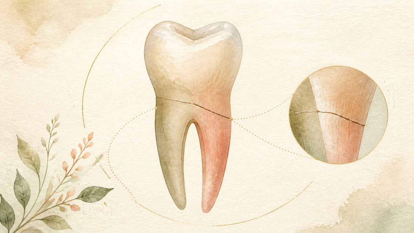
牙根斷裂會怎樣?一定要拔牙嗎?|證據鏈
本頁是〈牙根斷裂會怎樣?一定要拔牙嗎?〉的事實單元帳:每一條主張對應的來源、證據等級、地域與期間、原文引句與限制,逐條攤開。
牙根斷裂會怎樣?一定要拔牙嗎?|證據鏈
F-Units(事實單元帳)
- F1|本題選題依據=GSC 全量對帳:查詢詞「牙根斷裂」於之 web 明細共 2,753 列,曝光加總 19,322、點擊 594(2026-08-06 以 awk 對 `gsc-full-20260804/__web__full.tsv` 重跑)|來源 #16|confidence=verified|basis=internal_dataset|period=GSC 保留窗(2025-03-22 起)|geo: TW|caveat:曝光為屬性級數字、非去重流量;本欄為內部數據、非醫學宣稱,發布轉檔時整條剝除。
- F2[結構性整理]|本卡的三路分流骨幹(牙冠/牙尖斷裂、外傷性橫向或斜向根折、垂直性牙根斷裂)與「冠根斷裂為交界類型」的編排、「先分清哪一種比先問要不要拔重要」的定位、checklist 題目設計、紅旗清單的排序與家用觀察語言的轉寫,以及與 KM-DENTAL-50/33/30/18/35 的同族分工聲明,均為本站依 F3 至 F29 文獻整理的就醫溝通結構|來源 #18|confidence=n/a|basis=editorial_framework|period=2026-08-06|geo: universal|caveat:非診斷工具、非臨床分類、非決策流程圖,不得標為待驗;本欄不含任何療效或時程宣稱。
- F3|歐洲牙髓病學會(ESE)關於牙齒縱裂與斷裂之立場聲明(專家委員會共識,2025)之定義表(Table 1)與導論:垂直性牙根斷裂定義為涉及牙骨質、牙本質與根管腔之不完全縱向(軸向)根裂;裂根為完全之 VRF 且牙根可見地分離為不同節段;裂開的牙為整顆牙完全可見地分成兩部分,通常為近遠心方向;及時辨識與適當處置對延長患牙壽命為必要|來源 #1|confidence=verified|basis=clinical_guideline(ESE 立場聲明)|period=2025(線上 2025-01-22);檢索日 2026-08-06|geo: universal|取回方式(可復現):`efetch db=pmc id=11812625 rettype=full retmode=xml`(PMC 開放取用全文),去標籤後逐字比對;本欄之 span 取自全文而非 PubMed 摘要,故本行不加 PMID 前綴,以免 `--spans` 以摘要比對造成假不符|span:「An incomplete longitudinal (axial) root fracture, involving the cementum, dentine, and root canal space.」「Split Root A complete VRF with visible separation of the root into distinct segments.」「Complete, visible separation of the entire tooth into 2 parts, usually in a mesio‐distal direction」「Timely identification and appropriate management are essential to increase the life span of the affected tooth.」|caveat:專家共識層級;定義為分類用語,臨床歸類須由牙醫師判定;同 KM-DENTAL-50 卡 F3 部分同錨。
- F4|同一立場聲明之臨床與影像特徵表(Table 3)與 VRF 臨床特徵段:牙尖斷裂之進階期記載症狀可能於該牙尖斷掉後緩解、若斷裂牽涉牙髓可能發展牙髓炎或根尖周炎症狀;VRF 之臨床特徵包含類似牙周疾病之表現、孤立而深窄之牙周囊袋、動搖度、靠近牙齦邊緣之單一或多個廔管;早期 VRF 因病人可能無症狀而不易偵測;進階期 VRF 之典型特徵為孤立、狹窄、深之牙周囊袋(若於牙根兩側皆探測到則具病理特異性)與多個廔管|來源 #1|confidence=verified|basis=clinical_guideline(同 F3 來源)|period=2025;檢索日 2026-08-06|geo: universal|取回方式:同 F3(PMC 全文),本行不加 PMID 前綴|span:「Symptoms may be relieved when the cusp fractures off」「May develop pulpitis or apical periodontitis symptoms if fracture involves pulp」「Mimicking periodontal disease +/− isolated, deep, narrow periodontal pocket」「Presence of a sinus/multiple sinuses close to gingival margin」「Early‐stage VRFs are a challenge to detect as the patient may be asymptomatic」「Typical features of advanced VRF include an isolated, narrow, deep periodontal pocket (pathognomonic if detected on both sides of a root) and the presence of multiple sinus tracts」|caveat:表列項目為醫師檢查所見與病人症狀之混合列舉,本卡不將其重組為診斷準則;探測深度為醫師量測值,非病人可自行判斷之項目;原文表格內有拼寫誤植,本卡照錄原文不代為更正。
- F5|同一立場聲明之「根管治療牙齒之 VRF」節(盛行率與病因):VRF 較常與已做根管治療之牙齒相關;已報告之「已做根管治療且有 VRF 之牙齒拔除盛行率」介於 4% 至 32%;上顎(小)臼齒與下顎臼齒為較常受影響者;VRF 發生率隨年齡上升,40 歲以上較常見;VRF 為已做根管治療牙齒拔除之常見原因,佔 13% 至 21%;導論並記載 VRF 通常進展隱微,導致診斷延遲而結果為拔除|來源 #1|confidence=verified|basis=clinical_guideline(同 F3 來源)|period=2025;檢索日 2026-08-06|geo: universal|取回方式:同 F3(PMC 全文),本行不加 PMID 前綴|span:「VRF is more commonly associated with RFT than NFRT. The reported prevalence of extraction of RFT with VRF ranges from 4% to 32%」「The incidence of VRF increases with age, being more prevalent in 40+ year‐old patients」「VRF is a common cause of extraction (13–21%) of RFT」「A VRF commonly progresses subtly, leading to a delayed diagnosis that results in extraction.」|caveat:「4% to 32%」與「13–21%」之分母於原文皆未逐字界定(前者字面為 prevalence of extraction of RFT with VRF,後者為 a common cause of extraction of RFT 之括號值),本卡兩者皆照原文轉述、不代為解釋,亦不在正文作族群推論或個人風險推算;兩組數字皆為該聲明轉引之他人研究,非本聲明原始統計,本卡未取回該等原始文獻。
- F6|同一立場聲明之「根管治療牙齒之 VRF」節(臨床處置):預後不佳;一旦診斷成立建議及時處置,以降低急性根尖周炎症狀與進一步根側骨破壞之可能,因其可能使植體治療複雜化或延後;對單根牙之 VRF 建議拔除;於多根之根管治療牙齒,牙根切除或牙根截除可作為拔除之替代方案,並建議以多專科途徑進行治療計畫;目前無「以牙根切除或截除處理根管治療牙齒之 VRF」之存活率資料,文中 10 年 90.6%、15 年 96.8% 係自牙周病患牙齒之牙根切除結果外推|來源 #1|confidence=verified|basis=clinical_guideline(同 F3 來源)|period=2025;檢索日 2026-08-06|geo: universal|取回方式:同 F3(PMC 全文),本行不加 PMID 前綴|span:「The prognosis is poor」「Prompt management is recommended as soon as diagnosis is reached to reduce the likelihood of acute AP symptoms and/or further periradicular bone breakdown, as this may complicate and/or delay dental implant treatment.」「Extraction is recommended in a single‐rooted tooth with a VRF」「In multi‐rooted RFT, root resection or root amputation may be considered as an alternative to extraction. A multi‐disciplinary approach is recommended in the treatment planning. There is no data on survival rates of root resection or root amputation for VRF in RFT; however, extrapolation from root resection on periodontally affected teeth has been reported to be 90.6% and 96.8% over 10 and 15 years, respectively」|caveat:90.6% 與 96.8% 為牙周病患牙齒之牙根切除存活率、由原文明示為外推,禁作 VRF 之預後數字引用;本節建議之前提為診斷已成立,與「懷疑但未確認」階段不同。
- F7|同一立場聲明之「未做根管治療牙齒之 VRF」節:已報告之發生率介於 6% 至 37%,以男性與 50 歲以上族群較多;若上述檢查結果不明確、或需判斷處置與拔除孰為可行時,探查性手術可能有適應症;早期介入對限制 VRF 進展與鄰近患根之(進一步)骨破壞為必要;建議拔除單根牙,多根牙可視 VRF 之位置、高度與範圍考慮完整或部分之牙根切除;目前支持有意再植與口外黏著處理 VRF 之證據有限;結論為未做根管治療牙齒之 VRF 治療策略證據有限,需要更多長期追蹤之臨床研究|來源 #1|confidence=verified|basis=clinical_guideline(同 F3 來源)|period=2025;檢索日 2026-08-06|geo: universal|取回方式:同 F3(PMC 全文),本行不加 PMID 前綴|span:「The incidence of VRFs in non‐root‐filled teeth (NRFT) has been reported to be between 6% and 37%, with a predilection for males and patients over 50 years of age」「Exploratory surgery may be indicated if the above findings are inconclusive and/or the nature and feasibility of management versus extraction need to be determined」「Extraction of single‐rooted teeth is recommended. Root resection (complete or partial) of the affected root may be an option in multirooted teeth depending on the position, level, and extent of the VRF.」「At present there is limited evidence to support intentional replantation and extraoral bonding for VRF.」「The current evidence on treatment strategies for VRFs in NRFT is limited; more clinical studies with long‐term follow‐up are needed.」|caveat:「6% 至 37%」之分母於原文未界定(可能指 VRF 病例中發生於未做根管治療牙齒之比例,亦可能為其他口徑),本卡因此不在正文引用該數字,僅保留其性別與年齡之定性描述;此為本卡刻意採取之限制,非來源缺漏。
- F8|同一立場聲明之「裂開的牙」節:裂開的牙為冠部裂痕進展擴大導致牙冠與牙根機械性失敗、牙齒完全縱向分離之結果;臨床上斷片完全分離或受壓後分離,可能有根尖周炎症狀(含咬合痛、狹窄而孤立之牙周探測或牙齒動搖);預後不佳,應考慮及時拔除以減少急性症狀發展並限制骨喪失|來源 #1|confidence=verified|basis=clinical_guideline(同 F3 來源)|period=2025;檢索日 2026-08-06|geo: universal|取回方式:同 F3(PMC 全文),本行不加 PMID 前綴|span:「The tooth fragments are completely separated or separated with pressure. There may be symptoms of AP, including pain on biting, a narrow, isolated periodontal probing, or tooth mobility.」「ST has an unfavourable prognosis, and timely extraction should be considered to minimise the development of acute symptoms and limit bone loss.」|caveat:原文亦記載裂開的牙之盛行率與表現文獻稀少;本卡不對其發生頻率作陳述。
- F9|同一立場聲明之影像限制段與「CT 與 VRF 之通則考量」節:CBCT 無法可靠偵測牙根內之 VRF,因影像解析度不足以偵測寬度通常為 50 至 100 μm 之裂痕;過度咬合力量在裂齒、裂開的牙與 VRF 之病因中其影響再怎麼強調都不為過;應考慮對咬牙列(含其修復狀態與咬合形態);為處理異常功能習慣,應考慮製作咬合穩定咬合板與/或轉介相關專科|來源 #1|confidence=verified|basis=clinical_guideline(同 F3 來源)|period=2025;檢索日 2026-08-06|geo: universal|取回方式:同 F3(PMC 全文),本行不加 PMID 前綴|span:「CBCT cannot reliably detect VRFs within the root due to the image resolution being insufficient to detect fractures that are typically 50‐100 μm in width」「The impact of excessive occlusal forces cannot be overemphasised in the aetiology of CT, ST, and VRF.」「Fabrication of occlusal stabilisation splints and/or referral to relevant specialists should be considered for the management of parafunctional habits」|caveat:咬合板與轉介為對臨床醫師之建議、且以有適應症為前提,不得讀成對個別病人之處置指示;同 KM-DENTAL-50 卡 F5 部分同錨。
- F10|同一立場聲明之裂齒處置與預後結論段:現有證據顯示以覆蓋牙尖之修復處置的活髓與根管治療後裂齒結果令人鼓舞;早期處置、覆蓋牙尖修復與無非牙髓來源之深牙周探測深度可提高裂齒處置之存活率;5 mm 以上之牙周探測與較低存活率相關|來源 #1|confidence=verified|basis=clinical_guideline(同 F3 來源)|period=2025;檢索日 2026-08-06|geo: universal|取回方式:同 F3(PMC 全文),本行不加 PMID 前綴|span:「Current evidence suggests encouraging outcomes for vital as well as endodontically treated CT restored with cuspal coverage restorations. Early management, cuspal coverage restorations, and absence of deep periodontal probing depth of non‐endodontic origin increase the survival rate of CT management.」「A 5 + mm periodontal probing is associated with reduced survival rates」|caveat:本欄之對象為裂齒(CT)之修復決策,屬 KM-DENTAL-50 卡主題範圍,本卡僅摘述一句以連接「牙尖斷裂之修復方向」,不重寫該卡內容。
- F11|國際牙外傷學會(IADT)2020 指引第一篇(斷裂與脫位)Table 7 根折之定義與臨床所見:根折為牽涉牙本質、牙髓與牙骨質之牙根斷裂,斷裂方向可為水平、斜向或兩者組合;冠側斷片可能鬆動並可能移位、牙齒可能叩診會痛、牙齦溝可能出血、牙髓活性測試初期可能為陰性(代表暫時性或永久性神經損傷);斷裂可位於牙根任何高度;未加拍其他角度影像時根折可能未被偵測|來源 #2|confidence=verified|basis=clinical_guideline(IADT 2020 指引一,Dent Traumatol 2020;36(4):314-330,DOI 10.1111/edt.12578)|period=2020 現行版(2026-08-06 以 `International Association of Dental Traumatology guidelines[ti]` 檢索 PubMed,回傳最新之指引本體仍為 2020 年三篇,無更新版)|geo: universal|取回方式(可復現):以 ego-browser 開啟 Wiley 開放取用全文 `https://onlinelibrary.wiley.com/doi/10.1111/edt.12578`,取 `document.body.innerText` 後逐字比對;本欄之 span 取自全文而非 PubMed 摘要,故本行不加 PMID 前綴|span:「A fracture of the root involving dentin, pulp and cementum.」「The fracture may be horizontal, oblique or a combination of both.」「The coronal segment may be mobile and may be displaced」「Bleeding from the gingival sulcus may be seen」「Pulp sensibility testing may be negative initially, indicating transient or permanent neural damage」「Root fractures may be undetected without additional imaging」|caveat:共識指引,非隨機試驗結果;表列為醫師檢查所見,非病人自我判定項目。
- F12|同一指引 Table 7 之治療欄:若有移位,冠側斷片應儘快復位並以影像確認;以被動彈性固定裝置固定會晃之冠側斷片四週,若斷裂位於牙頸部則可能需要更長時間(最多四個月);牙頸部斷裂具癒合潛力,因此冠側斷片(尤其不會晃者)不應於急診當次移除;急診當次不應開始根管治療;建議至少監測斷裂癒合一年,並同時監測牙髓狀態|來源 #2|confidence=verified|basis=clinical_guideline(同 F11 來源)|period=2020 現行版|geo: universal|取回方式:同 F11(Wiley 全文),本行不加 PMID 前綴|span:「If displaced, the coronal fragment should be repositioned as soon as possible.」「Stabilize the mobile coronal segment with a passive and flexible splint for 4 wk. If the fracture is located cervically, stabilization for a longer period of time (up to 4 mo) may be needed」「Cervical fractures have the potential to heal. Thus, the coronal fragment, especially if not mobile, should not be removed at the emergency visit」「No endodontic treatment should be started at the emergency visit」「It is advisable to monitor healing of the fracture for at least one year.」|caveat:「4 wk」「up to 4 mo」為固定裝置配戴期間,不是療程總長,也不是對預後的任何承諾;復位與固定均為牙醫師之處置,本卡不描述其操作方式亦不建議自行嘗試。
- F13|同一指引 Table 7 之後續處置欄:牙髓壞死與感染可能於後來發生,且通常僅發生於冠側斷片,因此僅需對冠側斷片行根管治療;根折斷裂線常為斜向,根管長度判定具挑戰性,可能需要根尖誘導成形之作法;根側斷片鮮少發生需要治療之病理變化;於牙根成熟、牙頸部斷裂線位於齒槽脊之上且冠側斷片非常鬆動之牙齒,很可能需要移除冠側斷片,繼以根管治療與釘柱支持牙冠之修復,並可能需要矯正性拉出根側斷片、牙冠增長術、手術性拉出甚至拔除作為未來治療選項|來源 #2|confidence=verified|basis=clinical_guideline(同 F11 來源)|period=2020 現行版|geo: universal|取回方式:同 F11(Wiley 全文),本行不加 PMID 前綴|span:「Pulp necrosis and infection may develop later. It usually occurs in the coronal fragment only. Hence, endodontic treatment of the coronal segment only will be indicated.」「The apical segment rarely undergoes pathological changes that require treatment」「In mature teeth where the cervical fracture line is located above the alveolar crest and the coronal fragment is very mobile, removal of the coronal fragment, followed by root canal treatment and restoration with a post-retained crown will likely be required.」|caveat:原文之四個條件(牙根成熟+牙頸部斷裂+位於齒槽脊之上+冠側斷片非常鬆動)為同時成立之敘述,禁拆成單一條件推論「牙頸部斷裂即需拔除」。
- F14|同一指引 Table 7 之追蹤與結果欄:臨床與影像追蹤於四週(中段與根尖三分之一斷裂者拆除固定)、六至八週、四個月(牙頸三分之一斷裂者拆除固定)、六個月、一年,之後每年一次至少五年;有利結果為牙髓活性測試有反應(惟數個月內可能出現偽陰性,不應僅憑無反應即開始根管治療)、斷片間出現修復徵象、冠側斷片動度正常或略高於生理範圍;不利結果為有症狀、冠側斷片脫出與/或過度動搖、斷裂線出現透射影、牙髓壞死感染合併斷裂線發炎|來源 #2|confidence=verified|basis=clinical_guideline(同 F11 來源)|period=2020 現行版|geo: universal|取回方式:同 F11(Wiley 全文),本行不加 PMID 前綴|span:「Positive response to pulp sensibility testing; however, a false negative response is possible for several months. Endodontic treatment should not be started solely on the basis of no response to pulp sensibility testing」「Signs of repair between the fractured segments」「Extrusion and/or excessive mobility of the coronal segment」「Pulp necrosis and infection with inflammation in the fracture line」|caveat:追蹤時程為指引對牙醫師之排程建議,實際回診安排依個別狀況調整。
- F15|同一指引 Table 2 至 Table 4(牙冠斷裂):只斷琺瑯質時若斷片仍在可黏回,或依斷裂範圍與位置修整邊緣或以複合樹脂修復;琺瑯質與牙本質斷裂(未露髓)時,若斷片完整可先於水或生理食鹽水中回水 20 分鐘再黏回,並以玻璃離子體或黏著劑加複合樹脂覆蓋暴露之牙本質;露髓之複雜性牙冠斷裂,於牙根未成熟且根尖開放者應保留牙髓、建議部分牙髓切除術或蓋髓術以促進牙根繼續發育,於牙根已發育完成者保守之牙髓處置(如部分牙髓切除術)亦為優先治療;三張表皆載明應評估是否合併脫位性損傷或根折,尤其在有壓痛時|來源 #2|confidence=verified|basis=clinical_guideline(同 F11 來源)|period=2020 現行版|geo: universal|取回方式:同 F11(Wiley 全文),本行不加 PMID 前綴|span:「If the tooth fragment is available and intact, it can be bonded back on to the tooth. The fragment should be rehydrated by soaking in water or saline for 20 min before bonding」「In patients where teeth have immature roots and open apices, it is very important to preserve the pulp. Partial pulpotomy or pulp capping are recommended in order to promote further root development」「Conservative pulp treatment (eg, partial pulpotomy) is also the preferred treatment in teeth with completed root development」「Evaluate the tooth for a possible associated luxation injury or root fracture, especially if tenderness is present」|caveat:牙髓處置之材料與術式為醫師決策,本卡不描述操作方式、不建議自行處理斷片。
- F16|同一指引 Table 5 與 Table 6(冠根斷裂):冠根斷裂為牽涉琺瑯質、牙本質與牙骨質之斷裂,通常延伸至牙齦邊緣以下;在治療計畫確定前應嘗試將鬆動斷片暫時固定至鄰牙或不動之斷片;未來治療選項包含矯正性拉出根側或不動斷片後修復(可能另需牙周修形手術)、手術性拉出、牙髓壞死感染時之根管治療與修復、牙根埋入、有意再植(可含旋轉牙根)、拔除、自體牙移植;治療計畫部分取決於病人年齡與預期配合度|來源 #2|confidence=verified|basis=clinical_guideline(同 F11 來源)|period=2020 現行版|geo: universal|取回方式:同 F11(Wiley 全文),本行不加 PMID 前綴|span:「(Note: Crown-root fractures typically extend below the gingival margin)」「Until a treatment plan is finalized, temporary stabilization of the loose fragment to the adjacent tooth/teeth or to the non-mobile fragment should be attempted」「The treatment plan is dependent, in part, on the patient's age and anticipated co-operation.」「Orthodontic extrusion of the apical or non-mobile fragment, followed by restoration」|caveat:選項清單為指引對醫師之列舉,不代表任一選項適用於個別病人;本卡不對選項作優劣排序。
- F17|同一指引之影像章節:不同垂直與/或水平角度之影像有助於偵測根折;錐狀束電腦斷層可增強牙外傷之可視化,特別是根折、牙冠/牙根斷裂與側方脫位,可協助判定斷裂之位置、範圍與方向;考量使用游離輻射之指導原則為該影像是否可能改變處置|來源 #2|confidence=verified|basis=clinical_guideline(同 F11 來源)|period=2020 現行版|geo: universal|取回方式:同 F11(Wiley 全文),本行不加 PMID 前綴|span:「Cone beam computerized tomography (CBCT) provides enhanced visualization of TDIs, particularly root fractures, crown/root fractures, and lateral luxations. CBCT helps to determine the location, extent, and direction of a fracture.」「A guiding principle when considering exposing a patient to ionizing radiations (eg, either 2D or 3D radiographs) is whether the image is likely to change the management of the injury.」|caveat:本欄之 CBCT 效用陳述限於外傷性斷裂之可視化,與 F9/F21/F22 所述「CBCT 偵測垂直性牙根斷裂之限制」為不同臨床問題,禁互相套用。
- F18|同一指引之導論與牙髓保存章節與免責聲明:應盡一切努力保留牙髓,不論牙齒成熟與否;於未成熟恆牙尤為重要,以容許牙根繼續發育與根尖形成;IADT 並未、也無法對遵循指引可獲得良好結果作出擔保|來源 #2|confidence=verified|basis=clinical_guideline(同 F11 來源)|period=2020 現行版|geo: universal|取回方式:同 F11(Wiley 全文),本行不加 PMID 前綴|span:「Every effort should be made to preserve the pulp, in both mature and immature teeth.」「The IADT does not, and cannot, guarantee favorable outcomes from adherence to the Guidelines.」|caveat:免責聲明為指引原文,本卡於合規註記轉述時採「不作擔保」措辭,不作療效承諾。
- F19|系統性回顧與統合分析(已做根管治療牙齒之 VRF 相關臨床表現與風險因子;PROSPERO CRD42022354108;2022 年 10 月檢索 MEDLINE/EMBASE/Scopus/Web of Science;以 Newcastle-Ottawa 量表評估偏差風險):納入 14 個來源、2877 顆牙(489 顆有 VRF、2388 顆無 VRF);廔管(勝算比 4.87,95% 信賴區間 1.58 至 15.0)、牙周探測深度增加(13.24,5.44 至 32.22)、腫脹或膿瘍(2.86,1.74 至 4.70)、叩診壓痛(1.76,1.18 至 2.61)與 VRF 之存在顯著相關;所評估之風險因子(性別、牙齒類型、牙齒位置、釘柱、間接復形物、根管充填之根尖延伸)皆未與 VRF 顯著相關|來源 #3|confidence=verified|basis=peer_reviewed(PMID 37307871,系統性回顧與統合分析)|period=2023(檢索至 2022-10);版本時效已查——2026-08-06 以 `vertical root fracture[tiab] AND (systematic review[pt] OR meta-analysis[pt]) AND 2024:2026[dp]` 檢索回傳 2 筆,皆為體外抗斷裂力與斷片再黏著主題,非同題更新版;本篇為現行最新之同題系統性回顧;esummary 查 pubtype 無 Retracted Publication|geo: universal|span:「Fourteen sources reporting on 2877 teeth (489 with VRF and 2388 without VRF) were included in the meta-analyses.」「the presence of sinus tracts (OR = 4.87; 95% confidence interval [CI], 1.58-15.0), increased periodontal probing depths (OR = 13.24; 95% CI, 5.44-32.22), swelling/abscess (OR = 2.86; 95% CI, 1.74-4.70), and tenderness to percussion (OR = 1.76; 95% CI, 1.18-2.61) were significantly associated with the presence of a VRF」「None of the assessed risk factors (sex, type of teeth, tooth location, posts, indirect restoration, and apical extension of the root canal filling) were found to be significantly associated with the presence of a VRF」|caveat:勝算比為「有此表現者出現 VRF 的相對勝算」,非發生率、非陽性預測值,禁讀成「有廔管就是根裂」;納入研究為臨床研究之合併,異質性與偏差風險見原文。
- F20|病例對照研究(以手術診斷為基準之根管治療牙齒 VRF 盛行率與風險因子;2005 至 2022 年 411 顆接受牙髓外科手術之根管治療牙齒中,分析 130 例 VRF 與隨機選取之 260 例對照;以多重插補處理遺漏值、以穩健標準誤之邏輯斯迴歸處理同一病人多顆牙之聚集):VRF 盛行率為 31.63%;顯著風險因子包含年齡(校正勝算比 1.02)、牙周探測深度 ≥5 mm(2.37)、孤立之根側透射影(4.05)、光暈狀透射影(2.70)、根管充填過度(2.11)、根管空間與牙根寬度比大於三分之一(1.52);有釘柱之牙齒中釘柱空間與牙根寬度比大於三分之一者風險上升(未校正勝算比 2.46)|來源 #4|confidence=verified|basis=peer_reviewed(PMID 40769327,病例對照研究)|period=2026 年 1 月號(線上先行 2025-08-05);檢索日 2026-08-06;esummary 查 pubtype 無 Retracted Publication|geo: universal|span:「From 411 RFT that underwent endodontic surgery (2005-2022), 130 VRF cases and 260 randomly selected controls were analyzed.」「VRF prevalence was 31.63% among RFT undergoing endodontic surgery.」「Significant risk factors included age (adjusted odds ratio [aOR] = 1.02), probing depth ≥5 mm (aOR = 2.37), isolated perilateral radiolucency (aOR = 4.05), halo radiolucency (aOR = 2.70), overfilled root canal (aOR = 2.11), and root canal space-to-root-width ratio >1/3 (aOR = 1.52).」|caveat:31.63% 之分母為「接受牙髓外科手術之根管治療牙齒」,屬高度選擇之外科族群,禁外推至所有根管治療牙齒或一般族群;單一機構回溯資料,屬研究層級比例、非個人風險估計。
- F21|系統性回顧與統合分析(以直接目視為參考標準評估 CBCT 診斷根管充填牙齒 VRF 之準確度;檢索至 2020-06;以 QUADAS-2 評估偏差風險;納入 8 篇):合併敏感度 0.78(95% 信賴區間 0.64 至 0.88)、特異度 0.80(0.63 至 0.91)、準確度 0.86(0.83 至 0.89);GRADE 評估敏感度之證據品質為低、特異度為中;結論為與直接目視相比,CBCT 仍非診斷根管充填牙齒 VRF 之良好工具|來源 #5|confidence=verified|basis=peer_reviewed(PMID 33984375,系統性回顧與統合分析)|period=2021(檢索至 2020-06);版本時效已查——同題另有 2023 年之系統性回顧(F22),兩者納入標準不同(本篇限臨床研究且以直接目視為參考標準),本卡兩者並陳、不擇一;esummary 查 pubtype 無 Retracted Publication|geo: universal|span:「CBCT imaging had a pooled sensitivity and specificity of 0.78 (95% confidence interval [CI], 0.64-0.88) and 0.80 (95% CI, 0.63-0.91), respectively, and an accuracy of 0.86 (95% CI, 0.83-0.89).」「Evidence from this systematic review and meta-analysis indicates that CBCT imaging is still not a good tool for diagnosing VRFs in root-filled teeth compared with direct visualization.」|caveat:診斷準確度指標依受檢族群之盛行率而異,不可轉換為個人之檢查結果解讀。
- F22|系統性回顧(CBCT 偵測已做牙髓治療牙齒 VRF 之診斷效能;檢索 2000 至 2022 之 Web of Science/PubMed/Cochrane/SCOPUS/Embase;納入 20 篇):無釘柱且有根管充填材料時,CBCT 之敏感度與特異度平均值分別為 71.50 ± 22.19% 與 75.64 ± 19.41%;結論為由於敏感度偏低、研究間異質性顯著且缺乏活體研究,仍需進一步臨床研究以確認 CBCT 作為偵測工具的最適效能|來源 #6|confidence=verified|basis=peer_reviewed(PMID 37264339,系統性回顧)|period=2023(檢索 2000 至 2022);檢索日 2026-08-06;esummary 查 pubtype 無 Retracted Publication|geo: universal|span:「The final analysis included 20 papers that satisfied the eligibility requirements.」「Further clinical research is needed to validate the optimum efficiency of CBCT as a diagnostic technique for detecting VRFs in teeth that have had endodontic treatment, given the low sensitivity, significant heterogeneity of studies, and lack of in-vivo studies on the subject.」|caveat:該回顧納入者多為體外研究,其準確度數值不等於臨床情境之表現;本卡僅引用其結論方向與納入篇數。
- F23|回溯性長期臨床研究(恆牙外傷後橫向根折之初期癒合型態、預測因子與長期預後;以影像評估斷裂線之癒合與不癒合事件;癒合分為硬組織、結締組織、結締組織加骨,不癒合為肉芽組織介入;以競爭風險存活分析與 Fine & Gray 次分布迴歸評估):短期癒合 61.4%,強烈受冠側斷片是否合併損傷與合併型態影響;合併牙冠斷裂(次分布風險比 24.38,95% 信賴區間 3.16 至 188.3)、合併有移位之脫位性損傷(10.58,1.37 至 81.9)與亞脫位(9.66,1.14 至 81.7)者短期較易不癒合;長期癒合率 75.9%,多數為骨與結締組織介入;臨床意義段記載外傷後橫向根折具正向預後,因此支持在考慮更激進治療之前先採較保守之作法|來源 #7|confidence=verified|basis=peer_reviewed(PMID 38467867,回溯性長期臨床研究)|period=2024;版本時效已查——2026-08-06 以 `root fracture[tiab] AND healing[tiab] AND (systematic review[pt] OR meta-analysis[pt])` 檢索回傳 0 筆,本題目前無同題系統性回顧可用,本篇為可取得之較高層級臨床證據;esummary 查 pubtype 無 Retracted Publication|geo: universal|span:「Radiographic findings showed 61.4% of healing in the short-term being strongly influenced by the presence and type of concomitant injuries to the coronal fragment.」「The healing rate in the long-term was of 75.9%, most of them with interposition of bone and connective tissue.」「Post-traumatic transversal root fractures have a positive prognosis supporting therefore, a more conservative approach for these teeth before considering more radical treatments.」|caveat:單一機構回溯研究,61.4% 與 75.9% 為研究層級之影像癒合比例,非個人預後估計、亦非「牙齒保留率」;風險比之信賴區間極寬(如 3.16 至 188.3),代表估計不精確。
- F24|系統性回顧(矯正性強制拉出用於外傷後牙齦下齒質缺損之恆牙;檢索 MEDLINE/Cochrane Library/EMBASE 共 2757 篇符合初篩,納入 13 篇全文:1 篇隨機對照試驗、1 篇前瞻性臨床試驗、2 篇回溯性世代研究、9 篇病例系列):矯正性拉出之平均拉出速率為每週 1.5 mm、療程四至六週後進入保持期;結論為現有證據顯示矯正性拉出對於原判定無法修復之恆牙為可行之前置處置選項,且牙根吸收似乎不是相關之副作用|來源 #8|confidence=verified|basis=peer_reviewed(PMID 34886307,系統性回顧)|period=2021;檢索日 2026-08-06;esummary 查 pubtype 無 Retracted Publication|geo: universal|span:「Thirteen full-text papers were included: one RCT, one prospective clinical trial, two retrospective cohort studies, and nine case series.」「The mean extrusion rate of OFE was 1.5 mm a week within a four to six weeks treatment period followed by retention.」「The current state of the evidence suggests that OFE is a feasible pre-treatment option for non-restorable permanent teeth.」|caveat:納入 13 篇中有 9 篇為病例系列,證據層級偏低;「可行」為作者對現有證據之描述,非成功率;是否適用由牙醫師依個別狀況評估。
- F25|系統性回顧(前牙複雜性冠根斷裂之斷片再黏著方法與結果;檢索 PubMed/Web of Science/Embase/Scopus/Google Scholar):納入 12 篇病例報告與 2 篇病例系列;11 篇於再黏著前先暴露斷裂線;除 1 例採保守牙髓處置外皆行根管治療;85% 之病例使用釘柱作為修復之一部分;納入文章之治療結果皆為良好,追蹤期自三個月至七年;結論為在臨床條件有利時,斷片再黏著可視為可行之治療選項|來源 #9|confidence=verified|basis=peer_reviewed(PMID 32813931,系統性回顧)|period=2021(線上 2020-09-09);檢索日 2026-08-06;esummary 查 pubtype 無 Retracted Publication|geo: universal|span:「Twelve case reports and two case series were selected for this review after applying the inclusion and exclusion criteria.」「A post was used as part of the restoration in 85% of the cases.」「Treatment outcomes were favorable in all the included articles and the follow-up period ranged from three months to seven years.」「Fragment reattachment after complicated crown-root fractures of anterior teeth can be considered as a viable treatment option if the clinical conditions are favorable.」|caveat:納入者全為病例報告與病例系列,此類文獻具只報告成功案例之傾向,「結果皆為良好」禁讀成成功率。
- F26|前瞻性世代研究(瑞典公立牙科 20 家院所連續登錄拔牙原因,並以五年電子病歷追蹤是否進行補綴重建):共 133 位病人(61 男、72 女;平均年齡 54.0 歲,標準差 15.8)接受拔牙;最常見之拔牙原因為牙髓病理(36.8%)與斷裂(24.8%);61 位病人先前曾接受牙髓治療,三分之一之拔除牙齒為根管充填過之牙齒;35 顆牙進行補綴重建,其中最常見者為活動假牙(45.7%)|來源 #10|confidence=verified|basis=peer_reviewed(PMID 42034624,前瞻性世代研究)|period=2026(發表日 2026-04-25,早於本卡日期 2026-08-06);檢索日 2026-08-06;esummary 查 pubtype 無 Retracted Publication|geo: universal|span:「A total of 133 patients (61 men and 72 women; mean age 54.0 years, SD = ± 15.8) underwent extractions.」「Endodontic pathology (36.8%) and fractures (24.8%) were the most common reasons.」「Sixty-one patients had previous endodontic treatment, and one-third of extracted teeth were root-filled.」|caveat:單一瑞典郡之公立牙科族群、八週登錄期、樣本 133 人,屬該地之服務型態統計,禁外推為台灣或全球之拔牙原因分布;「斷裂」在該研究之定義涵蓋各類斷裂,未細分本卡之三種型態。
- F27|Cochrane 系統性回顧(成人症狀性根尖周炎與急性根尖膿瘍之全身性抗生素)之背景段:臨床指引建議其第一線治療應為以局部手術性措施移除發炎或感染來源,全身性抗生素目前僅建議用於有感染擴散跡象(蜂窩性組織炎、淋巴結侵犯、瀰漫性腫脹)或全身性影響(發燒、倦怠)之情形|來源 #11|confidence=verified|basis=peer_reviewed(PMID 38712714,Cochrane 系統性回顧 CD010136.pub4)|period=2024(檢索至 2022-11);版本鏈已查(CD010136 之 pub4 為現行版,原文標示更新自 2018 年 pub3);2026-08-06 以 esummary 複查 pubtype 無 Retracted Publication;同 KM-DENTAL-50/33 卡錨|geo: universal|span:「systemic antibiotics are currently only recommended for situations where there is evidence of spreading infection (cellulitis, lymph node involvement, diffuse swelling) or systemic involvement (fever, malaise)」|caveat:本卡僅引用此句作為「何時代表狀況已超出局部」之紅旗判準,不作任何用藥建議、不提供藥名與劑量。
- F28|教科書條目(深頸部感染):症狀常來自對呼吸道、神經或消化道之局部壓迫效應,包括頸部腫脹、吞嚥困難、發聲困難與張口受限;臨床表現常伴隨發燒、頸部疼痛與呼吸窘迫|來源 #12|confidence=verified|basis=textbook(PMID 30020634,StatPearls 條目)|period=條目版本 2026-01;檢索日 2026-08-06;同 KM-DENTAL-50 卡錨|geo: universal|span:「Symptoms often result from local pressure effects on the respiratory, nervous, or gastrointestinal tracts, including neck swelling, dysphagia, dysphonia, and trismus.」「often involving fever, neck pain, and respiratory distress」|caveat:深頸部感染之來源不限於牙齒;本欄僅用於界定「需立即就醫評估」之徵象範圍,不作診斷。
- F29|2017 年世界工作坊分類之共識報告(牙周炎工作組):牙周膿瘍定義為急性病灶,特徵為牙周囊袋/齦溝之牙齦壁內局部膿液堆積、組織快速破壞,並與全身散播之風險相關;牙髓-牙周聯合病灶定義為特定牙齒上牙髓與牙周組織之間的病理性交通,分為急性或慢性形式|來源 #13|confidence=verified|basis=clinical_guideline(PMID 29926951,共識報告/分類)|period=2018;檢索日 2026-08-06;同 KM-DENTAL-50 卡錨|geo: universal|span:「Periodontal abscesses are defined as acute lesions characterized by localized accumulation of pus within the gingival wall of the periodontal pocket/sulcus, rapid tissue destruction and are associated with risk for systemic dissemination.」「Endodontic-periodontal lesions, defined by a pathological communication between the pulpal and periodontal tissues at a given tooth, occur in either an acute or a chronic form」|caveat:屬臨床診斷架構,須由牙醫師依完整檢查判定,本卡不作分類判讀。
- F30|《全民健康保險法》第 51 條第 11 款:「義齒、義眼、眼鏡、助聽器、輪椅、拐杖及其他非具積極治療性之裝具。」不列入本保險給付範圍|來源 #14|confidence=verified(2026-08-06 以 ego-browser 開啟全國法規資料庫該條頁面,標題逐字為「全民健康保險法§51-全國法規資料庫」,條文逐字比對;同 本站的事實查核登錄表 既有錨)|basis=law|period=現行條文(頁面標示法規整編資料截止日:民國 115 年 07 月 31 日)|geo: TW|caveat:個別給付以健保署現行公告為準,本卡不作給付判定,亦不提供任何金額。
- F31|《醫療法》第 87 條:「廣告內容暗示或影射醫療業務者,視為醫療廣告。醫學新知或研究報告之發表、病人衛生教育、學術性刊物,未涉及招徠醫療業務者,不視為醫療廣告。」|來源 #15|confidence=verified(2026-08-06 以 ego-browser 實測,頁面標題逐字為「醫療法§87-全國法規資料庫」,條文逐字比對)|basis=law|period=現行條文|geo: TW|caveat:本條僅用於界定本站之發布身分(衛教而非廣告),不涉本卡任何醫學內容。
- F32|政府資料開放平臺資料集「臺北市醫療收費標準」,提供機關為臺北市政府衛生局;另據 本站的事實查核登錄表 已驗結論,健保署「醫材比價網」兩軌查詢類別皆不含牙科,牙科自費項目無法由該網查證|來源 #17|confidence=verified(2026-08-06 以 ego-browser 實測,頁面標題逐字為「臺北市醫療收費標準 | 政府資料開放平臺」,提供機關欄位逐字為「臺北市政府衛生局」,詮釋資料更新時間 2026-07-13)|basis=official_statement|period=資料集現況|geo: TW|caveat:單一縣市例證,其他縣市之收費標準頁面放量前逐一補驗;醫材比價網之結論為 OP 2026-08-05 親驗之跨卡錨,本卡沿用不重驗。
- F33|《醫療法》第 81 條:「醫療機構診治病人時,應向病人或其法定代理人、配偶、親屬或關係人告知其病情、治療方針、處置、用藥、預後情形及可能之不良反應。」|來源 #15|confidence=verified(沿用 本站的事實查核登錄表 已驗錨:全國法規資料庫實測 200+逐字;本卡未重驗該條頁面)|basis=law|period=現行條文|geo: TW|caveat:條文引述,非法律意見;本卡不提供任何法律或理賠見解。
- F34[結構性整理]|證據缺口聲明:本站於 2026-08-06 以 PubMed E-utilities 檢索,`root fracture[tiab] AND healing[tiab] AND (systematic review[pt] OR meta-analysis[pt])` 回傳 0 筆,即外傷性根折之癒合結果目前無同題系統性回顧可引,本卡改以可取得之回溯性長期研究(F23)陳述並標明其層級;`vertical root fracture[tiab] AND (systematic review[pt] OR meta-analysis[pt]) AND 2024:2026[dp]` 回傳 2 筆且皆非同題(體外抗斷裂力、斷片再黏著),故 F19 之 2023 年回顧為現行最新。另本卡之紅旗清單以 F27/F28/F29/F19/F4/F14 所列舉之徵象為限,來源未列舉者不自行增列;本卡不提供任何自我判定方法、不提供藥名與劑量、不列任何金額|來源 #18|confidence=n/a|basis=editorial_framework|period=2026-08-06|geo: universal|caveat:檢索侷限於 PubMed 與上列檢索式,未取得不等於已被推翻;本欄為編輯性陳述,不得標為待驗 claim。
來源清單
取用日期均為 2026-08-06。PubMed 條目以 E-utilities efetch 取得摘要原文並逐條對 span;ESE 立場聲明以 `efetch db=pmc id=11812625` 取得開放取用全文後去標籤逐字比對;IADT 指引為 Wiley 開放取用文章,以 ego-browser 開啟後取 innerText 逐字比對(此二者之 span 不在 PubMed 摘要內,故對應 F-Unit 不標 PMID 前綴,避免機器閘門拿錯文本比對);台灣法規與官方頁一律以 ego-browser 實載後逐字比對。
- Duncan HF, et al.(ESE 立場聲明)Position statement on longitudinal cracks and fractures of teeth. Int Endod J. 2025;58:379-390. PMID 39840523;全文 PMC11812625
- Bourguignon C, et al. International Association of Dental Traumatology guidelines for the management of traumatic dental injuries: 1. Fractures and luxations. Dent Traumatol. 2020;36(4):314-330. PMID 32475015
- Haupt F, Wiegand A, Kanzow P. Risk Factors for and Clinical Presentations Indicative of Vertical Root Fracture in Endodontically Treated Teeth: A Systematic Review and Meta-analysis. J Endod. 2023;49(8):940-952. PMID 37307871
- Promchouy P, Chotvorrarak K, Osiri S. Prevalence of Vertical Root Fractures in Root Filled Teeth and Their Clinical and Radiographic Risk Factors. J Endod. 2026;52(1):68-78. PMID 40769327
- PradeepKumar AR, et al. Diagnosis of Vertical Root Fractures by Cone-beam Computed Tomography in Root-filled Teeth with Confirmation by Direct Visualization: A Systematic Review and Meta-Analysis. J Endod. 2021;47(8):1198-1214. PMID 33984375
- Diagnostic efficacy of cone-beam computed tomography for detection of vertical root fractures in endodontically treated teeth: a systematic review. BMC Med Imaging. 2023;23:75. PMID 37264339
- Pedrosa NOM, et al. Healing and long-term prognosis of root-fractured permanent teeth: a retrospective longitudinal study. Clin Oral Investig. 2024;28(4):209. PMID 38467867
- Reichardt E, et al. Orthodontic Forced Eruption of Permanent Anterior Teeth with Subgingival Fractures: A Systematic Review. Int J Environ Res Public Health. 2021;18(23):12580. PMID 34886307
- Khandelwal P, et al. Fragment reattachment after complicated crown-root fractures of anterior teeth: A systematic review. Dent Traumatol. 2021;37(1):37-52. PMID 32813931
- Persson S, et al. Reasons for tooth extraction in a Swedish county dental service: a 5-year longitudinal cohort study with focus on endodontic pathology. BDJ Open. 2026;12(1):41. PMID 42034624
- Cope AL, et al. Systemic antibiotics for symptomatic apical periodontitis and acute apical abscess in adults. Cochrane Database Syst Rev. 2024;5(5):CD010136. PMID 38712714
- Almuqamam M, et al. Deep Neck Infections. StatPearls. PMID 30020634
- Papapanou PN, et al. Periodontitis: Consensus report of workgroup 2 of the 2017 World Workshop. J Periodontol. 2018;89 Suppl 1:S173-S182. PMID 29926951
- 全民健康保險法 第 51 條(全國法規資料庫)
- 醫療法 第 87 條(全國法規資料庫)
- 內部數據:`gsc-full-20260804/__web__full.tsv`(查詢詞「牙根斷裂」逐列可對帳)
- 政府資料開放平臺:臺北市醫療收費標準(提供機關:臺北市政府衛生局)
- 編輯框架與證據缺口:本站三路分流結構與 PubMed 檢索缺口紀錄(無外部來源,標示為結構性整理)
內部引用鏈
- 牙齒有裂痕但還沒斷、咬東西會痛:咬東西會痛,是牙裂還是咬合出問題?(KM-DENTAL-50)
- 牙齒痛的整體形態分流:牙齒痛怎麼辦?蛀牙痛是什麼感覺?(KM-DENTAL-33)
- 牙齒整顆掉出來的急症現場處置:牙齒突然掉了一顆怎麼辦?(KM-DENTAL-30)
- 根管治療是什麼、要跑幾次:根管治療(抽神經)是什麼?要跑幾次?(KM-DENTAL-18)
- 牙齒保不住之後補不補、怎麼決定:缺牙一定要植牙嗎?沒錢補怎麼辦?(KM-DENTAL-35)
- 牙冠重做的費用怎麼問、報價單怎麼讀:做一顆牙冠(假牙牙套)要多少錢?(KM-DENTAL-11)
FAQ
- 牙根斷了,是不是就一定要拔?
- **不一定,答案取決於是哪一種斷。** 外傷造成的橫向或斜向根折,IADT 的處置是復位加固定,並明寫不會晃的冠側斷片不應在急診當次移除 [F12];一份單一機構的回溯研究中,這類牙齒的長期影像癒合比例為 75.9%——那是研究層級的比例,不是任何一顆牙齒的個人預後估計 [F23]。冠根斷裂則有一整排未來治療選項,拔除只是其中之一 [F16]。垂直性牙根斷裂方向相反:ESE 寫它預後不佳,單根牙建議拔除,多根牙可考慮牙根切除或截除 [F6]。實際治療方式與效果因人而異,須由牙醫師依斷裂位置、範圍、剩餘齒質與牙周狀況評估 [F2]。
- 歯根が折れたら、必ず抜歯ですか? — **必ずではなく、どの型の破折かで答えが変わります。** 外傷性の横・斜め歯根破折に対する IADT の処置は整復と固定で、動揺しない歯冠側破折片を救急受診時に除去すべきでないと明記しています [F12]。単一施設後ろ向き研究でこの型の長期画像治癒割合は 75.9% でしたが、これは研究レベルの割合であり、個々の歯の予後推定ではありません [F23]。歯冠歯根破折には一組の将来治療選択肢があり、抜歯はその一つです [F16]。垂直性歯根破折は逆方向で、ESE は予後不良、単根歯には抜歯推奨、多根歯には根切除または根截除の考慮を記します [F6]。実際の治療と結果は個人により異なり、破折位置・範囲、残存歯質、歯周状態を歯科医師が評価します [F2]。
- If the root has fractured, does it definitely have to be extracted? — **Not necessarily; the answer depends on the type of fracture.** For a traumatic transverse or oblique root fracture, IADT management is repositioning and splinting, and it specifically says a non-mobile coronal fragment should not be removed at the emergency visit [F12]. In a single-center retrospective study, long-term radiographic healing in this type of tooth was 75.9%; that is a study-level proportion, not a personal prognosis for any particular tooth [F23]. A crown-root fracture has a whole range of future treatment options, of which extraction is only one [F16]. VRF points in the opposite direction: ESE says its prognosis is poor, recommends extraction for a single-rooted tooth, and allows consideration of root resection or amputation in a multi-rooted tooth [F6]. Actual treatment and outcomes vary by person and require a dentist's assessment of fracture location, extent, remaining tooth structure, and periodontal condition [F2].
- 什麼情況必須立即就醫、不能等?
- **若出現頸部腫脹、頸部疼痛、吞嚥困難、聲音改變、張口受到限制或呼吸窘迫,請不要等待,立即就醫評估 [F2][F28]。**
- どのような時は待たずに直ちに受診すべきですか? — **頸部腫脹、頸部痛、嚥下困難、声の変化、開口制限、呼吸窮迫がある場合は待たずに、直ちに医療評価を受けてください [F2][F28]。**
- When should I seek immediate care rather than wait? — **Do not wait if you develop neck swelling, neck pain, difficulty swallowing, voice change, restricted mouth opening, or respiratory distress; seek immediate medical assessment [F2][F28].**
- 為什麼 X 光片看不到我的根裂,醫師卻說很可能是?
- **因為影像本來就不容易照出這種裂痕。** ESE 立場聲明寫 CBCT 無法可靠偵測牙根內的 VRF,理由是解析度不足以偵測寬度通常只有 50 至 100 μm 的裂痕 [F9]。系統性回顧的合併結果也顯示 CBCT 對根管充填牙齒的 VRF 敏感度為 0.78、特異度為 0.80,作者結論是與直接目視相比它仍不是良好的診斷工具 [F21]。因此醫師會把臨床線索(廔管、孤立而深窄的牙周囊袋、叩診壓痛)與影像一起看 [F19][F4],必要時以探查性手術確認 [F7]。
- X 線に根の亀裂が写らないのに、歯科医師が可能性が高いと言うのはなぜ? — **画像はこの種の亀裂をそもそも捉えにくいからです。** ESE は CBCT が通常 50~100 μm の裂け目を捉えるには分解能不足で、歯根内 VRF を信頼して検出できないと記載します [F9]。系統的レビューの統合結果も、根管充填歯の VRF に対する CBCT の感度 0.78、特異度 0.80 を示し、直接視認と比べなお良い診断手段ではないとの結論です [F21]。そのため歯科医師は、瘻孔、孤立した深く狭い歯周ポケット、打診圧痛などの臨床手掛かりと画像を併せて判断し、必要なら探索的手術で確認します [F19][F4][F7]。
- Why does my X-ray not show a root crack when the dentist says it is likely? — **Because imaging is inherently poor at showing this kind of crack.** The ESE position statement says CBCT cannot reliably detect VRF within the root because its resolution cannot detect cracks typically 50 to 100 μm wide [F9]. The pooled systematic-review findings also give CBCT sensitivity of 0.78 and specificity of 0.80 for VRF in root-filled teeth, and the authors conclude that compared with direct visualization it is still not a good diagnostic tool [F21]. A dentist therefore considers clinical clues (sinus tract, an isolated deep narrow periodontal pocket, tenderness to percussion) together with imaging and may use exploratory surgery if needed [F19][F4][F7].
來源錨定
- Duncan HF, et al.(ESE 立場聲明)Position statement on longitudinal cracks and fractures of teeth. Int Endod J. 2025;58:379-390. PMID 39840523;全文 PMC11812625 · https://pmc.ncbi.nlm.nih.gov/articles/PMC11812625/ · 在 IDAEO 的其他引用
- Bourguignon C, et al. International Association of Dental Traumatology guidelines for the management of traumatic dental injuries: 1. Fractures and… · https://onlinelibrary.wiley.com/doi/10.1111/edt.12578 · 在 IDAEO 的其他引用
- Haupt F, Wiegand A, Kanzow P. Risk Factors for and Clinical Presentations Indicative of Vertical Root Fracture in Endodontically Treated Teeth: A Systematic… · https://pubmed.ncbi.nlm.nih.gov/37307871/ · 在 IDAEO 的其他引用
- Promchouy P, Chotvorrarak K, Osiri S. Prevalence of Vertical Root Fractures in Root Filled Teeth and Their Clinical and Radiographic Risk Factors. J Endod.… · https://pubmed.ncbi.nlm.nih.gov/40769327/ · 在 IDAEO 的其他引用
- PradeepKumar AR, et al. Diagnosis of Vertical Root Fractures by Cone-beam Computed Tomography in Root-filled Teeth with Confirmation by Direct Visualization:… · https://pubmed.ncbi.nlm.nih.gov/33984375/ · 在 IDAEO 的其他引用
- Diagnostic efficacy of cone-beam computed tomography for detection of vertical root fractures in endodontically treated teeth: a systematic review. BMC Med… · https://pubmed.ncbi.nlm.nih.gov/37264339/ · 在 IDAEO 的其他引用
- Pedrosa NOM, et al. Healing and long-term prognosis of root-fractured permanent teeth: a retrospective longitudinal study. Clin Oral Investig.… · https://pubmed.ncbi.nlm.nih.gov/38467867/ · 在 IDAEO 的其他引用
- Reichardt E, et al. Orthodontic Forced Eruption of Permanent Anterior Teeth with Subgingival Fractures: A Systematic Review. Int J Environ Res Public Health.… · https://pubmed.ncbi.nlm.nih.gov/34886307/ · 在 IDAEO 的其他引用
- Khandelwal P, et al. Fragment reattachment after complicated crown-root fractures of anterior teeth: A systematic review. Dent Traumatol. 2021;37(1):37-52.… · https://pubmed.ncbi.nlm.nih.gov/32813931/ · 在 IDAEO 的其他引用
- Persson S, et al. Reasons for tooth extraction in a Swedish county dental service: a 5-year longitudinal cohort study with focus on endodontic pathology. BDJ… · https://pubmed.ncbi.nlm.nih.gov/42034624/ · 在 IDAEO 的其他引用
- Cope AL, et al. Systemic antibiotics for symptomatic apical periodontitis and acute apical abscess in adults. Cochrane Database Syst Rev. 2024;5(5):CD010136.… · https://pubmed.ncbi.nlm.nih.gov/38712714/ · 在 IDAEO 的其他引用
- Almuqamam M, et al. Deep Neck Infections. StatPearls. PMID 30020634 · https://pubmed.ncbi.nlm.nih.gov/30020634/ · 在 IDAEO 的其他引用
- Papapanou PN, et al. Periodontitis: Consensus report of workgroup 2 of the 2017 World Workshop. J Periodontol. 2018;89 Suppl 1:S173-S182. PMID 29926951 · https://pubmed.ncbi.nlm.nih.gov/29926951/ · 在 IDAEO 的其他引用
- 全民健康保險法 第 51 條(全國法規資料庫 · https://law.moj.gov.tw/LawClass/LawSingle.aspx?pcode=L0060001&flno=51 · 在 IDAEO 的其他引用
- 醫療法 第 87 條(全國法規資料庫 · https://law.moj.gov.tw/LawClass/LawSingle.aspx?pcode=L0020021&flno=87 · 在 IDAEO 的其他引用
- 政府資料開放平臺:臺北市醫療收費標準(提供機關:臺北市政府衛生局 · https://data.gov.tw/dataset/121913 · 在 IDAEO 的其他引用
引用本文
Lucy・《牙根斷裂會怎樣?一定要拔牙嗎?|證據鏈》・IDAEO 知識庫・2026-08-13・https://km.idaeo.ai/post/reports/dental-root-fracture-evidence更新於 2026-08-27