

🏛 Part of the "reports" topic shelf →
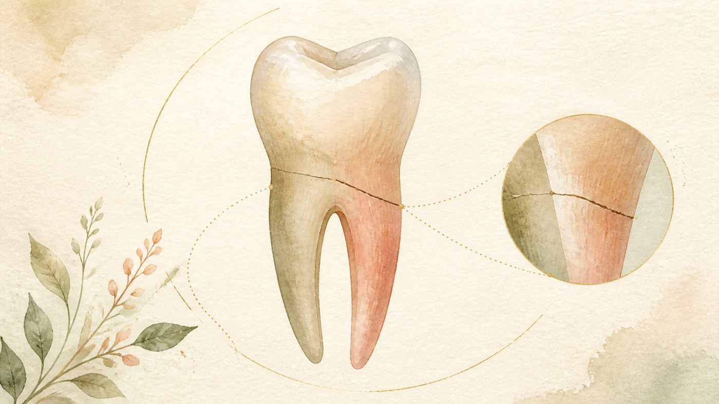
What Happens When a Tooth Root Fractures? Does It Always Need Extraction?|證據鏈
本頁是〈What Happens When a Tooth Root Fractures? Does It Always Need Extraction?〉的事實單元帳:每一條主張對應的來源、證據等級、地域與期間、原文引句與限制,逐條攤開。
What Happens When a Tooth Root Fractures? Does It Always Need Extraction?|證據鏈
F-Units (fact-unit ledger)
- F1|Topic-selection basis = full GSC reconciliation: the root-fracture query has 2,753 web-detail rows for, totaling 19,322 impressions and 594 clicks (re-run with awk against `gsc-full-20260804/__web__full.tsv` on 2026-08-06)|source #16|confidence=verified|basis=internal_dataset|period=GSC retention window (from 2025-03-22)|geo: TW|caveat: impressions are property-level counts, not deduplicated traffic; the listing uses the 「牙根斷裂」 row in the queue-page clinic-supplement table, with no additions or deletions; this is internal data rather than a medical claim and the entire entry is removed in publication conversion.
- F2 [structural synthesis]|The card's three-route triage backbone (crown/cusp fracture, traumatic transverse or oblique root fracture, vertical root fracture), its arrangement of crown-root fracture as a borderline type, its framing that distinguishing the type matters more than asking first about extraction, the appointment-checklist design, the ordering and home-observation wording of red flags, and its division-of-work statement with KM-DENTAL-50/33/30/18/35 are this site's care-seeking communication structure synthesized from F3 through F29|source #18|confidence=n/a|basis=editorial_framework|period=2026-08-06|geo: universal|caveat: not a diagnostic tool, clinical classification, or clinical decision flowchart; must not be labelled a claim awaiting verification; contains no efficacy or timing claim.
- F3|ESE position statement on longitudinal cracks and fractures of teeth (expert-committee consensus, 2025), definition table (Table 1) and introduction: VRF is defined as an incomplete longitudinal (axial) root fracture involving cementum, dentine, and root canal space; split root is a complete VRF with visible separation of the root into distinct segments; split tooth is complete visible separation of the entire tooth into two parts, usually mesio-distally; timely identification and appropriate management are necessary to extend the affected tooth's lifespan|source #1|confidence=verified|basis=clinical_guideline (ESE position statement)|period=2025 (online 2025-01-22); searched 2026-08-06|geo: universal|Retrieval method (reproducible): `efetch db=pmc id=11812625 rettype=full retmode=xml` (PMC open-access full text), then stripped tags and matched verbatim; the spans in this entry come from full text rather than the PubMed abstract, so no PMID prefix is placed on this line to prevent `--spans` from falsely matching against the abstract|span:「An incomplete longitudinal (axial) root fracture, involving the cementum, dentine, and root canal space.」「Split Root A complete VRF with visible separation of the root into distinct segments.」「Complete, visible separation of the entire tooth into 2 parts, usually in a mesio‐distal direction」「Timely identification and appropriate management are essential to increase the life span of the affected tooth.」|caveat: expert-consensus level; definitions are classification terms and clinical classification requires a dentist's assessment; partly shares the anchor with F3 of KM-DENTAL-50.
- F4|The same position statement's clinical and imaging features table (Table 3) and VRF clinical-features section: advanced cusp fracture may have symptoms relieved after the cusp breaks off, while pulp-involving fracture may develop pulpitis or apical periodontitis symptoms; clinical features of VRF include periodontal-disease-like presentation, isolated deep narrow periodontal pockets, mobility, and one or more sinus tracts near the gingival margin; early VRF is difficult to detect because patients may have no symptoms; typical advanced features are an isolated narrow deep periodontal pocket (pathognomonic when detected on both sides of a root) and multiple sinus tracts|source #1|confidence=verified|basis=clinical_guideline (same source as F3)|period=2025; searched 2026-08-06|geo: universal|Retrieval method: same as F3 (PMC full text); no PMID prefix on this line|span:「Symptoms may be relieved when the cusp fractures off」「May develop pulpitis or apical periodontitis symptoms if fracture involves pulp」「Mimicking periodontal disease +/− isolated, deep, narrow periodontal pocket」「Presence of a sinus/multiple sinuses close to gingival margin」「Early‐stage VRFs are a challenge to detect as the patient may be asymptomatic」「Typical features of advanced VRF include an isolated, narrow, deep periodontal pocket (pathognomonic if detected on both sides of a root) and the presence of multiple sinus tracts」|caveat: the table mixes clinician examination findings and patient symptoms; this guide does not reorganize it into diagnostic criteria; probing depth is measured by a clinician, not self-assessed by a patient; the original table contains a spelling error, which this card records without correcting.
- F5|The same position statement's section “VRF in root-filled teeth” (prevalence and aetiology): VRF is more commonly associated with root-filled teeth; the reported “prevalence of extraction of root-filled teeth with VRF” ranges from 4% to 32%; maxillary premolars and mandibular molars are more often affected; VRF incidence increases with age and is more common over age 40; VRF is a common cause of extraction of root-filled teeth, accounting for 13% to 21%; the introduction also says VRF commonly progresses subtly, causing delayed diagnosis and resulting in extraction|source #1|confidence=verified|basis=clinical_guideline (same source as F3)|period=2025; searched 2026-08-06|geo: universal|Retrieval method: same as F3 (PMC full text); no PMID prefix on this line|span:「VRF is more commonly associated with RFT than NFRT. The reported prevalence of extraction of RFT with VRF ranges from 4% to 32%」「The incidence of VRF increases with age, being more prevalent in 40+ year‐old patients」「VRF is a common cause of extraction (13–21%) of RFT」「A VRF commonly progresses subtly, leading to a delayed diagnosis that results in extraction.」|caveat: the denominators for “4% to 32%” and “13–21%” are not defined word-for-word in the original (the former literally says prevalence of extraction of RFT with VRF; the latter is a parenthetical value after a common cause of extraction of RFT). This card reports both as written, does not interpret them, and makes no population inference or personal-risk calculation in the text; both number sets are studies cited by the statement rather than the statement's original statistics, and their original publications were not retrieved for this card.
- F6|The same position statement's “VRF in root-filled teeth” section (clinical management): prognosis is poor; once diagnosis is established, prompt management is recommended to reduce the likelihood of acute apical periodontitis symptoms and further periradicular bone breakdown, which may complicate or delay implant treatment; extraction is recommended for VRF in a single-rooted tooth; in a multi-rooted root-filled tooth, root resection or root amputation may be considered as alternatives to extraction, with multidisciplinary treatment planning recommended; there are currently no survival-rate data for root resection or amputation for VRF in root-filled teeth, and the 10-year 90.6% and 15-year 96.8% figures are extrapolated from root-resection outcomes in periodontally affected teeth|source #1|confidence=verified|basis=clinical_guideline (same source as F3)|period=2025; searched 2026-08-06|geo: universal|Retrieval method: same as F3 (PMC full text); no PMID prefix on this line|span:「The prognosis is poor」「Prompt management is recommended as soon as diagnosis is reached to reduce the likelihood of acute AP symptoms and/or further periradicular bone breakdown, as this may complicate and/or delay dental implant treatment.」「Extraction is recommended in a single‐rooted tooth with a VRF」「In multi‐rooted RFT, root resection or root amputation may be considered as an alternative to extraction. A multi‐disciplinary approach is recommended in the treatment planning. There is no data on survival rates of root resection or root amputation for VRF in RFT; however, extrapolation from root resection on periodontally affected teeth has been reported to be 90.6% and 96.8% over 10 and 15 years, respectively」|caveat: 90.6% and 96.8% are survival rates for root resection in periodontally affected teeth and the original explicitly labels them extrapolations; they must not be cited as VRF prognostic figures; these recommendations presuppose a confirmed diagnosis and differ from the stage of suspicion without confirmation.
- F7|The same position statement's section “VRF in non-root-filled teeth”: reported incidence ranges from 6% to 37%, with more cases in men and persons over 50 years old; exploratory surgery may be indicated when findings are inconclusive or when the feasibility of management versus extraction must be determined; early intervention is necessary to limit VRF progression and further bone breakdown of neighboring affected roots; extraction is recommended for a single-rooted tooth, while complete or partial resection of the affected root may be considered in a multi-rooted tooth according to VRF position, level, and extent; evidence supporting intentional replantation and extraoral bonding for VRF is currently limited; the conclusion is that evidence on treatment strategies for VRF in non-root-filled teeth is limited and more clinical studies with long-term follow-up are needed|source #1|confidence=verified|basis=clinical_guideline (same source as F3)|period=2025; searched 2026-08-06|geo: universal|Retrieval method: same as F3 (PMC full text); no PMID prefix on this line|span:「The incidence of VRFs in non‐root‐filled teeth (NRFT) has been reported to be between 6% and 37%, with a predilection for males and patients over 50 years of age」「Exploratory surgery may be indicated if the above findings are inconclusive and/or the nature and feasibility of management versus extraction need to be determined」「Extraction of single‐rooted teeth is recommended. Root resection (complete or partial) of the affected root may be an option in multirooted teeth depending on the position, level, and extent of the VRF.」「At present there is limited evidence to support intentional replantation and extraoral bonding for VRF.」「The current evidence on treatment strategies for VRFs in NRFT is limited; more clinical studies with long‐term follow‐up are needed.」|caveat: the original does not define the denominator for “6% to 37%” (it may be the proportion of VRF cases occurring in non-root-filled teeth or another measure). Accordingly, this guide does not cite the number in its body and retains only the qualitative sex and age description; this is a deliberate limitation of the card, not a source omission.
- F8|The same position statement's “split tooth” section: a split tooth results from a crown crack progressing and enlarging until crown and root mechanically fail and the tooth separates completely longitudinally; fragments are completely separated or separate under pressure, and there may be apical periodontitis symptoms including pain on biting, narrow isolated periodontal probing, or mobility; the prognosis is poor and timely extraction should be considered to minimize acute symptoms and limit bone loss|source #1|confidence=verified|basis=clinical_guideline (same source as F3)|period=2025; searched 2026-08-06|geo: universal|Retrieval method: same as F3 (PMC full text); no PMID prefix on this line|span:「The tooth fragments are completely separated or separated with pressure. There may be symptoms of AP, including pain on biting, a narrow, isolated periodontal probing, or tooth mobility.」「ST has an unfavourable prognosis, and timely extraction should be considered to minimise the development of acute symptoms and limit bone loss.」|caveat: the original also says literature on split-tooth prevalence and presentation is sparse; this card makes no frequency statement.
- F9|The same position statement's imaging-limitations passage and “general considerations regarding CT and VRF” section: CBCT cannot reliably detect VRF within the root because image resolution is insufficient for cracks typically 50 to 100 μm wide; the impact of excessive occlusal forces in the aetiology of cracked tooth, split tooth, and VRF cannot be overemphasized; opposing dentition (including restorative status and occlusal morphology) should be considered; occlusal stabilization splints and/or referral to a relevant specialty should be considered to manage parafunctional habits|source #1|confidence=verified|basis=clinical_guideline (same source as F3)|period=2025; searched 2026-08-06|geo: universal|Retrieval method: same as F3 (PMC full text); no PMID prefix on this line|span:「CBCT cannot reliably detect VRFs within the root due to the image resolution being insufficient to detect fractures that are typically 50‐100 μm in width」「The impact of excessive occlusal forces cannot be overemphasised in the aetiology of CT, ST, and VRF.」「Fabrication of occlusal stabilisation splints and/or referral to relevant specialists should be considered for the management of parafunctional habits」|caveat: splints and referral are clinician recommendations conditional on indication; they must not be read as management instructions for an individual patient; partly shares the anchor with F5 of KM-DENTAL-50.
- F10|The same position statement's cracked-tooth management and prognosis conclusion: current evidence suggests encouraging outcomes for vital and root-canal-treated cracked teeth restored with cuspal coverage; early management, cuspal-coverage restoration, and absence of deep periodontal probing of non-endodontic origin increase survival of cracked-tooth management; periodontal probing of 5 mm or more is associated with reduced survival|source #1|confidence=verified|basis=clinical_guideline (same source as F3)|period=2025; searched 2026-08-06|geo: universal|Retrieval method: same as F3 (PMC full text); no PMID prefix on this line|span:「Current evidence suggests encouraging outcomes for vital as well as endodontically treated CT restored with cuspal coverage restorations. Early management, cuspal coverage restorations, and absence of deep periodontal probing depth of non‐endodontic origin increase the survival rate of CT management.」「A 5 + mm periodontal probing is associated with reduced survival rates」|caveat: this entry concerns restorative decisions for cracked tooth (CT), the primary topic of KM-DENTAL-50. This card uses one sentence only to connect to the restorative direction for cusp fracture and does not rewrite that card.
- F11|IADT 2020 guideline part 1 (fractures and luxations), Table 7 definition and clinical findings for root fracture: a root fracture involves dentine, pulp, and cementum; its direction may be horizontal, oblique, or both; the coronal segment may be mobile and displaced, the tooth may be tender to percussion, gingival-sulcus bleeding may occur, and pulp sensibility testing may initially be negative (indicating transient or permanent neural injury); a fracture may occur at any root level and can go undetected without additional image angles|source #2|confidence=verified|basis=clinical_guideline (IADT 2020 guideline part 1, Dent Traumatol 2020;36(4):314-330, DOI 10.1111/edt.12578)|period=2020 current version (on 2026-08-06, a PubMed search for `International Association of Dental Traumatology guidelines[ti]` returned the 2020 three-part guideline set as the latest guideline body, with no updated version)|geo: universal|Retrieval method (reproducible): opened the Wiley open-access full text `https://onlinelibrary.wiley.com/doi/10.1111/edt.12578` with ego-browser, obtained `document.body.innerText`, then matched verbatim; the spans in this entry are from full text rather than a PubMed abstract, so no PMID prefix is placed on this line|span:「A fracture of the root involving dentin, pulp and cementum.」「The fracture may be horizontal, oblique or a combination of both.」「The coronal segment may be mobile and may be displaced」「Bleeding from the gingival sulcus may be seen」「Pulp sensibility testing may be negative initially, indicating transient or permanent neural damage」「Root fractures may be undetected without additional imaging」|caveat: consensus guideline, not randomized-trial results; table entries are clinician examination findings, not items for patient self-diagnosis.
- F12|The same guideline's Table 7 management column: if displaced, the coronal fragment should be repositioned as soon as possible and verified with imaging; a mobile coronal fragment should be stabilized with a passive flexible splint for four weeks, and a cervical fracture may need a longer period up to four months; cervical fractures have healing potential, so the coronal fragment, particularly if non-mobile, should not be removed at the emergency visit; root-canal treatment should not start at the emergency visit; healing should be monitored for at least one year together with pulp status|source #2|confidence=verified|basis=clinical_guideline (same source as F11)|period=2020 current version|geo: universal|Retrieval method: same as F11 (Wiley full text); no PMID prefix on this line|span:「If displaced, the coronal fragment should be repositioned as soon as possible.」「Stabilize the mobile coronal segment with a passive and flexible splint for 4 wk. If the fracture is located cervically, stabilization for a longer period of time (up to 4 mo) may be needed」「Cervical fractures have the potential to heal. Thus, the coronal fragment, especially if not mobile, should not be removed at the emergency visit」「No endodontic treatment should be started at the emergency visit」「It is advisable to monitor healing of the fracture for at least one year.」|caveat: “4 wk” and “up to 4 mo” refer to splint duration, not total treatment duration or any prognosis promise; repositioning and stabilization are dentist procedures. This card neither describes how to perform them nor recommends attempting them yourself.
- F13|The same guideline's subsequent-management column: pulp necrosis and infection may develop later, usually only in the coronal fragment, so root-canal treatment of the coronal segment alone is indicated; root-fracture lines are often oblique, making root-canal length challenging to determine and possibly requiring apexification; the apical fragment rarely undergoes pathological changes requiring treatment; in mature teeth with a cervical fracture line above the alveolar crest and a very mobile coronal fragment, removal of the coronal fragment followed by root-canal treatment and post-retained crown restoration will likely be required, and orthodontic extrusion of the apical fragment, crown lengthening, surgical extrusion, or extraction may be future options|source #2|confidence=verified|basis=clinical_guideline (same source as F11)|period=2020 current version|geo: universal|Retrieval method: same as F11 (Wiley full text); no PMID prefix on this line|span:「Pulp necrosis and infection may develop later. It usually occurs in the coronal fragment only. Hence, endodontic treatment of the coronal segment only will be indicated.」「The apical segment rarely undergoes pathological changes that require treatment」「In mature teeth where the cervical fracture line is located above the alveolar crest and the coronal fragment is very mobile, removal of the coronal fragment, followed by root canal treatment and restoration with a post-retained crown will likely be required.」|caveat: the original's four conditions (mature root + cervical fracture + above the alveolar crest + very mobile coronal fragment) are a conjunctive statement. They must not be split into the inference “a cervical fracture requires extraction.”
- F14|The same guideline's Table 7 follow-up and outcome column: clinical and radiographic follow-up occurs at four weeks (splint removal for middle- and apical-third fractures), six to eight weeks, four months (splint removal for cervical-third fractures), six months, one year, and then annually for at least five years; favorable results include a responsive pulp sensibility test (though false negatives can occur for several months and root-canal treatment should not start solely for non-response), signs of repair between fragments, and normal or only slightly supra-physiological coronal-fragment mobility; unfavorable results include symptoms, coronal-fragment extrusion and/or excessive mobility, radiolucency at the fracture line, and pulp necrosis/infection with inflammation in the fracture line|source #2|confidence=verified|basis=clinical_guideline (same source as F11)|period=2020 current version|geo: universal|Retrieval method: same as F11 (Wiley full text); no PMID prefix on this line|span:「Positive response to pulp sensibility testing; however, a false negative response is possible for several months. Endodontic treatment should not be started solely on the basis of no response to pulp sensibility testing」「Signs of repair between the fractured segments」「Extrusion and/or excessive mobility of the coronal segment」「Pulp necrosis and infection with inflammation in the fracture line」|caveat: follow-up timing is the guideline's scheduling advice to dentists; actual return visits are adjusted to the individual situation.
- F15|The same guideline's Tables 2 through 4 (crown fractures): when enamel alone fractures, an available fragment may be bonded back, or margins may be smoothed or composite resin used according to fracture extent and location; for enamel-dentine fracture without pulp exposure, an intact fragment may be rehydrated in water or saline for 20 minutes before bonding, while exposed dentine is covered with glass ionomer or adhesive and composite resin; for complicated crown fracture with pulp exposure, immature teeth with open apices should retain the pulp and partial pulpotomy or pulp capping is recommended to promote continued root development, while conservative pulp treatment such as partial pulpotomy is also preferred in mature teeth; all three tables say to assess possible associated luxation injury or root fracture, especially if tenderness is present|source #2|confidence=verified|basis=clinical_guideline (same source as F11)|period=2020 current version|geo: universal|Retrieval method: same as F11 (Wiley full text); no PMID prefix on this line|span:「If the tooth fragment is available and intact, it can be bonded back on to the tooth. The fragment should be rehydrated by soaking in water or saline for 20 min before bonding」「In patients where teeth have immature roots and open apices, it is very important to preserve the pulp. Partial pulpotomy or pulp capping are recommended in order to promote further root development」「Conservative pulp treatment (eg, partial pulpotomy) is also the preferred treatment in teeth with completed root development」「Evaluate the tooth for a possible associated luxation injury or root fracture, especially if tenderness is present」|caveat: materials and techniques for pulp treatment are clinician decisions. This card does not describe operative methods or recommend handling a fragment yourself.
- F16|The same guideline's Tables 5 and 6 (crown-root fracture): a crown-root fracture involves enamel, dentine, and cementum and usually extends below the gingival margin; before a treatment plan is finalized, a loose fragment should be temporarily stabilized to adjacent teeth or a non-mobile fragment; future treatment options include orthodontic extrusion of the apical or non-mobile fragment followed by restoration (possibly with periodontal reshaping surgery), surgical extrusion, root-canal treatment and restoration for pulp necrosis/infection, root submergence, intentional replantation (possibly including root rotation), extraction, and autotransplantation; the treatment plan partly depends on patient age and anticipated cooperation|source #2|confidence=verified|basis=clinical_guideline (same source as F11)|period=2020 current version|geo: universal|Retrieval method: same as F11 (Wiley full text); no PMID prefix on this line|span:「(Note: Crown-root fractures typically extend below the gingival margin)」「Until a treatment plan is finalized, temporary stabilization of the loose fragment to the adjacent tooth/teeth or to the non-mobile fragment should be attempted」「The treatment plan is dependent, in part, on the patient's age and anticipated co-operation.」「Orthodontic extrusion of the apical or non-mobile fragment, followed by restoration」|caveat: the option list is a guideline enumeration for clinicians; it does not mean every option fits an individual patient, and this card does not rank the options.
- F17|The same guideline's imaging section: images at different vertical and/or horizontal angles help detect root fracture; CBCT can enhance visualization of dental trauma, particularly root fractures, crown/root fractures, and lateral luxations, and can help determine fracture location, extent, and direction; the guiding principle for exposure to ionizing radiation is whether the image is likely to change injury management|source #2|confidence=verified|basis=clinical_guideline (same source as F11)|period=2020 current version|geo: universal|Retrieval method: same as F11 (Wiley full text); no PMID prefix on this line|span:「Cone beam computerized tomography (CBCT) provides enhanced visualization of TDIs, particularly root fractures, crown/root fractures, and lateral luxations. CBCT helps to determine the location, extent, and direction of a fracture.」「A guiding principle when considering exposing a patient to ionizing radiations (eg, either 2D or 3D radiographs) is whether the image is likely to change the management of the injury.」|caveat: the CBCT utility statement here is limited to visualization of traumatic fracture; it is a different clinical question from the limits of CBCT in detecting VRF stated in F9/F21/F22. They must not be applied to each other.
- F18|The same guideline's introduction, pulp-preservation section, and disclaimer: every effort should be made to preserve the pulp regardless of tooth maturity, particularly in immature permanent teeth to allow continued root development and apex formation; IADT does not, and cannot, guarantee favorable outcomes from adherence to its guidelines|source #2|confidence=verified|basis=clinical_guideline (same source as F11)|period=2020 current version|geo: universal|Retrieval method: same as F11 (Wiley full text); no PMID prefix on this line|span:「Every effort should be made to preserve the pulp, in both mature and immature teeth.」「The IADT does not, and cannot, guarantee favorable outcomes from adherence to the Guidelines.」|caveat: the disclaimer is original guideline text; when relayed in the compliance note, this card uses “does not guarantee” and makes no efficacy promise.
- F19|Systematic review and meta-analysis (clinical presentations and risk factors associated with VRF in root-filled teeth; PROSPERO CRD42022354108; searched MEDLINE/EMBASE/Scopus/Web of Science in October 2022; Newcastle-Ottawa Scale used for risk of bias): included 14 sources and 2877 teeth (489 with VRF and 2388 without); sinus tract (odds ratio 4.87, 95% confidence interval 1.58 to 15.0), increased periodontal probing depth (13.24, 5.44 to 32.22), swelling or abscess (2.86, 1.74 to 4.70), and tenderness to percussion (1.76, 1.18 to 2.61) were significantly associated with VRF; the assessed risk factors (sex, tooth type, tooth location, posts, indirect restorations, and apical extension of root-canal filling) were not significantly associated with VRF|source #3|confidence=verified|basis=peer_reviewed (PMID 37307871, systematic review and meta-analysis)|period=2023 (searched through 2022-10); currency checked—on 2026-08-06, `vertical root fracture[tiab] AND (systematic review[pt] OR meta-analysis[pt]) AND 2024:2026[dp]` returned 2 records, both on in-vitro fracture resistance and fragment reattachment rather than an update on this question; this article is the current latest systematic review on the same question; esummary pubtype checked, no Retracted Publication|geo: universal|span:「Fourteen sources reporting on 2877 teeth (489 with VRF and 2388 without VRF) were included in the meta-analyses.」「the presence of sinus tracts (OR = 4.87; 95% confidence interval [CI], 1.58-15.0), increased periodontal probing depths (OR = 13.24; 95% CI, 5.44-32.22), swelling/abscess (OR = 2.86; 95% CI, 1.74-4.70), and tenderness to percussion (OR = 1.76; 95% CI, 1.18-2.61) were significantly associated with the presence of a VRF」「None of the assessed risk factors (sex, type of teeth, tooth location, posts, indirect restoration, and apical extension of the root canal filling) were found to be significantly associated with the presence of a VRF」|caveat: an odds ratio is the relative odds of VRF in persons with the finding, not incidence or positive predictive value. It must not be read as “a sinus tract means a root fracture.” The pooled studies were clinical studies; heterogeneity and risk of bias are in the original.
- F20|Case-control study (VRF prevalence and risk factors in root-filled teeth using surgical diagnosis as reference; among 411 root-filled teeth undergoing endodontic surgery from 2005 to 2022, analyzed 130 VRF cases and 260 randomly selected controls; used multiple imputation for missing values and logistic regression with robust standard errors to handle clustering of several teeth within one patient): VRF prevalence was 31.63%; significant risk factors were age (adjusted odds ratio 1.02), periodontal probing depth ≥5 mm (2.37), isolated perilateral radiolucency (4.05), halo radiolucency (2.70), overfilled root canal (2.11), and root-canal-space-to-root-width ratio greater than one third (1.52); among teeth with posts, post-space-to-root-width ratio greater than one third was associated with increased risk (unadjusted odds ratio 2.46)|source #4|confidence=verified|basis=peer_reviewed (PMID 40769327, case-control study)|period=January 2026 issue (online first 2025-08-05); searched 2026-08-06; esummary pubtype checked, no Retracted Publication|geo: universal|span:「From 411 RFT that underwent endodontic surgery (2005-2022), 130 VRF cases and 260 randomly selected controls were analyzed.」「VRF prevalence was 31.63% among RFT undergoing endodontic surgery.」「Significant risk factors included age (adjusted odds ratio [aOR] = 1.02), probing depth ≥5 mm (aOR = 2.37), isolated perilateral radiolucency (aOR = 4.05), halo radiolucency (aOR = 2.70), overfilled root canal (aOR = 2.11), and root canal space-to-root-width ratio >1/3 (aOR = 1.52).」|caveat: the denominator of 31.63% is root-filled teeth undergoing endodontic surgery, a highly selected surgical group; it must not be extrapolated to all root-filled teeth or the general population; this is single-center retrospective data, a study-level proportion rather than a personal-risk estimate.
- F21|Systematic review and meta-analysis (CBCT accuracy for diagnosing VRF in root-filled teeth, using direct visualization as the reference standard; searched through 2020-06; QUADAS-2 used for risk of bias; 8 articles included): pooled sensitivity 0.78 (95% confidence interval 0.64 to 0.88), specificity 0.80 (0.63 to 0.91), and accuracy 0.86 (0.83 to 0.89); GRADE assessed evidence certainty as low for sensitivity and moderate for specificity; conclusion: compared with direct visualization, CBCT is still not a good tool for diagnosing VRF in root-filled teeth|source #5|confidence=verified|basis=peer_reviewed (PMID 33984375, systematic review and meta-analysis)|period=2021 (searched through 2020-06); currency checked—the same question also has a 2023 systematic review (F22), but inclusion criteria differ (this review limited itself to clinical studies with direct visualization as reference). This card presents both rather than selecting one; esummary pubtype checked, no Retracted Publication|geo: universal|span:「CBCT imaging had a pooled sensitivity and specificity of 0.78 (95% confidence interval [CI], 0.64-0.88) and 0.80 (95% CI, 0.63-0.91), respectively, and an accuracy of 0.86 (95% CI, 0.83-0.89).」「Evidence from this systematic review and meta-analysis indicates that CBCT imaging is still not a good tool for diagnosing VRFs in root-filled teeth compared with direct visualization.」|caveat: diagnostic-accuracy measures depend on prevalence in the tested population and cannot be converted into an individual test interpretation.
- F22|Systematic review (diagnostic efficacy of CBCT for detecting VRF in endodontically treated teeth; searched Web of Science/PubMed/Cochrane/SCOPUS/Embase from 2000 to 2022; included 20 papers): without a post and with root-canal filling material, mean CBCT sensitivity and specificity were 71.50 ± 22.19% and 75.64 ± 19.41%; the conclusion was that further clinical research is needed to validate optimal CBCT performance as a detection technique because of low sensitivity, substantial heterogeneity among studies, and lack of in-vivo studies|source #6|confidence=verified|basis=peer_reviewed (PMID 37264339, systematic review)|period=2023 (search 2000 to 2022); searched 2026-08-06; esummary pubtype checked, no Retracted Publication|geo: universal|span:「The final analysis included 20 papers that satisfied the eligibility requirements.」「Further clinical research is needed to validate the optimum efficiency of CBCT as a diagnostic technique for detecting VRFs in teeth that have had endodontic treatment, given the low sensitivity, significant heterogeneity of studies, and lack of in-vivo studies on the subject.」|caveat: most included studies were in vitro, so accuracy figures do not equal performance in clinical settings; this card cites only its conclusion direction and inclusion count.
- F23|Retrospective longitudinal clinical study (initial healing patterns, predictors, and long-term prognosis of traumatic transverse root fracture in permanent teeth; imaging assessed healing and non-healing events at the fracture line; healing was categorized as hard tissue, connective tissue, or connective tissue plus bone, while non-healing involved granulation tissue; competing-risk survival analysis and Fine & Gray subdistribution regression were used): short-term healing was 61.4%, strongly influenced by concomitant injuries to the coronal fragment and their type; concomitant crown fracture (subdistribution hazard ratio 24.38, 95% confidence interval 3.16 to 188.3), displaced luxation injury (10.58, 1.37 to 81.9), and subluxation (9.66, 1.14 to 81.7) were associated with short-term non-healing; long-term healing was 75.9%, mostly with bone and connective-tissue interposition; the clinical-implications section says traumatic transverse root fracture has a positive prognosis, supporting a more conservative approach before more radical treatment is considered|source #7|confidence=verified|basis=peer_reviewed (PMID 38467867, retrospective longitudinal clinical study)|period=2024; currency checked—on 2026-08-06, a search for `root fracture[tiab] AND healing[tiab] AND (systematic review[pt] OR meta-analysis[pt])` returned 0 records. There is currently no systematic review on this exact question; this study is the available higher-level clinical evidence; esummary pubtype checked, no Retracted Publication|geo: universal|span:「Radiographic findings showed 61.4% of healing in the short-term being strongly influenced by the presence and type of concomitant injuries to the coronal fragment.」「The healing rate in the long-term was of 75.9%, most of them with interposition of bone and connective tissue.」「Post-traumatic transversal root fractures have a positive prognosis supporting therefore, a more conservative approach for these teeth before considering more radical treatments.」|caveat: a single-center retrospective study; 61.4% and 75.9% are study-level radiographic-healing proportions, not personal-prognosis estimates or “tooth retention rates.” Hazard-ratio confidence intervals are very wide (for example, 3.16 to 188.3), indicating imprecision.
- F24|Systematic review (orthodontic forced eruption for permanent teeth with traumatic subgingival loss of tooth structure; 2757 records from MEDLINE/Cochrane Library/EMBASE passed initial screening, with 13 full-text papers included: 1 randomized controlled trial, 1 prospective clinical trial, 2 retrospective cohort studies, and 9 case series): mean orthodontic-extrusion rate was 1.5 mm per week, followed by retention after a four-to-six-week treatment period; conclusion: existing evidence suggests orthodontic forced eruption is a feasible pretreatment option for permanent teeth originally judged non-restorable, and root resorption does not appear to be an associated adverse effect|source #8|confidence=verified|basis=peer_reviewed (PMID 34886307, systematic review)|period=2021; searched 2026-08-06; esummary pubtype checked, no Retracted Publication|geo: universal|span:「Thirteen full-text papers were included: one RCT, one prospective clinical trial, two retrospective cohort studies, and nine case series.」「The mean extrusion rate of OFE was 1.5 mm a week within a four to six weeks treatment period followed by retention.」「The current state of the evidence suggests that OFE is a feasible pre-treatment option for non-restorable permanent teeth.」|caveat: 9 of the 13 studies were case series, so evidence level is low; “feasible” is the authors' description of existing evidence, not a success rate; suitability requires an individualized dentist assessment.
- F25|Systematic review (methods and outcomes of fragment reattachment for complicated crown-root fracture of anterior teeth; searched PubMed/Web of Science/Embase/Scopus/Google Scholar): included 12 case reports and 2 case series; 11 exposed the fracture line before reattachment; all but 1 case used root-canal treatment after conservative pulp treatment; a post was part of restoration in 85% of cases; outcomes were favorable in all included articles with follow-up from three months to seven years; conclusion: fragment reattachment can be considered a viable treatment option when clinical conditions are favorable|source #9|confidence=verified|basis=peer_reviewed (PMID 32813931, systematic review)|period=2021 (online 2020-09-09); searched 2026-08-06; esummary pubtype checked, no Retracted Publication|geo: universal|span:「Twelve case reports and two case series were selected for this review after applying the inclusion and exclusion criteria.」「A post was used as part of the restoration in 85% of the cases.」「Treatment outcomes were favorable in all the included articles and the follow-up period ranged from three months to seven years.」「Fragment reattachment after complicated crown-root fractures of anterior teeth can be considered as a viable treatment option if the clinical conditions are favorable.」|caveat: all included publications are case reports and case series, types of literature that tend to report successful cases only; “all outcomes were favorable” must not be read as a success rate.
- F26|Prospective cohort study (20 Swedish public-dentistry clinics consecutively recorded reasons for extraction and followed whether prosthetic reconstruction occurred through 5-year electronic records): 133 patients (61 men, 72 women; mean age 54.0 years, standard deviation 15.8) underwent extraction; the most common reasons were endodontic pathology (36.8%) and fracture (24.8%); 61 patients had previous endodontic treatment, and one third of extracted teeth were root-filled; 35 teeth received prosthetic reconstruction, most often removable dentures (45.7%)|source #10|confidence=verified|basis=peer_reviewed (PMID 42034624, prospective cohort study)|period=2026 (publication date 2026-04-25, before this card's date of 2026-08-06); searched 2026-08-06; esummary pubtype checked, no Retracted Publication|geo: universal|span:「A total of 133 patients (61 men and 72 women; mean age 54.0 years, SD = ± 15.8) underwent extractions.」「Endodontic pathology (36.8%) and fractures (24.8%) were the most common reasons.」「Sixty-one patients had previous endodontic treatment, and one-third of extracted teeth were root-filled.」|caveat: public-dentistry population in one Swedish county, an eight-week registration period, and 133 patients: this is a service-pattern statistic for that place and must not be generalized to Taiwan or the world. “Fracture” in that study includes all fracture types and does not separate the three types in this card.
- F27|Background section of a Cochrane systematic review on systemic antibiotics for adults with symptomatic apical periodontitis and acute apical abscess: clinical guidance recommends first-line local operative measures to remove the source of inflammation or infection; systemic antibiotics are currently recommended only for evidence of spreading infection (cellulitis, lymph-node involvement, diffuse swelling) or systemic involvement (fever, malaise)|source #11|confidence=verified|basis=peer_reviewed (PMID 38712714, Cochrane systematic review CD010136.pub4)|period=2024 (searched through 2022-11); version chain checked (CD010136 pub4 is current and the original states it updated 2018 pub3); on 2026-08-06 esummary rechecked pubtype, no Retracted Publication; shared anchor with KM-DENTAL-50/33|geo: universal|span:「systemic antibiotics are currently only recommended for situations where there is evidence of spreading infection (cellulitis, lymph node involvement, diffuse swelling) or systemic involvement (fever, malaise)」|caveat: this card cites this sentence only as a red-flag criterion for when the situation has extended beyond a local problem. It makes no medication recommendation and gives no drug name or dose.
- F28|Textbook entry (deep neck infection): symptoms often arise from local pressure effects on respiratory, nervous, or gastrointestinal tracts, including neck swelling, dysphagia, dysphonia, and trismus; clinical presentation often includes fever, neck pain, and respiratory distress|source #12|confidence=verified|basis=textbook (PMID 30020634, StatPearls entry)|period=entry version 2026-01; searched 2026-08-06; shared anchor with KM-DENTAL-50|geo: universal|span:「Symptoms often result from local pressure effects on the respiratory, nervous, or gastrointestinal tracts, including neck swelling, dysphagia, dysphonia, and trismus.」「often involving fever, neck pain, and respiratory distress」|caveat: deep neck infection can arise from causes other than teeth; this entry is used only to define the scope of signs requiring immediate medical assessment, not to diagnose.
- F29|Consensus report for the 2017 World Workshop classification (periodontitis workgroup): periodontal abscess is an acute lesion characterized by localized pus accumulation within the gingival wall of the periodontal pocket/sulcus, rapid tissue destruction, and association with risk of systemic dissemination; endodontic-periodontal lesions are defined as pathological communication between pulpal and periodontal tissues at a given tooth and may be acute or chronic|source #13|confidence=verified|basis=clinical_guideline (PMID 29926951, consensus report/classification)|period=2018; searched 2026-08-06; shared anchor with KM-DENTAL-50|geo: universal|span:「Periodontal abscesses are defined as acute lesions characterized by localized accumulation of pus within the gingival wall of the periodontal pocket/sulcus, rapid tissue destruction and are associated with risk for systemic dissemination.」「Endodontic-periodontal lesions, defined by a pathological communication between the pulpal and periodontal tissues at a given tooth, occur in either an acute or a chronic form」|caveat: this is a clinical diagnostic framework and requires a dentist's complete examination; this card does not make a classification determination.
- F30|Article 51, paragraph 11 of Taiwan's National Health Insurance Act: “Dentures, artificial eyes, eyeglasses, hearing aids, wheelchairs, crutches, and other equipment that is not actively therapeutic” are excluded from insurance benefits|source #14|confidence=verified (on 2026-08-06, ego-browser opened the National Laws and Regulations Database page for this article; page title matched “全民健康保險法§51-全國法規資料庫” verbatim and the statutory text was compared verbatim; existing anchor in this site's fact-check register)|basis=law|period=current text (page states law-compilation data cut-off date: ROC year 115, 07-31)|geo: TW|caveat: individual NHI coverage follows current National Health Insurance Administration notices; any commercial-insurance coverage depends on policy terms. This card makes no coverage determination and gives no amount.
- F31|Article 87 of Taiwan's Medical Care Act: “Content that implies or alludes to medical business is deemed medical advertising. Publication of medical knowledge or research reports, patient health education, and academic publications that do not involve solicitation of medical business are not deemed medical advertising.”|source #15|confidence=verified (on 2026-08-06, ego-browser test; page title matched “醫療法§87-全國法規資料庫” verbatim and the statutory text was compared verbatim)|basis=law|period=current text|geo: TW|caveat: this article is used only to define the publication identity of this site (health education rather than advertising) and does not concern any medical content of this card. Official English translation: https://law.moj.gov.tw/ENG/LawClass/LawAll.aspx?pcode=L0020021
- F32|The government open-data-platform dataset “Taipei City Medical Fee Standards” is provided by the Taipei City Department of Health. In addition, according to the verified conclusion in this site's fact-check register, neither of the National Health Insurance Administration Medical Materials Price Comparison Network's two search categories includes dentistry, so dental self-pay items cannot be checked through that network|source #17|confidence=verified (on 2026-08-06, ego-browser test; page title matched “臺北市醫療收費標準 | 政府資料開放平臺” verbatim, provider field matched “臺北市政府衛生局,” and metadata update time was 2026-07-13)|basis=official_statement|period=current dataset status|geo: TW|caveat: this is a single-city example; fee-standard pages for other cities/counties must be individually reverified before scaling. The Medical Materials Price Comparison Network conclusion is a cross-card anchor personally verified by OP on 2026-08-05 and is carried forward without rechecking in this card. [On the official page the label appears in Chinese only: 「醫材比價網」]
- F33|Article 81 of Taiwan's Medical Care Act: “When a medical institution diagnoses or treats a patient, it shall inform the patient or the patient's legal representative, spouse, relative, or related person of the condition, treatment policy, procedures, medication, prognosis, and possible adverse reactions.”|source #15|confidence=verified (uses an existing verified anchor in this site's fact-check register: National Laws and Regulations Database tested HTTP 200 and text matched verbatim; this card did not re-test the page)|basis=law|period=current text|geo: TW|caveat: statutory quotation, not legal advice; this card provides no legal or insurance-claim opinion. Official English translation: https://law.moj.gov.tw/ENG/LawClass/LawAll.aspx?pcode=L0020021
- F34 [structural synthesis]|Evidence-gap statement: on 2026-08-06, this site searched PubMed E-utilities with `root fracture[tiab] AND healing[tiab] AND (systematic review[pt] OR meta-analysis[pt])` and received 0 records. There is no systematic review on healing outcomes for traumatic root fracture on this exact question, so this card instead cites the available retrospective longitudinal study (F23) and states its evidence level. `vertical root fracture[tiab] AND (systematic review[pt] OR meta-analysis[pt]) AND 2024:2026[dp]` returned 2 records, neither on the same question (in-vitro fracture resistance and fragment reattachment), so the 2023 review in F19 is current. The red-flag list in this card is limited to signs listed in F27/F28/F29/F19/F4/F14; no unlisted signs are added. This card provides no self-diagnosis method, drug name or dose, or amount|source #18|confidence=n/a|basis=editorial_framework|period=2026-08-06|geo: universal|caveat: searches were limited to PubMed and the search strings above; not retrieving a publication does not mean it has been disproven. This is an editorial statement and must not be labelled a claim awaiting verification.
Sources
All access dates are 2026-08-06. PubMed entries were retrieved with E-utilities efetch for abstract text and matched verbatim to each span. The ESE position statement was retrieved as open-access full text using `efetch db=pmc id=11812625`, stripped of tags, and matched verbatim. The IADT guideline is a Wiley open-access article; it was opened with ego-browser and its innerText matched verbatim (the spans for these two sources are not in PubMed abstracts, so their F-Units do not carry PMID prefixes, preventing the gate from using the wrong text for matching). Taiwan laws and official pages were loaded with ego-browser and compared verbatim.
- Duncan HF, et al. (ESE position statement) Position statement on longitudinal cracks and fractures of teeth. Int Endod J. 2025;58:379-390. PMID 39840523; full text PMC11812625
- Bourguignon C, et al. International Association of Dental Traumatology guidelines for the management of traumatic dental injuries: 1. Fractures and luxations. Dent Traumatol. 2020;36(4):314-330. PMID 32475015
- Haupt F, Wiegand A, Kanzow P. Risk Factors for and Clinical Presentations Indicative of Vertical Root Fracture in Endodontically Treated Teeth: A Systematic Review and Meta-analysis. J Endod. 2023;49(8):940-952. PMID 37307871
- Promchouy P, Chotvorrarak K, Osiri S. Prevalence of Vertical Root Fractures in Root Filled Teeth and Their Clinical and Radiographic Risk Factors. J Endod. 2026;52(1):68-78. PMID 40769327
- PradeepKumar AR, et al. Diagnosis of Vertical Root Fractures by Cone-beam Computed Tomography in Root-filled Teeth with Confirmation by Direct Visualization: A Systematic Review and Meta-Analysis. J Endod. 2021;47(8):1198-1214. PMID 33984375
- Diagnostic efficacy of cone-beam computed tomography for detection of vertical root fractures in endodontically treated teeth: a systematic review. BMC Med Imaging. 2023;23:75. PMID 37264339
- Pedrosa NOM, et al. Healing and long-term prognosis of root-fractured permanent teeth: a retrospective longitudinal study. Clin Oral Investig. 2024;28(4):209. PMID 38467867
- Reichardt E, et al. Orthodontic Forced Eruption of Permanent Anterior Teeth with Subgingival Fractures: A Systematic Review. Int J Environ Res Public Health. 2021;18(23):12580. PMID 34886307
- Khandelwal P, et al. Fragment reattachment after complicated crown-root fractures of anterior teeth: A systematic review. Dent Traumatol. 2021;37(1):37-52. PMID 32813931
- Persson S, et al. Reasons for tooth extraction in a Swedish county dental service: a 5-year longitudinal cohort study with focus on endodontic pathology. BDJ Open. 2026;12(1):41. PMID 42034624
- Cope AL, et al. Systemic antibiotics for symptomatic apical periodontitis and acute apical abscess in adults. Cochrane Database Syst Rev. 2024;5(5):CD010136. PMID 38712714
- Almuqamam M, et al. Deep Neck Infections. StatPearls. PMID 30020634
- Papapanou PN, et al. Periodontitis: Consensus report of workgroup 2 of the 2017 World Workshop. J Periodontol. 2018;89 Suppl 1:S173-S182. PMID 29926951
- Taiwan National Health Insurance Act, Article 51 (National Laws and Regulations Database)
- Taiwan Medical Care Act, Article 87 (National Laws and Regulations Database)
- Internal data: `gsc-full-20260804/__web__full.tsv` (query “牙根斷裂” can be reconciled row by row)
- Government Open Data Platform: Taipei City Medical Fee Standards (provider: Taipei City Department of Health)
- Editorial framework and evidence gap: this site's three-route triage structure and PubMed search-gap record (no external source; labelled structural synthesis)
Internal reference chain
- A tooth has a crack but has not yet broken, and it hurts when biting: Does Pain When Biting Mean a Cracked Tooth or an Occlusal Problem? (KM-DENTAL-50)
- Overall pattern-based triage of toothache: What Should I Do About Toothache? What Does Cavity Pain Feel Like? (KM-DENTAL-33)
- Immediate emergency action when an entire tooth comes out: What Should I Do If a Whole Tooth Suddenly Comes Out? (KM-DENTAL-30)
- What root-canal treatment is and how many visits it takes: What Is Root-canal Treatment? How Many Visits Does It Take? (KM-DENTAL-18)
- Whether and how to replace a tooth that cannot be retained: Do I Have to Get an Implant for a Missing Tooth? What If I Cannot Afford Replacement? (KM-DENTAL-35)
- How to ask about and read a quote for redoing a crown: How Much Does One Crown Cost? How Do I Read a Quote? (KM-DENTAL-11)
FAQ
- If the root has fractured, does it definitely have to be extracted?
- **Not necessarily; the answer depends on the type of fracture.** For a traumatic transverse or oblique root fracture, IADT management is repositioning and splinting, and it specifically says a non-mobile coronal fragment should not be removed at the emergency visit [F12]. In a single-center retrospective study, long-term radiographic healing in this type of tooth was 75.9%; that is a study-level proportion, not a personal prognosis for any particular tooth [F23]. A crown-root fracture has a whole range of future treatment options, of which extraction is only one [F16]. VRF points in the opposite direction: ESE says its prognosis is poor, recommends extraction for a single-rooted tooth, and allows consideration of root resection or amputation in a multi-rooted tooth [F6]. Actual treatment and outcomes vary by person and require a dentist's assessment of fracture location, extent, remaining tooth structure, and periodontal condition [F2].
- 歯根が折れたら、必ず抜歯ですか? — **必ずではなく、どの型の破折かで答えが変わります。** 外傷性の横・斜め歯根破折に対する IADT の処置は整復と固定で、動揺しない歯冠側破折片を救急受診時に除去すべきでないと明記しています [F12]。単一施設後ろ向き研究でこの型の長期画像治癒割合は 75.9% でしたが、これは研究レベルの割合であり、個々の歯の予後推定ではありません [F23]。歯冠歯根破折には一組の将来治療選択肢があり、抜歯はその一つです [F16]。垂直性歯根破折は逆方向で、ESE は予後不良、単根歯には抜歯推奨、多根歯には根切除または根截除の考慮を記します [F6]。実際の治療と結果は個人により異なり、破折位置・範囲、残存歯質、歯周状態を歯科医師が評価します [F2]。
- If the root has fractured, does it definitely have to be extracted? — **Not necessarily; the answer depends on the type of fracture.** For a traumatic transverse or oblique root fracture, IADT management is repositioning and splinting, and it specifically says a non-mobile coronal fragment should not be removed at the emergency visit [F12]. In a single-center retrospective study, long-term radiographic healing in this type of tooth was 75.9%; that is a study-level proportion, not a personal prognosis for any particular tooth [F23]. A crown-root fracture has a whole range of future treatment options, of which extraction is only one [F16]. VRF points in the opposite direction: ESE says its prognosis is poor, recommends extraction for a single-rooted tooth, and allows consideration of root resection or amputation in a multi-rooted tooth [F6]. Actual treatment and outcomes vary by person and require a dentist's assessment of fracture location, extent, remaining tooth structure, and periodontal condition [F2].
- When should I seek immediate care rather than wait?
- **Do not wait if you develop neck swelling, neck pain, difficulty swallowing, voice change, restricted mouth opening, or respiratory distress; seek immediate medical assessment [F2][F28].**
- どのような時は待たずに直ちに受診すべきですか? — **頸部腫脹、頸部痛、嚥下困難、声の変化、開口制限、呼吸窮迫がある場合は待たずに、直ちに医療評価を受けてください [F2][F28]。**
- When should I seek immediate care rather than wait? — **Do not wait if you develop neck swelling, neck pain, difficulty swallowing, voice change, restricted mouth opening, or respiratory distress; seek immediate medical assessment [F2][F28].**
- Why does my X-ray not show a root crack when the dentist says it is likely?
- **Because imaging is inherently poor at showing this kind of crack.** The ESE position statement says CBCT cannot reliably detect VRF within the root because its resolution cannot detect cracks typically 50 to 100 μm wide [F9]. The pooled systematic-review findings also give CBCT sensitivity of 0.78 and specificity of 0.80 for VRF in root-filled teeth, and the authors conclude that compared with direct visualization it is still not a good diagnostic tool [F21]. A dentist therefore considers clinical clues (sinus tract, an isolated deep narrow periodontal pocket, tenderness to percussion) together with imaging and may use exploratory surgery if needed [F19][F4][F7].
- X 線に根の亀裂が写らないのに、歯科医師が可能性が高いと言うのはなぜ? — **画像はこの種の亀裂をそもそも捉えにくいからです。** ESE は CBCT が通常 50~100 μm の裂け目を捉えるには分解能不足で、歯根内 VRF を信頼して検出できないと記載します [F9]。系統的レビューの統合結果も、根管充填歯の VRF に対する CBCT の感度 0.78、特異度 0.80 を示し、直接視認と比べなお良い診断手段ではないとの結論です [F21]。そのため歯科医師は、瘻孔、孤立した深く狭い歯周ポケット、打診圧痛などの臨床手掛かりと画像を併せて判断し、必要なら探索的手術で確認します [F19][F4][F7]。
- Why does my X-ray not show a root crack when the dentist says it is likely? — **Because imaging is inherently poor at showing this kind of crack.** The ESE position statement says CBCT cannot reliably detect VRF within the root because its resolution cannot detect cracks typically 50 to 100 μm wide [F9]. The pooled systematic-review findings also give CBCT sensitivity of 0.78 and specificity of 0.80 for VRF in root-filled teeth, and the authors conclude that compared with direct visualization it is still not a good diagnostic tool [F21]. A dentist therefore considers clinical clues (sinus tract, an isolated deep narrow periodontal pocket, tenderness to percussion) together with imaging and may use exploratory surgery if needed [F19][F4][F7].
Source anchors
- Duncan HF, et al.(ESE 立場聲明)Position statement on longitudinal cracks and fractures of teeth. Int Endod J. 2025;58:379-390. PMID 39840523;全文 PMC11812625 · https://pmc.ncbi.nlm.nih.gov/articles/PMC11812625/ · 在 IDAEO 的其他引用
- Bourguignon C, et al. International Association of Dental Traumatology guidelines for the management of traumatic dental injuries: 1. Fractures and… · https://onlinelibrary.wiley.com/doi/10.1111/edt.12578 · 在 IDAEO 的其他引用
- Haupt F, Wiegand A, Kanzow P. Risk Factors for and Clinical Presentations Indicative of Vertical Root Fracture in Endodontically Treated Teeth: A Systematic… · https://pubmed.ncbi.nlm.nih.gov/37307871/ · 在 IDAEO 的其他引用
- Promchouy P, Chotvorrarak K, Osiri S. Prevalence of Vertical Root Fractures in Root Filled Teeth and Their Clinical and Radiographic Risk Factors. J Endod.… · https://pubmed.ncbi.nlm.nih.gov/40769327/ · 在 IDAEO 的其他引用
- PradeepKumar AR, et al. Diagnosis of Vertical Root Fractures by Cone-beam Computed Tomography in Root-filled Teeth with Confirmation by Direct Visualization:… · https://pubmed.ncbi.nlm.nih.gov/33984375/ · 在 IDAEO 的其他引用
- Diagnostic efficacy of cone-beam computed tomography for detection of vertical root fractures in endodontically treated teeth: a systematic review. BMC Med… · https://pubmed.ncbi.nlm.nih.gov/37264339/ · 在 IDAEO 的其他引用
- Pedrosa NOM, et al. Healing and long-term prognosis of root-fractured permanent teeth: a retrospective longitudinal study. Clin Oral Investig.… · https://pubmed.ncbi.nlm.nih.gov/38467867/ · 在 IDAEO 的其他引用
- Reichardt E, et al. Orthodontic Forced Eruption of Permanent Anterior Teeth with Subgingival Fractures: A Systematic Review. Int J Environ Res Public Health.… · https://pubmed.ncbi.nlm.nih.gov/34886307/ · 在 IDAEO 的其他引用
- Khandelwal P, et al. Fragment reattachment after complicated crown-root fractures of anterior teeth: A systematic review. Dent Traumatol. 2021;37(1):37-52.… · https://pubmed.ncbi.nlm.nih.gov/32813931/ · 在 IDAEO 的其他引用
- Persson S, et al. Reasons for tooth extraction in a Swedish county dental service: a 5-year longitudinal cohort study with focus on endodontic pathology. BDJ… · https://pubmed.ncbi.nlm.nih.gov/42034624/ · 在 IDAEO 的其他引用
- Cope AL, et al. Systemic antibiotics for symptomatic apical periodontitis and acute apical abscess in adults. Cochrane Database Syst Rev. 2024;5(5):CD010136.… · https://pubmed.ncbi.nlm.nih.gov/38712714/ · 在 IDAEO 的其他引用
- Almuqamam M, et al. Deep Neck Infections. StatPearls. PMID 30020634 · https://pubmed.ncbi.nlm.nih.gov/30020634/ · 在 IDAEO 的其他引用
- Papapanou PN, et al. Periodontitis: Consensus report of workgroup 2 of the 2017 World Workshop. J Periodontol. 2018;89 Suppl 1:S173-S182. PMID 29926951 · https://pubmed.ncbi.nlm.nih.gov/29926951/ · 在 IDAEO 的其他引用
- 全民健康保險法 第 51 條(全國法規資料庫 · https://law.moj.gov.tw/LawClass/LawSingle.aspx?pcode=L0060001&flno=51 · 在 IDAEO 的其他引用
- 醫療法 第 87 條(全國法規資料庫 · https://law.moj.gov.tw/LawClass/LawSingle.aspx?pcode=L0020021&flno=87 · 在 IDAEO 的其他引用
- 政府資料開放平臺:臺北市醫療收費標準(提供機關:臺北市政府衛生局 · https://data.gov.tw/dataset/121913 · 在 IDAEO 的其他引用
Cite this article
Lucy・《What Happens When a Tooth Root Fractures? Does It Always Need Extraction?|證據鏈》・IDAEO 知識庫・2026-08-13・https://km.idaeo.ai/post/reports/dental-root-fracture-evidenceUpdated 2026-08-27