

🏛 Part of the "reports" topic shelf →
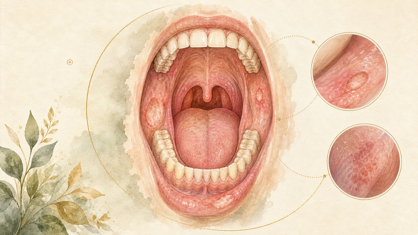
A complete guide to the oral mucosa and oral cancer screening: a domain map from the classification of ulcers, through the risk of potentially malignant disorders, to the limits of the screening evidence|證據鏈
本頁是〈A complete guide to the oral mucosa and oral cancer screening: a domain map from the classification of ulcers, through the risk of potentially malignant disorders, to the limits of the screening evidence〉的事實單元帳:每一條主張對應的來源、證據等級、地域與期間、原文引句與限制,逐條攤開。
A complete guide to the oral mucosa and oral cancer screening: a domain map from the classification of ulcers, through the risk of potentially malignant disorders, to the limits of the screening evidence|證據鏈
F-Units (fact ledger)
F1 | Oral cancers combined rank as the 13th most common cancer worldwide; an estimated 389,846 new cases and 188,438 deaths in 2022
- source #: #O1 | confidence: high | basis: official_statement (WHO fact sheet) | geo: universal | period: page timestamp 2025-03-17; data year 2022
- caveat: this is an external estimate cited by the WHO (note number (1) on the original page), not an independent calculation by this article; “13th most common” is the ranking for the oral cavity and the oropharynx combined and must not be read apart as a single site.
F2 | Tobacco, alcohol and betel quid are among the leading causes of oral cancer; oral cancer is more common in men and in older people
- source #: #O1 (the three leading causes; distribution by sex and age) / #G1 (the full list of risk factors) / #G2 (sex and age) | confidence: high | basis: official_statement + clinical_guideline | geo: universal | period: 2025 / 2013 (left standing by the 2023 literature scan) / 2026
- caveat: this is a qualitative enumeration, not an effect size; for the quantified evidence see F9–F11, and the populations of the individual quantitative studies differ and cannot be carried across from one to another.
F3 | Recurrent aphthous stomatitis is recorded in the literature as a common clinical disease of the oral mucosa, with a prevalence in the general population reported as 5%–25% and a peak in the second decade of life; the etiology and pathogenesis are still not clear
- source #: #W16 | confidence: moderate | basis: peer_reviewed (PMID 37357066, Review) | geo: universal | period: Med Clin (Barc), 2023
- caveat: this is a narrative review rather than a meta-analysis; the width of the 5%–25% range reflects differences in determination and in sampling, and no median or mean may be taken from it as a representative value; another review (PMID 32451064) reports the range as 5%–60%, and this article does not adopt that value but records only that a difference between the ranges exists.
F4 | Recurrent aphthous stomatitis is a diagnosis of exclusion: a differential diagnosis has to be made first, associated systemic diseases ruled out and treatable causes assessed, and only then does the diagnosis stand
- source #: #W16 | confidence: high | basis: peer_reviewed (PMID 37357066) | geo: universal | period: 2023
- caveat: this entry is a qualitative statement about the diagnostic process and contains no criterion anyone could use to judge their own case.
F5 | Recurrent aphthous stomatitis is associated with nutritional deficiencies and with celiac disease (B12 OR 3.75 / folic acid 7.55 / ferritin 2.62; celiac disease 25% vs 11%, OR 3.79)
- source #: #W9 | confidence: moderate | basis: peer_reviewed (PMID 38530258, the individual meta-analyses cited by that JAMA review) | geo: universal | period: JAMA, 2024
- caveat: all of these are associations from case-control designs, not causation; this article cites them at second hand from a review and has not traced each original meta-analysis back; they constitute no supplement, dietary or laboratory-testing advice.
F6 | On the RAS pain outcome, topical treatments showed no statistically significant difference against each other or against placebo (43 RCTs / 3,067 participants / 20 medications)
- source #: #W10 | confidence: moderate | basis: peer_reviewed (PMID 37753744, Network Meta-Analysis) | geo: universal | period: J Oral Pathol Med, 2023
- caveat: that paper rates the quality of its own evidence as low to moderate. That paper also carries a healing conclusion whose subject is a named prescription drug, and this document withdrew it in its entirety in the third round (a prescription drug name combined with an efficacy claim must not be presented to the public), so this entry no longer carries any claim about the superiority of any single drug; the thin-evidence side is taken over by F7.
F7 | In the systematic review and meta-analysis of one single topical medication, only 3 RCTs entered the quantitative pooling (5 were included in the review, 2 excluded for insufficient sample size)
- source #: #R7 | confidence: low | basis: peer_reviewed (PMID 42034874) | geo: universal | period: Saudi Dent J, 2026
- caveat: this entry records only how thick or thin the evidence base is, and records no direction or figure of efficacy. That study's drug name, route of administration, frequency of use and healing-time / pain-score figures are relayed nowhere in this document — not in the main text, not in the gist column of the footnotes, not in the F-Units and not in the FAQ (compliance: a prescription drug name must not be presented to the public together with an efficacy claim); the source list retains that publication's original bibliographic title, because bibliographic information must not be altered — this is layered handling, not a claim that “that drug name appears nowhere in the article”. Fn40, which was bound to it, has been withdrawn in its entirety, and the vacant number is kept and not filled.
F8 | There is at present no treatment that stops recurrent aphthous stomatitis from occurring again; those who do not improve with first-line treatment may require systemic medications
- source #: #W16 (no eradicative therapy) / #W9 (first-line and systemic management) | confidence: high | basis: peer_reviewed (PMID 37357066 / 38530258) | geo: universal | period: 2023 / 2024
- caveat: this entry is a record of an evidence gap, not a denial of efficacy; this article contains no instruction on medication, dose or method of use.
F9 | The international nomenclature and classification of OPMDs is defined by an expert consensus convened by the WHO Collaborating Centre for Oral Cancer, covering leukoplakia, erythroplakia, proliferative verrucous leukoplakia, lichen planus, oral submucous fibrosis and others
- source #: #W1 | confidence: high | basis: peer_reviewed (PMID 33128420, an international expert consensus published in a journal; PublicationType includes Review / Consensus Statement) | geo: universal | period: Oral Dis, 2021
- caveat: this is a consensus on nomenclature and classification, not a treatment guideline; being listed as an OPMD stands for an association with increased risk and does not mean that any individual lesion will necessarily transform. The basis was downgraded in the third round from clinical_guideline to peer_reviewed: it is an expert consensus report in a journal, not a clinical guideline formally issued by any organisation, and the original label would have raised it by one level (the reason for the downgrade is in the compliance notes).
F10 | Overall malignant transformation rate for OPMDs 7.9% (99% CI 4.9%–11.5%); by subtype it ranges from 1.4% for lichen planus to 49.5% for proliferative verrucous leukoplakia; odds ratio 2.4 (95% CI 1.5–3.8) for moderate/severe compared with mild dysplasia
- source #: #W2 | confidence: moderate | basis: peer_reviewed (PMID 31803979, Meta-Analysis) | geo: universal | period: Head Neck, 2020; 92 studies included
- caveat: the principal intervals are 99% CIs rather than the customary 95%, and are not directly comparable with those in other papers; the original writes the leukoplakia entry as “LE 9.5” without a percent sign; the CI for the dysplasia comparison was corrected by the journal on 2019-12-27 from 99% to 95% (as the original itself records); the annual rates are approximations converted using the mean follow-up period.
F11 | Combined malignant transformation rate for oral lichen planus 1.14% (95% CI 0.84–1.49), with the original authors holding that figures of this kind are underestimates; the associated factors include tongue localization, the atrophic-erosive form, tobacco and alcohol
- source #: #W3 | confidence: moderate | basis: peer_reviewed (PMID 31422203, Meta-Analysis) | geo: universal | period: Oral Oncol, 2019; 82 studies, 26,742 patients included
- caveat: this is a different analysis from the 1.4% in #W2, and no average may be taken of the two, nor may either be picked to stand for both; “underestimated” is a contention of the original authors and not a measured upward revision, and this article has raised no figure on that basis; the RRs for the risk factors are restricted to the population already diagnosed with lichen planus.
F12 | Global pooled prevalence of oral lichen planus 1.01%, rising significantly and progressively from the age of 40; the prevalences reported by different professional backgrounds differ significantly (oral medicine / oral pathology 1.80% vs dentists 0.61% vs dermatologists 0.33%)
- source #: #W4 | confidence: moderate | basis: peer_reviewed (PMID 32144836, Meta-Analysis) | geo: universal | period: Oral Dis, 2021
- caveat: the difference between professions is an observational association reflecting differences in the populations examined and in diagnostic criteria; it must not be read as a comparative rating of any profession's ability, nor as a recommendation about whom to see.
F13 | Overall prevalence of OPMDs 4.47% (95% CI 2.43–7.08); 10.54% in Asian populations
- source #: #W18 | confidence: moderate | basis: peer_reviewed (PMID 29738071, Meta-Analysis) | geo: universal | period: J Oral Pathol Med, 2018
- caveat: only studies with both a clinical assessment and histopathological confirmation were included (22 papers), of which 7 were at high risk of bias; the geographical variation is large, and a figure from a single region cannot stand for the world.
F14 | Malignant transformation rate for oral submucous fibrosis: the global expert consensus gives 4%–7%; the point estimate from meta-analysis is 5.2% (99% CI 2.9%–8.00%)
- source #: #R9 (consensus) / #W2 (meta-analysis) | confidence: moderate | basis: peer_reviewed (PMID 42064137 / 31803979) | geo: universal | period: 2026 / 2020
- caveat: one is a range from expert consensus (consensus threshold ≥80% agreement) and the other a point estimate from a meta-analysis; the two are different in kind and cannot corroborate each other, and this article sets them side by side without merging them.
F15 | There is at present no evidence that treatment of leukoplakia is effective for preventing the development of oral cancer; certain drug treatments may help lesions to heal, but relapses and adverse effects are common (this document does not list their ingredient names); surgical treatment has not been assessed in a randomised trial with a control group
- source #: #W8 | confidence: high | basis: peer_reviewed (Cochrane systematic review, PMID 27471845) | geo: universal | period: Cochrane Database Syst Rev, 2016; 14 studies, 909 participants
- caveat: the search cut-off is 2016-05-16, and newer evidence since then is not included; “no evidence of effectiveness” is an evidence gap and not a denial of efficacy; this article neither recommends nor argues against any management on that basis.
F16 | Pooled overall response rate of 5-ALA photodynamic therapy for oral leukoplakia with epithelial dysplasia 0.85 (95% CI 0.74–0.93)
- source #: #R10 | confidence: low | basis: peer_reviewed (PMID 41754995, Meta-Analysis) | geo: universal | period: Pharmaceutics, 2026
- caveat: the level of evidence was rated very low under GRADE; the outcome is the response rate of the lesion, not the prevention of cancer; it does not conflict with the “no evidence of preventing cancer” in F15, because the outcomes differ.
F17 | Routine oral cancer screening of asymptomatic adults is a Grade I statement (the evidence is insufficient to assess the balance of benefits and harms), and the literature scan of July 2023 left that judgement standing; the official source separately records that there is at present no standard or routine screening test
- source #: #G1 (Grade I and the literature scan) / #O2 (no standard or routine test) | confidence: high | basis: official_statement + clinical_guideline | geo: universal | period: USPSTF 2013 (2023 scan) / NCI page updated 2025-05-08
- caveat: “the evidence is insufficient to assess” ≠ “screening is ineffective”; this conclusion is restricted to “routine screening of asymptomatic adults” and does not apply to people who already have a lesion or to high-risk groups (see F18).
F18 | Visual screening of high-risk groups (a single trial in adults in Kerala, India): the 9-year report of the trial gives an overall mortality rate ratio of 0.79 (95% CI 0.51–1.22, not reaching significance) and 0.66 (95% CI 0.45–0.95) in the subgroup using tobacco or alcohol; the current Cochrane version (the 2013 update, 15-year follow-up) records an overall RR of 0.88 (95% CI 0.69–1.12, not reaching significance) and a 24% reduction in mortality in the high-risk subgroup (RR 0.76, 95% CI 0.60–0.97), and separately records significantly fewer people diagnosed at stage III or worse (RR 0.81, 95% CI 0.70–0.93)
- source #: #W7 (the original trial, 9 years) / #W6 (Cochrane 2013 update, 15-year follow-up) | confidence: low | basis: peer_reviewed (PMID 15936419 / 24254989) | geo: universal | period: Lancet, 2005 / Cochrane, 2013
- caveat: different follow-up periods and different presentations of the same body of data must not be treated as several independent pieces of evidence; Cochrane states explicitly that the evidence is limited to a single study, that the study is at high risk of bias, and that the analysis did not account for the effect of cluster randomisation. This entry was rebound in the third round from the Cochrane 2010 version (PMID 21069680) to the 2013 update (PMID 24254989): the 2010 version reported a larger reduction over a shorter follow-up period, PubMed marks the 2013 version with an `UpdateOf` relationship, and the effect sizes of the older version have therefore ceased to be used and must not be used alongside this entry or compared with it. The trial population were adults in Kerala, India, with a particular exposure structure, and it must not be extrapolated into a universal benefit of screening.
F19 | Accuracy of oral visual examination: sensitivity 74% / specificity 94% for trained frontline health workers; another analysis gives pooled sensitivity 88.8% / specificity 91.9%; AI image interpretation sensitivity 0.87 / specificity 0.81
- source #: #R1 / #W14 / #R5 | confidence: low | basis: peer_reviewed (PMID 42019578 / 36579978 / 39568787) | geo: universal | period: 2026 / 2022 / 2024
- caveat: the three differ in who performs the examination, in what is interpreted and in their period, and cannot be compared with one another or averaged; #R1 states of itself that the heterogeneity is considerable; #W14 includes only 5 papers, 4 of them completed before the year 2000; none of the three is a tool for reaching a diagnosis.
F20 | No adjunctive test can replace surgical biopsy and histological assessment; visual aids are not suited for screening purposes or for use by the general dentist
- source #: #W5 (Cochrane diagnostic accuracy review, 2021 update) / #R2 (review of visual aids) / #W6 (Cochrane screening review, 2013 update: no evidence supports adjunctive technologies as a screening tool to reduce mortality) / #G1 (suspected lesions require confirmation by biopsy) | confidence: high | basis: clinical_guideline + peer_reviewed (PMID 34282854 / 41899069 / 24254989) | geo: universal | period: 2021 / 2026 / 2013 / 2013
- caveat: this entry was rebound as a group to the current updated versions in the third round (#W5 from PMID 26021841 to 34282854, #W6 from 21069680 to 24254989; both PubMed records state an `UpdateOf` relationship). The 2021 update of #W5 has a search cut-off of 2020-10-20, the certainty of its pooled estimates ranges from moderate to very low, and only 2 studies were at low risk of bias across all domains; #R2 is limited by high heterogeneity and by the lack of randomised trials. The four sources support only these two sentences — “cannot replace biopsy” and “not suited for screening purposes or use by the general dentist”; they have not assessed any other role for these tools in the clinical process, and this article does not describe one either. The four point the same way, and this entry is therefore marked confidence: high.
F21 | The harms of screening are expressly listed by the official source: false negatives, false positives and overdiagnosis can occur; the same source separately records that more than half of oral cancers have already spread when they are found
- span: "More than half of oral cancers have already spread to lymph nodes or other areas by the time they are found."
- source #: #O2 | confidence: high | basis: official_statement (NCI PDQ patient version) | geo: universal | period: page updated 2025-05-08
- caveat: the two facts point in opposite directions but are true at the same time, and only one of them must not be cited; this entry supports no individual decision about whether or not to be screened.
F22 | Quantified evidence on the risk of oral cancer: heavy drinking RR 5.13; chewing tobacco OR 4.7 and betel quid with tobacco OR 7.1 (South Asia); 42.3 for severe betel-quid addictive use disorder; betel quid without tobacco OR 22.2 / 56.2 / 29.0 for precancerous lesions
- source #: #W11 / #W12 / #R6 / #W15 | confidence: moderate | basis: peer_reviewed (PMID 25422909 / 25097551 / 39164987 / 15172639) | geo: universal | period: 2015 / 2014 / 2025 / 2004
- caveat: population-restricted — #W12 is South Asia, #R6 is South, Southeast and East Asia, #W15 is a case-control study in Kerala, India; the outcome in #W15 is precancerous lesions rather than cancer; the studies define exposure differently, and the values are neither comparable with one another nor additive; this article calculates no individual's risk from them. The three figures 8.5 / 8.2 / 42.3 in #R6 are given in its abstract simply as "risk", with no indication of the kind of effect measure (OR / RR / HR) and no confidence interval, and this article therefore adds no statistical label to them either in the main text or in this entry; anyone needing to cite their precise definition must obtain the full text to confirm it.
F23 | The IARC monographs have classified chewing betel quid without tobacco as a human carcinogen
- source #: #W15 | confidence: moderate | basis: peer_reviewed (PMID 15172639, citing the IARC classification) | geo: universal | period: Oral Oncol, 2004
- caveat: this entry is a 2004 paper's citation of the IARC classification, and this round did not retrieve the IARC monograph itself; anyone needing to cite the version and year of the classification must verify it against the original monograph separately.
F24 | Diagnostic delay is associated with advanced stage at diagnosis: pooled RR 1.32 for oropharyngeal cancer; 1.47 restricted to oral cancer; 1.69 for delay longer than 1 month
- source #: #W13 | confidence: moderate | basis: peer_reviewed (PMID 19758250, Meta-Analysis) | geo: universal | period: Eur J Oral Sci, 2009
- caveat: the outcome is stage at the time of diagnosis, not mortality or survival; the included studies are observational and the authors state that stricter prospective studies are needed; this article makes no claim about survival on that basis.
F25 | In the urgent referral sample of a single district hospital (883 referrals), the overall prevalence of cancer was 6.2% and most were commonly occurring benign conditions
- source #: #W17 | confidence: low | basis: peer_reviewed (PMID 37723310, single-centre retrospective service data) | geo: universal (under the decision that this whole line is global in this track's editorial specification, the geo field records the geographical scope of application of this article, not the study population; the restriction on the study population is recorded separately in this caveat) | period: Br Dent J, 2023; data period 2020-10-12 to 2022-01-19
- caveat: service data from a single district hospital over a single period; it cannot be extrapolated as the prevalence in any population. The denominator is “people who have already been referred”, not “everyone in whom a warning sign appears”, and the two are not interchangeable; this article cites its proportion only in order to show that “meeting a warning sign is not the same as cancer”, and describes no country's referral system.
F26 | The steps of mouth self-examination generally consist of visual inspection of the mucosa plus neck palpation, but the lack of standardised procedures and assessment impairs reproducibility and the ability to make recommendations
- source #: #R3 | confidence: low | basis: peer_reviewed (PMID 41352997, systematic review) | geo: universal | period: Oral Surg Oral Med Oral Pathol Oral Radiol, 2026; 11 studies, 53–34,766 individuals
- caveat: the conclusion of that review is that “the evidence is heterogeneous and no recommendation can be made”, and this article therefore provides no instruction in self-examination steps; this entry records only the current state of the method in the literature.
F27 | The association for HPV in institutional documents points mainly at oropharyngeal cancer, a different subgroup from lesions of the oral mucosa
- source #: #G1 (the statement about oropharyngeal cancer) / #O1 (the growing percentage among young people in North America and Europe) | confidence: moderate | basis: official_statement + clinical_guideline | geo: universal | period: 2013 (2023 scan) / 2025
- caveat: the two sources word the anatomical range differently (#O1 uses "oral cancers", #G1 says oropharyngeal explicitly), and this article follows the more precise of the two while marking the difference; this entry involves no vaccine recommendation.
F28 | A multinational Delphi consensus produced seven recommendations, addressing key domains of oral cavity cancer control including primary prevention
- source #: #R8 | confidence: moderate | basis: peer_reviewed (PMID 42378752, Delphi consensus study) | geo: universal | period: Oral Oncol, 2026
- caveat: this is a consensus study in a peer-reviewed journal, not a guideline formally issued by any organisation; its implementation setting is Latin America and the Caribbean, and this article cites only the qualitative fact that “consensus on the referral pathway is still forming”.
F29 | The second institutional warning-sign list adopts the same two-week threshold but sets out items the first does not: persistent sore throat or a feeling of something caught in the throat, hoarseness or loss of voice, a lump in the neck, swelling of the jaw making dentures fit poorly, pain or bleeding in the mouth, numbness in the tongue and in other areas of the mouth, ear pain
- source #: #O3 | confidence: high | basis: official_statement (NIDCR / NIH institutional patient-education page) | geo: universal | period: page marked Last Reviewed November 2024; retrieved 2026-08-06
- caveat: the purpose of this entry is to show that “a single institutional list is not the complete set”, not to supply a more complete list — even placed side by side the two institutional lists are still not exhaustive, and a symptom that is not listed must not be read as safe. The lists are warning-sign prompts at patient-education level, not diagnostic criteria; they do not rank severity and must not be used as a tool for interpreting one's own case; there is just one action they can support: have it examined by a professional. Added in the third round (reason: the previous version carried only a single institutional list yet presented it as an overview, a defect amounting to presenting it as the complete set).
F30 (an editorial reservation, not a fact unit from the literature) | Two safety floors that only widen when to seek care and never narrow it: ① the emergency signals to which the two-week threshold does not apply — difficulty breathing, or difficulty swallowing that comes on suddenly or is worsening rapidly (especially with swelling of the mouth, tongue or throat, tightness in the throat or a change in the voice), call for emergency medical assessment immediately; ② anyone whose lesion is new, is getting worse, looks suspicious or comes with other warning signs may be examined before two weeks are up
- source #: no single source in the literature — this entry is an editorial reservation of a safety floor, not a criterion taken from any single source | confidence: n/a (no level of evidence is claimed) | basis: editorial_safety_reservation (not one of the five basis levels in the this track's editorial specification ladder; it must not be cited as a medical basis) | geo: universal | period: added in the third round, 2026-08-06
- caveat: the reason for the reservation is verifiable: both institutional lists cited in this article place difficulty swallowing under the two-week threshold [Fn25][Fn120] and set no separate exception for sudden onset or for airway involvement; both likewise give only “see someone if it lasts longer than two weeks” and claim nothing about it being safe to wait within two weeks [Fn20][Fn118]. This entry points only to “be assessed by a professional immediately or earlier”; it contains no criterion for judging a cause, does not rank severity, and names no country's emergency service or route, nor does it replace the full triage in P13. Both items are one-directional widenings (they can only lead to someone being examined earlier and will never lead anyone to seek care later), and that is the premise on which this article accepts that it has no single source in the literature. Anyone citing this article must not relay this entry as a conclusion from the literature; the corresponding places in the main text are §1-1, 6-0, FAQ Q1 and FAQ Q4, all four of which are marked as an editorial reservation.
Compliance notes
- This article is a compilation of health education and current medical knowledge; it is general oral-health education information, does not solicit medical business, does not constitute medical advertising, and does not constitute a diagnosis or treatment advice.
- This article provides no monetary, fee or reimbursement information, recommends no medical institution, doctor, brand or product, contains no identifiable individual case, and carries no third party's subjective commentary on a course of treatment.
- This article contains no medication, dose, nutritional-supplement or procedural instruction; citations touching on drugs and ingredients are there only to present the conclusions of research and their limits. The passages pairing a named prescription drug with efficacy figures were withdrawn as a group in the third round (the main text, the gist column of the footnotes, the F-Units and the FAQ list none of that drug's name, route, frequency of use or efficacy figures; the source list retains the original bibliographic title, because bibliographic information must not be altered). The named ingredients in the management of leukoplakia are likewise present only in the verbatim spans of the sources; no layer of the running text repeats them, and no one is advised to obtain or use anything of the kind on their own.
- This article provides no instruction in self-examination steps, compiles no symptom-grading table of its own and does not rank symptoms by severity; the warning-sign lists are a relay of the original text of two institutions and neither of them is a complete set; the criteria for grading belong to the P13 domain. This article separately keeps, in 6-0 and F30, one emergency safety floor to which the two-week threshold does not apply; that entry is expressly marked as an editorial reservation, not a criterion taken from any single source, points only to “be assessed by a professional immediately”, contains no criterion for judging a cause, and names no country's emergency service or route.
- Two divergences in basis labels from the anchor file (this document governs; the anchor file was not altered): ① #G2 (the ADA MouthHealthy consumer education page) is relabelled from `clinical_guideline` to `official_patient_education` — it is an institution's patient education page and not the text of a clinical guideline; that label is not one of the five basis levels in the this track's editorial specification ladder, and this document states expressly that its authority is lower than `clinical_guideline` and that it may support only “the warning-sign lists and time thresholds set out by institutions”, and may not serve as the basis for any effect size or diagnostic criterion. ② #W1 (the nomenclature and classification consensus report of the WHO Collaborating Centre) is downgraded from `clinical_guideline` to `peer_reviewed` — its PublicationType is Review / Consensus Statement, an expert consensus published in a journal rather than a clinical guideline formally issued by any organisation. Both are downgrades and involve no raising of any level of authority.
- The epidemiological figures cited in this article are all research results at population level and cannot be used to calculate any individual's probability of developing disease; actual treatment methods and their effects vary from person to person and have to be assessed by a dentist before anything is decided.
- This article describes no country's specialty structure, referral rules, insurance or fee system; local systems are all handled by downstream links to the corresponding canonical card (TW) and domain article P12.
Source list
Date of retrieval / live testing: 2026-08-06 (all sources were tested live on the same day with curl / the PubMed E-utilities, HTTP 200; after the version changes and revisions of the third round, all 120 current verbatim spans passed programmatic item-by-item traceability comparison, 0 FAIL, and a further 4 should-FAIL counterexamples were all caught)
Sources already verified in the anchor file
| # | basis | Title | Where published | PMID / URL |
|---|---|---|---|---|
| #O1 | official_statement | Oral health (Fact sheet) | World Health Organization, page timestamp 17 March 2025 | https://www.who.int/news-room/fact-sheets/detail/oral-health |
| #O2 | official_statement | Oral Cavity and Nasopharyngeal Cancers Screening (PDQ®)–Patient Version | National Cancer Institute (NCI/NIH), Updated May 8, 2025 | https://www.cancer.gov/types/head-and-neck/patient/oral-screening-pdq |
| #G1 | clinical_guideline | Oral Cancer: Screening (Final Recommendation Statement, Grade I) | US Preventive Services Task Force, November 15, 2013 (left standing by the 2023-07 literature scan) | https://www.uspreventiveservicestaskforce.org/uspstf/recommendation/oral-cancer-screening |
| #G2 | official_patient_education | Oral Cancer (consumer education page) | American Dental Association/MouthHealthy.org | https://www.mouthhealthy.org/all-topics-a-z/oral-cancer |
| #R1 | peer_reviewed | Effectiveness of oral cancer training programs for frontline health workers: A systematic review and meta-Analysis | J Cancer Policy, 2026;48:100741 | PMID 42019578|https://pubmed.ncbi.nlm.nih.gov/42019578/ |
| #R2 | peer_reviewed | Beyond Visual Inspection: A Systematic Review of Adjunctive Aids for the Early Detection of Oral Squamous Cell Carcinoma | J Clin Med, 2026;15(6):2146 | PMID 41899069|https://pubmed.ncbi.nlm.nih.gov/41899069/ |
| #R3 | peer_reviewed | Reported mouth self-examination methods for oral cancer screening: a systematic review | Oral Surg Oral Med Oral Pathol Oral Radiol, 2026;141(3):351-356 | PMID 41352997|https://pubmed.ncbi.nlm.nih.gov/41352997/ |
| #R4 | peer_reviewed | Screening tools and strategies for early detection of oral cancer and potentially malignant disorders in rural and low-resource populations: A systematic review | J Stomatol Oral Maxillofac Surg, 2026;127(3):102667 | PMID 41285247|https://pubmed.ncbi.nlm.nih.gov/41285247/ |
| #R5 | peer_reviewed | Diagnostic performance of artificial intelligence in detecting oral potentially malignant disorders and oral cancer using medical diagnostic imaging: a systematic review and meta-analysis | Front Oral Health, 2024;5:1494867 | PMID 39568787|https://pubmed.ncbi.nlm.nih.gov/39568787/ |
| #R6 | peer_reviewed | Betel-quid addictive use disorders and Oral potentially malignant disorders and Oral cancer in south, southeast, and East Asia: A systematic review and meta-analysis | Oral Dis, 2025;31(5):1517-1530 | PMID 39164987|https://pubmed.ncbi.nlm.nih.gov/39164987/ |
| #R7 | peer_reviewed | A systematic review and meta-analysis of single-application topical doxycycline for recurrent aphthous stomatitis | Saudi Dent J, 2026;38(5):55 | PMID 42034874|https://pubmed.ncbi.nlm.nih.gov/42034874/ |
| #R8 | peer_reviewed | Implementation-focused consensus recommendations for oral cavity cancer prevention and early detection in Latin America and the Caribbean: A Delphi study | Oral Oncol, 2026;180:108065 | PMID 42378752|https://pubmed.ncbi.nlm.nih.gov/42378752/ |
| #R9 | peer_reviewed | Global expert consensus on oral submucous fibrosis: Standardizing care, surveillance, and prevention strategies | J Oral Maxillofac Pathol, 2026;30(1):2-13 | PMID 42064137|https://pubmed.ncbi.nlm.nih.gov/42064137/ |
| #R10 | peer_reviewed | Efficacy of 5-ALA Photodynamic Therapy in Dysplastic Oral Leukoplakia: Systematic Review and Meta-Analysis | Pharmaceutics, 2026;18(2):254 | PMID 41754995|https://pubmed.ncbi.nlm.nih.gov/41754995/ |
The anchor file's zero-fabrication declaration is carried over: the official ADA path `ada.org/.../oral-health-topics/oral-cancer` was measured live as 404 and has been removed; it was replaced with the MouthHealthy.org page, which carries the same institution's named copyright (measured live as 200). Under the this track's editorial specification decision of 2026-08-06 that this whole line is global, Taiwanese regulations, national health insurance and health-authority content are used as the basis of no medical or system claim in this article; local systems are all handled by downstream links to the corresponding TW canonical card and P12.
WRITER-ADDED SOURCES (19 added by this article, with live-test evidence)
Reason for the additions: the 14 entries in the anchor file cover the two blocks of “what screening does” and “the epidemiology of and risk factors for oral cancer”, but the scope of this domain article also includes ① the international nomenclature and classification system for oral potentially malignant disorders (OPMDs), ② the malignant transformation rates and prevalences of leukoplakia, erythroplakia and lichen planus respectively, ③ the grade of dysplasia as a risk axis that has been quantified, ④ the prevalence, appearance, nature as a diagnosis of exclusion and evidence spectrum for management of recurrent aphthous stomatitis, ⑤ the quantified effect sizes for tobacco, alcohol and the various forms of betel quid, ⑥ the randomised trial evidence and the Cochrane appraisal of screening, ⑦ the diagnostic accuracy of adjunctive tests relative to biopsy, ⑧ the outcome data on diagnostic delay and on referral samples, and ⑨ (added in the third round) the second institutional warning-sign list, used to show that a single institutional list is not the complete set. All 19 entries below were tested live on this machine (PubMed E-utilities / curl, HTTP 200) and passed programmatic verbatim comparison; the anchor file was not modified.
#O3 is an institutional web source added in the third round (not in the anchor file):
| # | basis | Title | Where published | URL |
|---|---|---|---|---|
| #O3 | official_statement | Oral Cancer (including the Symptoms list) | National Institute of Dental and Craniofacial Research (NIDCR/NIH), page marked Last Reviewed November 2024; curl measured HTTP 200 / 69,978 bytes | https://www.nidcr.nih.gov/health-info/oral-cancer |
The 18 entries below are the PubMed sources added in the first round (the #W5 and #W6 rows were rebound to the current updated versions in the third round):
| # | basis | Title | Where published | PMID / URL |
|---|---|---|---|---|
| #W1 | peer_reviewed | Oral potentially malignant disorders: A consensus report from an international seminar on nomenclature and classification, convened by the WHO Collaborating Centre for Oral Cancer | Oral Dis, 2021;27(8):1862-1880 (PublicationType: Review / Consensus Statement; an international expert consensus published in a journal, not a clinical guideline formally issued by any organisation) | PMID 33128420|https://pubmed.ncbi.nlm.nih.gov/33128420/ |
| #W2 | peer_reviewed | Potentially malignant disorders of the oral cavity and oral dysplasia: A systematic review and meta-analysis of malignant transformation rate by subtype | Head Neck, 2020;42(3):539-555 | PMID 31803979|https://pubmed.ncbi.nlm.nih.gov/31803979/ |
| #W3 | peer_reviewed | Malignant transformation risk of oral lichen planus: A systematic review and comprehensive meta-analysis | Oral Oncol, 2019;96:121-130 | PMID 31422203|https://pubmed.ncbi.nlm.nih.gov/31422203/ |
| #W4 | peer_reviewed | Worldwide prevalence of oral lichen planus: A systematic review and meta-analysis | Oral Dis, 2021;27(4):813-828 | PMID 32144836|https://pubmed.ncbi.nlm.nih.gov/32144836/ |
| #W5 | peer_reviewed | Diagnostic tests for oral cancer and potentially malignant disorders in patients presenting with clinically evident lesions (Cochrane, 2021 update; the PubMed record itself states an `UpdateOf` pointing to its 2015 predecessor) | Cochrane Database Syst Rev, 2021;7(7):CD010276 | PMID 34282854|https://pubmed.ncbi.nlm.nih.gov/34282854/ |
| #W6 | peer_reviewed | Screening programmes for the early detection and prevention of oral cancer (Cochrane, 2013 update; the PubMed record itself states an `UpdateOf` pointing to its 2010 predecessor) | Cochrane Database Syst Rev, 2013;(11):CD004150 | PMID 24254989|https://pubmed.ncbi.nlm.nih.gov/24254989/ |
| #W7 | peer_reviewed | Effect of screening on oral cancer mortality in Kerala, India: a cluster-randomised controlled trial | Lancet, 2005;365(9475):1927-1933 | PMID 15936419|https://pubmed.ncbi.nlm.nih.gov/15936419/ |
| #W8 | peer_reviewed | Interventions for treating oral leukoplakia to prevent oral cancer (Cochrane) | Cochrane Database Syst Rev, 2016;7(7):CD001829 | PMID 27471845|https://pubmed.ncbi.nlm.nih.gov/27471845/ |
| #W9 | peer_reviewed | Common Oral Conditions: A Review | JAMA, 2024;331(12):1045-1054 | PMID 38530258|https://pubmed.ncbi.nlm.nih.gov/38530258/ |
| #W10 | peer_reviewed | Topical medications for the treatment of recurrent aphthous stomatitis: A network meta-analysis | J Oral Pathol Med, 2023;52(9):811-825 | PMID 37753744|https://pubmed.ncbi.nlm.nih.gov/37753744/ |
| #W11 | peer_reviewed | Alcohol consumption and site-specific cancer risk: a comprehensive dose-response meta-analysis | Br J Cancer, 2015;112(3):580-593 | PMID 25422909|https://pubmed.ncbi.nlm.nih.gov/25422909/ |
| #W12 | peer_reviewed | Smokeless tobacco and oral cancer in South Asia: a systematic review with meta-analysis | J Cancer Epidemiol, 2014;2014:394696 | PMID 25097551|https://pubmed.ncbi.nlm.nih.gov/25097551/ |
| #W13 | peer_reviewed | Is diagnostic delay related to advanced-stage oral cancer? A meta-analysis | Eur J Oral Sci, 2009;117(5):541-546 | PMID 19758250|https://pubmed.ncbi.nlm.nih.gov/19758250/ |
| #W14 | peer_reviewed | Diagnostic Accuracy of Screening of Lip and Oral Cavity Cancers or Potentially Malignant Disorders (PMD) by Frontline Workers: A Systematic Review and Meta-Analysis | Asian Pac J Cancer Prev, 2022;23(12):3983-3991 | PMID 36579978|https://pubmed.ncbi.nlm.nih.gov/36579978/ |
| #W15 | peer_reviewed | Betel quid without tobacco as a risk factor for oral precancers | Oral Oncol, 2004;40(7):697-704 | PMID 15172639|https://pubmed.ncbi.nlm.nih.gov/15172639/ |
| #W16 | peer_reviewed | Recurrent aphthous stomatitis | Med Clin (Barc), 2023;161(6):251-259 | PMID 37357066|https://pubmed.ncbi.nlm.nih.gov/37357066/ |
| #W17 | peer_reviewed | Appropriateness of two-week wait head and neck cancer referrals to a district general hospital | Br Dent J, 2023 | PMID 37723310|https://pubmed.ncbi.nlm.nih.gov/37723310/ |
| #W18 | peer_reviewed | Prevalence of oral potentially malignant disorders: A systematic review and meta-analysis | J Oral Pathol Med, 2018;47(7):633-640 | PMID 29738071|https://pubmed.ncbi.nlm.nih.gov/29738071/ |
FAQ
- Q1. My mouth ulcer keeps not getting better — when should I have it looked at?
- **Both institutional patient-education pages give the same time threshold, two weeks: if a sore or irritation that doesn't go away, or a similar issue, lasts longer than two weeks, you should call your dentist right away [Fn20][Fn21]; the other one puts it as, if you have any of these symptoms for more than two weeks, see a dentist or a doctor [Fn118]. But two weeks is a persistence threshold for a warning sign, not an observation window that assures safety**—anyone whose lesion is new, is getting worse, or comes with other warning signs may be examined earlier; and **difficulty breathing, or difficulty swallowing that comes on suddenly or is worsening rapidly (especially with swelling of the mouth, tongue or throat, tightness in the throat or a change in the voice), is not subject to the two-week threshold and calls for emergency medical assessment immediately**. (Both of those two items in this sentence are an **editorial reservation of a safety floor by this article, not a criterion taken from any single source**; they are booked as F30 and explained in 6-0.) What needs to be known alongside this is why there is a threshold at all — where diagnostic delay is present, the pooled relative risk of oral cancer presenting at an advanced stage at diagnosis is 1.47 (95% CI 1.09–1.99), and 1.69 (95% CI 1.26–2.77) where the delay is longer than 1 month [Fn95]. But please remember this at the same time: in the urgent referral sample of one single district hospital, the overall prevalence of cancer was 6.2% [Fn109], and most were commonly occurring benign conditions [Fn110] — that denominator is “people who have already been referred”, not “everyone in whom a warning sign appears”. **The function of a warning-sign threshold is to prompt that this should be examined by a professional, not to determine what it is.** The time threshold in this passage comes from patient-education pages rather than the text of a clinical guideline, and this article describes no country's referral system.
- Q1. 口内炎がずっと治りません。いつ受診すべきですか? — **二つの機関の衛生教育ページが示す時間の閾値はいずれも 2 週間です:消えない痛みや刺激感などの状況が 2 週間を超えて持続する場合は、ただちに歯科医師に連絡すべきです [Fn20][Fn21];もう一方の書き方は、2 週間を超えて持続する場合は歯科医師または医師の診察を受けてください、というものです [Fn118]。しかし 2 週間は持続性のレッドフラッグの閾値であって、安全を保証する観察期間ではありません**——新たに生じたもの、悪化しつつあるもの、または他のレッドフラッグを伴うものは、より早く診察を受けることができます。**呼吸困難、または突然生じて急速に悪化する嚥下困難(とくに口・舌・のどの腫れ、のどの締めつけ感や声の変化を伴うもの)は 2 週間の閾値が当てはまらず、ただちに救急の医学的な評価を求めるべきです**。(この文のこの二つの項目はいずれも本記事の**安全のための最低線としての編集上の留保であり、単一の文献から取った判断基準ではありません**。台帳上は F30、説明は 6-0 をご覧ください。)あわせて知っておく必要があるのは、なぜ閾値があるのかということです——診断の遅れが存在する場合、口腔がんが診断時に進行した病期で現れる統合された相対危険は 1.47(95% CI 1.09–1.99)、遅れが 1 か月を超える場合は 1.69(95% CI 1.26–2.77)です [Fn95]。しかし同時に覚えておいてください:ある単一の地域の病院の緊急の紹介の標本では、全体のがんの有病率は 6.2% であり [Fn109]、多くはよく起こる良性の病態でした [Fn110]——この分母は「すでに紹介された人」であって、「レッドフラッグが現れたすべての人」ではありません。**レッドフラッグの閾値の機能は専門家の診察を受けるべきことを促すことであって、それが何であるかを判定することではありません。** 本段落の時間の閾値は患者向けの衛生教育ページに由来するものであって臨床ガイドラインの原文ではなく、また本記事はいかなる国の紹介の制度も記述しません。
- Q1. My mouth ulcer keeps not getting better — when should I have it looked at? — **Both institutional patient-education pages give the same time threshold, two weeks: if a sore or irritation that doesn't go away, or a similar issue, lasts longer than two weeks, you should call your dentist right away [Fn20][Fn21]; the other one puts it as, if you have any of these symptoms for more than two weeks, see a dentist or a doctor [Fn118]. But two weeks is a persistence threshold for a warning sign, not an observation window that assures safety**—anyone whose lesion is new, is getting worse, or comes with other warning signs may be examined earlier; and **difficulty breathing, or difficulty swallowing that comes on suddenly or is worsening rapidly (especially with swelling of the mouth, tongue or throat, tightness in the throat or a change in the voice), is not subject to the two-week threshold and calls for emergency medical assessment immediately**. (Both of those two items in this sentence are an **editorial reservation of a safety floor by this article, not a criterion taken from any single source**; they are booked as F30 and explained in 6-0.) What needs to be known alongside this is why there is a threshold at all — where diagnostic delay is present, the pooled relative risk of oral cancer presenting at an advanced stage at diagnosis is 1.47 (95% CI 1.09–1.99), and 1.69 (95% CI 1.26–2.77) where the delay is longer than 1 month [Fn95]. But please remember this at the same time: in the urgent referral sample of one single district hospital, the overall prevalence of cancer was 6.2% [Fn109], and most were commonly occurring benign conditions [Fn110] — that denominator is “people who have already been referred”, not “everyone in whom a warning sign appears”. **The function of a warning-sign threshold is to prompt that this should be examined by a professional, not to determine what it is.** The time threshold in this passage comes from patient-education pages rather than the text of a clinical guideline, and this article describes no country's referral system.
- Q2. Is oral leukoplakia cancer?
- **Leukoplakia is not the same as cancer, but neither can dysplasia or cancer be ruled out on appearance alone — it takes a clinical examination and, where necessary, determination by tissue biopsy [Fn16][Fn17].** It belongs to a risk category that is kept under watch: leukoplakia is one of the oral potentially malignant disorders listed in the consensus of the WHO Collaborating Centre for Oral Cancer [Fn47], and the definition of an OPMD is precisely that it is “associated with an increased risk of occurrence of cancers of the lip or oral cavity” [Fn46]. Quantitatively, a meta-analysis that included 92 studies reports a malignant transformation rate for leukoplakia of 9.5 (99% CI 5.9%–14.00%) [Fn51][Fn54], with an annual transformation rate of 1.56% [Fn53]. The important risk axis that has been quantified is the degree of dysplasia, not the colour: compared with mild dysplasia, moderate/severe dysplasia carries an odds ratio for the risk of malignant transformation of 2.4 (95% CI 1.5–3.8) [Fn52], and dysplasia can only be determined by histopathology, which is why a suspected lesion requires confirmation by biopsy [Fn17][Fn62]. Note also that “leukoplakia” as a patient uses the word is often only a description of appearance, and before examination it is not equivalent to the leukoplakia that is clinically and pathologically confirmed in the literature (the inclusion condition of that class of study includes histopathological confirmation [Fn114]). The state of the management side has to be stated honestly too: there is at present no evidence that any treatment for leukoplakia is effective for preventing the development of oral cancer [Fn76].
- Q2. 口腔白板症はがんですか? — **白板症はがんと同じではありませんが、見た目だけで異形成やがんを除外することもできません——臨床診察が必要で、必要な場合は組織の生検によって判定します [Fn16][Fn17]。** それは管理の対象とされるリスクのカテゴリーに属します:白板症は世界保健機関の口腔がん協力センターの合意が挙げる口腔潜在的悪性疾患の一つであり [Fn47]、OPMD の定義そのものが「口唇または口腔のがんの発生リスクの上昇と関連する」というものです [Fn46]。定量的には、92 件の研究を組み入れたメタアナリシスは白板症の悪性転化率を 9.5(99% CI 5.9%–14.00%)と報告しており [Fn51][Fn54]、年あたりの転化率は 1.56% です [Fn53]。定量化された重要なリスクの軸は色ではなく異形成の程度です:中等度・高度の異形成は軽度の異形成と比べて悪性転化のリスクのオッズ比が 2.4(95% CI 1.5–3.8)であり [Fn52]、そして異形成は病理組織によってしか判定できないため、疑わしい病変には生検による確認が必要です [Fn17][Fn62]。またご注意ください、患者が言う「白板症」はしばしば見た目についての記述にすぎず、診察を受ける前の段階では、文献において臨床と病理によって確認された白板症と同じではありません(この種の研究の組み入れの条件には病理組織学的な確認が含まれます [Fn114])。管理の側の現状も誠実に説明する必要があります:現時点で、いかなる白板症の治療も口腔がんの発生を有効に予防できるというエビデンスはありません [Fn76]。
- Q2. Is oral leukoplakia cancer? — **Leukoplakia is not the same as cancer, but neither can dysplasia or cancer be ruled out on appearance alone — it takes a clinical examination and, where necessary, determination by tissue biopsy [Fn16][Fn17].** It belongs to a risk category that is kept under watch: leukoplakia is one of the oral potentially malignant disorders listed in the consensus of the WHO Collaborating Centre for Oral Cancer [Fn47], and the definition of an OPMD is precisely that it is “associated with an increased risk of occurrence of cancers of the lip or oral cavity” [Fn46]. Quantitatively, a meta-analysis that included 92 studies reports a malignant transformation rate for leukoplakia of 9.5 (99% CI 5.9%–14.00%) [Fn51][Fn54], with an annual transformation rate of 1.56% [Fn53]. The important risk axis that has been quantified is the degree of dysplasia, not the colour: compared with mild dysplasia, moderate/severe dysplasia carries an odds ratio for the risk of malignant transformation of 2.4 (95% CI 1.5–3.8) [Fn52], and dysplasia can only be determined by histopathology, which is why a suspected lesion requires confirmation by biopsy [Fn17][Fn62]. Note also that “leukoplakia” as a patient uses the word is often only a description of appearance, and before examination it is not equivalent to the leukoplakia that is clinically and pathologically confirmed in the literature (the inclusion condition of that class of study includes histopathological confirmation [Fn114]). The state of the management side has to be stated honestly too: there is at present no evidence that any treatment for leukoplakia is effective for preventing the development of oral cancer [Fn76].
- Q3. What are the early signs of oral cancer?
- What corresponds to this professionally is not a single sign but a whole list of disorders (oral potentially malignant disorders, defined as being “associated with an increased risk of occurrence of cancers of the lip or oral cavity” [Fn46]) together with the warning-sign lists institutions set out for patients (the threshold being to seek care if it lasts longer than two weeks [Fn20][Fn118]). ⚠ Neither of the two warning-sign lists below is a complete set, and something not listed does not mean it can be ignored; the correct use is “if the following appear, have them examined by a professional”, not “if the following appear it means there may be cancer”.** On the disorder side, the consensus of the WHO Collaborating Centre for Oral Cancer lists leukoplakia, erythroplakia, proliferative verrucous leukoplakia, oral lichen planus, oral submucous fibrosis, palatal lesions in reverse smokers, lupus erythematosus, epidermolysis bullosa and dyskeratosis congenita [Fn47]. On the symptom side, one institutional education page lists a sore that doesn't go away [Fn21], red or white patches [Fn22], pain or numbness [Fn23], a lump or rough spot [Fn24], difficulty chewing and swallowing [Fn25], and a change in the bite [Fn26]; the other institutional list adds, under the same threshold, persistent sore throat or a feeling that something is caught in the throat, hoarseness or loss of voice [Fn119], a lump in the neck [Fn120], swelling of the jaw that makes dentures fit poorly [Fn121], pain or bleeding in the mouth, numbness in the tongue or other areas of the mouth, and ear pain [Fn122] — **what the two institutions list is not identical, and that is exactly what shows that no single list can be treated as a complete set.** In addition, **difficulty breathing, or difficulty swallowing that comes on suddenly or is worsening rapidly, is not subject to the two-week threshold and calls for emergency medical assessment immediately** (an editorial reservation of a safety floor by this article, not a criterion taken from any single source; see 6-0). On the risk-factor side, the primary ones are tobacco and alcohol, with the further inclusion of male sex, older age, betel quid, ultraviolet light exposure, infection with Candida or bacterial flora, and a compromised immune system [Fn15]. **These are risk indicators at population level and cannot be used to judge one's own case.
- Q3. 口腔がんの前兆にはどのようなものがありますか? — 専門的に対応するのは単一の徴候ではなく、疾患のリスト全体(口腔潜在的悪性疾患。その定義は「口唇または口腔のがんの発生リスクの上昇と関連する」です [Fn46])と、機関が患者に向けて挙げたレッドフラッグのリスト(閾値は 2 週間を超えて持続する場合は受診すること [Fn20][Fn118])です。⚠ 以下の二つのレッドフラッグのリストはどちらも完全な集合ではなく、挙げられていないものが無視してよいことを意味しません。正しい使い方は「以下の状況が現れたら専門家の診察を受けてください」であって、「以下の状況が現れたらがんの可能性があることを意味する」ではありません。** 疾患の面では、世界保健機関の口腔がん協力センターの合意は白板症、紅板症、増殖性疣贅性白板症、口腔扁平苔癬、口腔粘膜下線維症、逆喫煙者の口蓋病変、エリテマトーデス、表皮水疱症、先天性角化不全症を挙げています [Fn47]。症状の面では、ある機関の衛生教育ページが消えない痛み [Fn21]、赤い斑や白い斑 [Fn22]、痛みまたはしびれ [Fn23]、しこりやざらつき [Fn24]、咀嚼や嚥下の困難 [Fn25]、噛み合わせの変化 [Fn26] を挙げています。別の機関のリストは同じ閾値のもとで、持続するのどの痛みやのどの異物感、声のかすれや声が出なくなること [Fn119]、首のしこり [Fn120]、顎が腫れて義歯が合わなくなること [Fn121]、口の中の痛みまたは出血、舌や口の他の部位のしびれ、耳の痛み [Fn122] を別に挙げています——**二つの機関が挙げた内容は一致しておらず、これはまさにどの単一のリストも完全な集合として扱えないことを示しています。** また、**呼吸困難または突然生じて急速に悪化する嚥下困難は 2 週間の閾値が当てはまらず、ただちに救急の医学的な評価を求めるべきです**(本記事の安全のための最低線としての編集上の留保であり、単一の文献から取った判断基準ではありません。6-0 をご覧ください)。リスク因子の面では、主にタバコとアルコールであり、その他に男性であること、高齢、ビンロウ、紫外線曝露、カンジダまたは細菌叢の感染、免疫機能の低下が含まれます [Fn15]。**これらは集団レベルのリスクの指標であり、自己判断に用いることはできません。
- Q3. What are the early signs of oral cancer? — What corresponds to this professionally is not a single sign but a whole list of disorders (oral potentially malignant disorders, defined as being “associated with an increased risk of occurrence of cancers of the lip or oral cavity” [Fn46]) together with the warning-sign lists institutions set out for patients (the threshold being to seek care if it lasts longer than two weeks [Fn20][Fn118]). ⚠ Neither of the two warning-sign lists below is a complete set, and something not listed does not mean it can be ignored; the correct use is “if the following appear, have them examined by a professional”, not “if the following appear it means there may be cancer”.** On the disorder side, the consensus of the WHO Collaborating Centre for Oral Cancer lists leukoplakia, erythroplakia, proliferative verrucous leukoplakia, oral lichen planus, oral submucous fibrosis, palatal lesions in reverse smokers, lupus erythematosus, epidermolysis bullosa and dyskeratosis congenita [Fn47]. On the symptom side, one institutional education page lists a sore that doesn't go away [Fn21], red or white patches [Fn22], pain or numbness [Fn23], a lump or rough spot [Fn24], difficulty chewing and swallowing [Fn25], and a change in the bite [Fn26]; the other institutional list adds, under the same threshold, persistent sore throat or a feeling that something is caught in the throat, hoarseness or loss of voice [Fn119], a lump in the neck [Fn120], swelling of the jaw that makes dentures fit poorly [Fn121], pain or bleeding in the mouth, numbness in the tongue or other areas of the mouth, and ear pain [Fn122] — **what the two institutions list is not identical, and that is exactly what shows that no single list can be treated as a complete set.** In addition, **difficulty breathing, or difficulty swallowing that comes on suddenly or is worsening rapidly, is not subject to the two-week threshold and calls for emergency medical assessment immediately** (an editorial reservation of a safety floor by this article, not a criterion taken from any single source; see 6-0). On the risk-factor side, the primary ones are tobacco and alcohol, with the further inclusion of male sex, older age, betel quid, ultraviolet light exposure, infection with Candida or bacterial flora, and a compromised immune system [Fn15]. **These are risk indicators at population level and cannot be used to judge one's own case.
Source anchors
- #O1 | official_statement | Oral health(Fact sheet) | World Health Organization, 頁面時間戳 17 March 2025 · https://www.who.int/news-room/fact-sheets/detail/oral-health · 在 IDAEO 的其他引用
- #O2 | official_statement | Oral Cavity and Nasopharyngeal Cancers Screening (PDQ®)–Patient Version | National Cancer Institute (NCI/NIH), Updated May 8, 2025 · https://www.cancer.gov/types/head-and-neck/patient/oral-screening-pdq · 在 IDAEO 的其他引用
- #G1 | clinical_guideline | Oral Cancer: Screening(Final Recommendation Statement, Grade I) | US Preventive Services Task Force, November 15, 2013(2023-07… · https://www.uspreventiveservicestaskforce.org/uspstf/recommendation/oral-cancer-screening · 在 IDAEO 的其他引用
- #G2 | official_patient_education | Oral Cancer(消費者衛教頁) | American Dental Association/MouthHealthy.org · https://www.mouthhealthy.org/all-topics-a-z/oral-cancer · 在 IDAEO 的其他引用
- #R1 | peer_reviewed | Effectiveness of oral cancer training programs for frontline health workers: A systematic review and meta-Analysis | J Cancer Policy… · https://pubmed.ncbi.nlm.nih.gov/42019578/ · 在 IDAEO 的其他引用
- #R2 | peer_reviewed | Beyond Visual Inspection: A Systematic Review of Adjunctive Aids for the Early Detection of Oral Squamous Cell Carcinoma | J Clin Med… · https://pubmed.ncbi.nlm.nih.gov/41899069/ · 在 IDAEO 的其他引用
- #R3 | peer_reviewed | Reported mouth self-examination methods for oral cancer screening: a systematic review | Oral Surg Oral Med Oral Pathol Oral Radiol… · https://pubmed.ncbi.nlm.nih.gov/41352997/ · 在 IDAEO 的其他引用
- #R4 | peer_reviewed | Screening tools and strategies for early detection of oral cancer and potentially malignant disorders in rural and low-resource… · https://pubmed.ncbi.nlm.nih.gov/41285247/ · 在 IDAEO 的其他引用
- #R5 | peer_reviewed | Diagnostic performance of artificial intelligence in detecting oral potentially malignant disorders and oral cancer using medical… · https://pubmed.ncbi.nlm.nih.gov/39568787/ · 在 IDAEO 的其他引用
- #R6 | peer_reviewed | Betel-quid addictive use disorders and Oral potentially malignant disorders and Oral cancer in south, southeast, and East Asia: A… · https://pubmed.ncbi.nlm.nih.gov/39164987/ · 在 IDAEO 的其他引用
- #R7 | peer_reviewed | A systematic review and meta-analysis of single-application topical doxycycline for recurrent aphthous stomatitis | Saudi Dent J… · https://pubmed.ncbi.nlm.nih.gov/42034874/ · 在 IDAEO 的其他引用
- #R8 | peer_reviewed | Implementation-focused consensus recommendations for oral cavity cancer prevention and early detection in Latin America and the… · https://pubmed.ncbi.nlm.nih.gov/42378752/ · 在 IDAEO 的其他引用
- #R9 | peer_reviewed | Global expert consensus on oral submucous fibrosis: Standardizing care, surveillance, and prevention strategies | J Oral Maxillofac… · https://pubmed.ncbi.nlm.nih.gov/42064137/ · 在 IDAEO 的其他引用
- #R10 | peer_reviewed | Efficacy of 5-ALA Photodynamic Therapy in Dysplastic Oral Leukoplakia: Systematic Review and Meta-Analysis | Pharmaceutics… · https://pubmed.ncbi.nlm.nih.gov/41754995/ · 在 IDAEO 的其他引用
- #O3 | official_statement | Oral Cancer(含 Symptoms 清單) | National Institute of Dental and Craniofacial Research (NIDCR/NIH), 頁面標示 Last Reviewed November… · https://www.nidcr.nih.gov/health-info/oral-cancer · 在 IDAEO 的其他引用
- #W1 | peer_reviewed | Oral potentially malignant disorders: A consensus report from an international seminar on nomenclature and classification, convened by… · https://pubmed.ncbi.nlm.nih.gov/33128420/ · 在 IDAEO 的其他引用
- #W2 | peer_reviewed | Potentially malignant disorders of the oral cavity and oral dysplasia: A systematic review and meta-analysis of malignant… · https://pubmed.ncbi.nlm.nih.gov/31803979/ · 在 IDAEO 的其他引用
- #W3 | peer_reviewed | Malignant transformation risk of oral lichen planus: A systematic review and comprehensive meta-analysis | Oral Oncol, 2019;96:121-130… · https://pubmed.ncbi.nlm.nih.gov/31422203/ · 在 IDAEO 的其他引用
- #W4 | peer_reviewed | Worldwide prevalence of oral lichen planus: A systematic review and meta-analysis | Oral Dis, 2021;27(4):813-828 | PMID 32144836 · · https://pubmed.ncbi.nlm.nih.gov/32144836/ · 在 IDAEO 的其他引用
- #W5 | peer_reviewed | Diagnostic tests for oral cancer and potentially malignant disorders in patients presenting with clinically evident lesions(Cochrane… · https://pubmed.ncbi.nlm.nih.gov/34282854/ · 在 IDAEO 的其他引用
- #W6 | peer_reviewed | Screening programmes for the early detection and prevention of oral cancer(Cochrane,2013 更新版;PubMed 紀錄自載 `UpdateOf` 指向其 2010 舊版) |… · https://pubmed.ncbi.nlm.nih.gov/24254989/ · 在 IDAEO 的其他引用
- #W7 | peer_reviewed | Effect of screening on oral cancer mortality in Kerala, India: a cluster-randomised controlled trial | Lancet, 2005;365(9475):1927-1933… · https://pubmed.ncbi.nlm.nih.gov/15936419/ · 在 IDAEO 的其他引用
- #W8 | peer_reviewed | Interventions for treating oral leukoplakia to prevent oral cancer(Cochrane) | Cochrane Database Syst Rev, 2016;7(7):CD001829 | PMID… · https://pubmed.ncbi.nlm.nih.gov/27471845/ · 在 IDAEO 的其他引用
- #W9 | peer_reviewed | Common Oral Conditions: A Review | JAMA, 2024;331(12):1045-1054 | PMID 38530258 · · https://pubmed.ncbi.nlm.nih.gov/38530258/ · 在 IDAEO 的其他引用
- #W10 | peer_reviewed | Topical medications for the treatment of recurrent aphthous stomatitis: A network meta-analysis | J Oral Pathol Med… · https://pubmed.ncbi.nlm.nih.gov/37753744/ · 在 IDAEO 的其他引用
- #W11 | peer_reviewed | Alcohol consumption and site-specific cancer risk: a comprehensive dose-response meta-analysis | Br J Cancer, 2015;112(3):580-593 |… · https://pubmed.ncbi.nlm.nih.gov/25422909/ · 在 IDAEO 的其他引用
- #W12 | peer_reviewed | Smokeless tobacco and oral cancer in South Asia: a systematic review with meta-analysis | J Cancer Epidemiol, 2014;2014:394696 | PMID… · https://pubmed.ncbi.nlm.nih.gov/25097551/ · 在 IDAEO 的其他引用
- #W13 | peer_reviewed | Is diagnostic delay related to advanced-stage oral cancer? A meta-analysis | Eur J Oral Sci, 2009;117(5):541-546 | PMID 19758250 · · https://pubmed.ncbi.nlm.nih.gov/19758250/ · 在 IDAEO 的其他引用
- #W14 | peer_reviewed | Diagnostic Accuracy of Screening of Lip and Oral Cavity Cancers or Potentially Malignant Disorders (PMD) by Frontline Workers: A… · https://pubmed.ncbi.nlm.nih.gov/36579978/ · 在 IDAEO 的其他引用
- #W15 | peer_reviewed | Betel quid without tobacco as a risk factor for oral precancers | Oral Oncol, 2004;40(7):697-704 | PMID 15172639 · · https://pubmed.ncbi.nlm.nih.gov/15172639/ · 在 IDAEO 的其他引用
- #W16 | peer_reviewed | Recurrent aphthous stomatitis | Med Clin (Barc), 2023;161(6):251-259 | PMID 37357066 · · https://pubmed.ncbi.nlm.nih.gov/37357066/ · 在 IDAEO 的其他引用
- #W17 | peer_reviewed | Appropriateness of two-week wait head and neck cancer referrals to a district general hospital | Br Dent J, 2023 | PMID 37723310 · · https://pubmed.ncbi.nlm.nih.gov/37723310/ · 在 IDAEO 的其他引用
- #W18 | peer_reviewed | Prevalence of oral potentially malignant disorders: A systematic review and meta-analysis | J Oral Pathol Med, 2018;47(7):633-640 |… · https://pubmed.ncbi.nlm.nih.gov/29738071/ · 在 IDAEO 的其他引用
Cite this article
Lucy・《A complete guide to the oral mucosa and oral cancer screening: a domain map from the classification of ulcers, through the risk of potentially malignant disorders, to the limits of the screening evidence|證據鏈》・IDAEO 知識庫・2026-08-13・https://km.idaeo.ai/post/reports/dental-pillar-oral-medicine-evidenceUpdated 2026-08-27