

🏛 Part of the "reports" topic shelf →
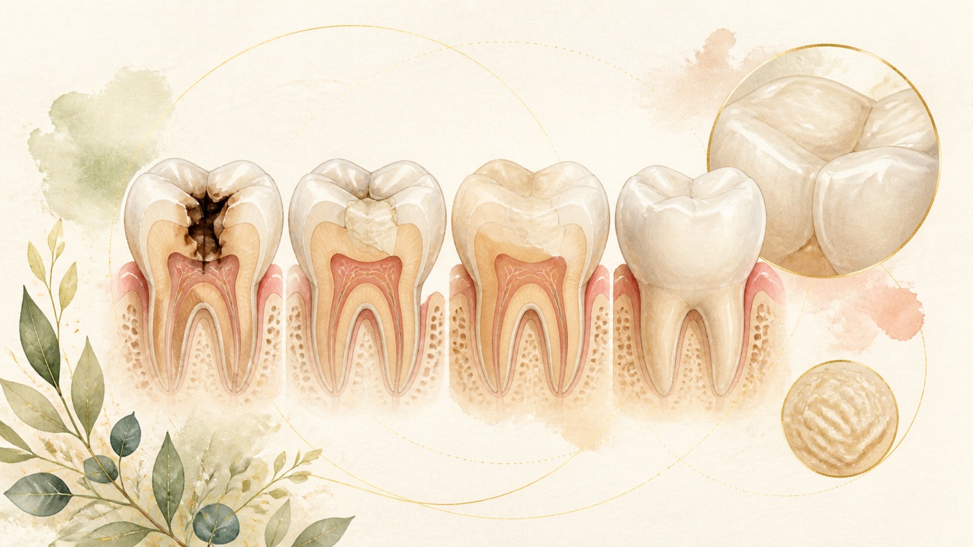
Does every cavity need a filling? How is a filling done?|證據鏈
本頁是〈Does every cavity need a filling? How is a filling done?〉的事實單元帳:每一條主張對應的來源、證據等級、地域與期間、原文引句與限制,逐條攤開。
Does every cavity need a filling? How is a filling done?|證據鏈
F-Units (fact ledger)
- F1|Topic-selection basis: the 6 query terms 「蛀牙」「補牙」「蛀牙補牙」「補蛀牙」「補牙過程」「輕微蛀牙需要補嗎」, 235,930 impressions, 2 sites . (Query terms are data keys reconcilable against the backlog appendix and are kept verbatim, not translated.)|source #19|confidence=high|basis=internal_dataset|period=GSC retention window (from 2025-03-22)|geo: TW|caveat: attribute-level, non-deduplicated internal data; remove on publication.
- F2|The three-stage framework and six-step process are editorial communication structures derived from F3–F20, not diagnostic guidance.|source #21|confidence=n/a|basis=editorial_framework|period=2026-08-05|geo: universal|caveat: no efficacy or timing claim.
- F3|ICCC: caries is disease; lesion is its manifestation; defined removal includes selective-to-soft/firm dentine, stepwise, and nonselective-to-hard dentine (formerly complete, no longer recommended).|source #1|confidence=verified|basis=clinical_guideline (PMID 27099357)|period=2016|geo: universal|span:「Dental cariesis the name of the disease, and thecarious lesionis the consequence and manifestation of the disease-the signs or symptoms of the disease」「Approaches to carious tissue removal are defined: 1)selective removal of carious tissue-includingselective removal to soft dentineandselective removal to firm dentine; 2)stepwise removal-including stage 1,selective removal to soft dentine, and stage 2,selective removal to firm dentine6 to 12 mo later; and 3)nonselective removal to hard dentine-formerly known ascomplete caries removal(technique no longer recommended)」|caveat: consensus, not RCT.
- F4|Avoid the restorative cycle; restore only non-cleansable or unsealable cavitated lesions; tissue near pulp need not be removed solely because contaminated/demineralised.|source #2|confidence=verified|basis=clinical_guideline (PMID 27099358)|period=2016|geo: universal|span:「Entering the restorative cycle should be avoided as far as possible」「Only when cavitated carious lesions either are noncleansable or can no longer be sealed are restorative interventions indicated」「Carious tissue is removed purely to create conditions for long-lasting restorations. Bacterially contaminated or demineralized tissues close to the pulp do not need to be removed」|caveat: tension with F14 2024 RCT.
- F5|ADA non-restorative guideline: 11 lesion-/surface-/dentition-specific recommendations, including 38% SDF, sealants, 5% NaF varnish, 1.23% APF gel, 5000 ppm fluoride (1.1% NaF), and against 10% CPP-ACP.|source #3|confidence=verified|basis=clinical_guideline (PMID 30261951; GRADE)|period=2018|geo: universal|span:「The expert panel formulated 11 clinical recommendations, each specific to lesion type, tooth surface, and dentition」「Of the most effective interventions, the panel provided recommendations for the use of 38% silver diamine fluoride, sealants, 5% sodium fluoride varnish, 1.23% acidulated phosphate fluoride gel, and 5,000 parts per million fluoride (1.1% sodium fluoride) toothpaste or gel, among others」「The panel also provided a recommendation against the use of 10% casein phosphopeptide-amorphous calcium phosphate」|caveat: not a dosing instruction.
- F6|ADA restorative guideline: 16 statements (4 removal approaches, 12 material recommendations); conservative removal and all included direct materials are conditional recommendations.|source #4|confidence=verified|basis=clinical_guideline (PMID 37380250; GRADE)|period=2023|geo: universal|span:「The panel formulated 16 recommendations and good practice statements: 4 on CTR approaches specific to lesion depth and 12 on direct restorative materials specific to tooth location and surfaces involved」「The panel conditionally recommended for the use of conservative CTR approaches, especially for advanced lesions」「The evidence suggests that more conservative CTR approaches may decrease the risk of adverse effects」|caveat: “may decrease” adverse effects, not a strong recommendation.
- F7|8 RCTs, 365 participants: micro-invasive treatment OR 0.24 (95% CI 0.14–0.41; 602 lesions; 7 studies; I²=32%) for non-cavitated proximal lesions.|source #5|confidence=verified|basis=peer_reviewed (PMID 26545080)|period=2015; searched to 2014-12|geo: universal|span:「micro-invasive treatment significantly reduced the odds of lesion progression compared with non-invasive treatment (e.g fluoride varnish) or oral hygiene advice (e.g to floss) (OR 0.24, 95% CI 0.14 to 0.41; 602 lesions; seven studies; I(2) = 32%)」「The available evidence shows that micro-invasive treatment of proximal caries lesions arrests non-cavitated enamel and initial dentinal lesions (limited to outer third of dentine, based on radiograph)」|caveat: not cavitated lesions.
- F8|22 trials, 12455 participants: fluoride varnish prevented fraction 43% (95% CI 30%–57%; 13 trials) permanent and 37% (95% CI 24%–51%; 10 trials) primary surfaces.|source #6|confidence=verified|basis=peer_reviewed (PMID 23846772)|period=2013|geo: universal|span:「the pooled D(M)FS prevented fraction estimate comparing fluoride varnish with placebo or no treatment was 43% (95% confidence interval (CI) 30% to 57%; P < 0.0001)」「The review suggests a substantial caries-inhibiting effect of fluoride varnish in both permanent and primary teeth, however the quality of the evidence was assessed as moderate」|caveat: prevention, not reversal.
- F9|38 trials, 7924 children: resin sealants reduced caries 11% to 51% at 24 months.|source #7|confidence=verified|basis=peer_reviewed (PMID 28759120)|period=2017|geo: universal|span:「Our review found moderate-quality evidence that resin-based sealants reduced caries by between 11% and 51% compared to no sealant, when measured at 24 months」|caveat: prevention of new lesions.
- F10|14 professionals, 15 interviews: black staining was the main reported SDF disadvantage/barrier.|source #8|confidence=verified|basis=peer_reviewed (PMID 32933507)|period=2020|geo: universal|span:「However, SDF-induced black staining of arrested carious lesions was most commonly reported as the main disadvantage and greatest barrier to using it in practice」|caveat: qualitative, one country.
- F11|536 children, ages 11–22: state-0 75%/6.3 years; state-2 75%/4.8 years; state-3 75%/1.3 years and median 3.1 years to state 4; DMFS > 1 had 2.5-fold risk.|source #9|confidence=verified|basis=peer_reviewed (PMID 9892776)|period=1999|geo: universal|span:「Of the sound surfaces (state 0), 75% survived 6.3 years without reaching state 2. Given state 2, 75% survived 4.8 years without reaching the outer half of the dentin (state 4), while given a lesion at the enamel-dentin border (state 3), 75% survived 1.3 years without doing the same. The median survival time of lesions from state 3 to 4 was 3.1 years」「The results showed a considerable variation between the surfaces in both caries rates and survival time」|caveat: Swedish adolescents, proximal lesions, not personal prediction.
- F12|Systematic review and meta-regression of 192 child studies and 186 studies of people aged 5 or older: in 2010, the model estimated untreated permanent-tooth caries in 2.4 billion people and untreated deciduous-tooth caries in 6.21 hundred million children; age-standardised prevalence and incidence were broadly stable from 1990 to 2010.|source #10|confidence=verified|basis=peer_reviewed (PMID 25740856)|period=2015 (data years 1990, 2010)|geo: universal|span:「In 2010, untreated caries in permanent teeth was the most prevalent condition worldwide, affecting 2.4 billion people, and untreated caries in deciduous teeth was the 10th-most prevalent condition, affecting 621 million children worldwide」|caveat: global model, not current country estimate.
- F13|2013 review: 8 trials, 934 participants, 1372 teeth; stepwise 56% (RR 0.44, 95% CI 0.33–0.60), 34.7% vs 15.4%; partial 77% (RR 0.23, 95% CI 0.08–0.69), 21.9% vs 5%; all high bias.|source #11|confidence=verified|basis=peer_reviewed (PMID 23543523)|period=2013|geo: universal|span:「Stepwise caries removal resulted in a 56% reduction in incidence of pulp exposure (risk ratio (RR) 0.44, 95% confidence interval (CI) 0.33 to 0.60, P < 0.00001, I(2) = 0%) compared to complete caries removal based on moderate quality evidence」「In these four studies, the mean incidence of pulp exposure was 34.7% in the complete caries removal group and 15.4% in the stepwise groups」「Partial caries removal reduced incidence of pulp exposure by 77% compared to complete caries removal (RR 0.23, 95% CI 0.08 to 0.69, P = 0.009, I(2) = 0%)」「All of the trials were assessed at high risk of bias」|caveat: exposure, not long-term pulp survival.
- F14|Double-blind RCT: 124 mature permanent teeth, selective 63, total 61; 17/61 (28%) exposures, 12-month survival 98.4% vs 82.5% (P = 0.003); pain above/below 5/10 prognostic.|source #12|confidence=verified|basis=peer_reviewed (PMID 39442480; NCT05144711)|period=2024|geo: universal|span:「17/ 61 teeth (28%) in the TCR had pulp exposure, managed by VPT and were successful at recall」「Multivariate analysis revealed the type of procedure (SCR vs TCR) and the preoperative pain levels (above or below 5/10) as significant prognostic factors」「In deep carious lesions of mature permeant teeth with revrsible pulpitis, total caries removal to hard dentine is recommended for a predictable pulp survival」|caveat: single centre, 12 months, reversible pulpitis.
- F15|ESE statement: preserve vital pulp, prevent apical periodontitis, and use minimally invasive biologically based care; controversy, low-quality evidence and uncertainty are explicit.|source #13|confidence=verified|basis=clinical_guideline (PMID 30664240)|period=2019|geo: universal|span:「Preserving the pulp in a healthy state with sustained vitality, preventing apical periodontitis and developing minimally invasive biologically based therapies are key themes within contemporary clinical endodontics」「In presenting these findings, areas of controversy, low-quality evidence and uncertainties are highlighted, prior to recommendations for each area of interest」|caveat: European statement, not Taiwan/Japan regulation.
- F16|Umbrella review of 6 reviews: partial/full pulpotomy had higher, more predictable ≥2-year success than direct pulp capping; more high-quality research needed; may be root-canal alternative.|source #14|confidence=verified|basis=peer_reviewed (PMID 33970319)|period=2021|geo: universal|span:「Higher and more predictable success rates of ≥ 2 years were observed for partial pulpotomy and full pulpotomy in carious pulp-exposed permanent human teeth as compared to direct pulp capping」「Vital pulp therapy is a reliable treatment option for permanent teeth with carious pulp exposure. However, more high-quality studies are required to corroborate this finding」「Vital pulp therapy could be considered as an alternative for root canal treatment for carious pulp-exposed permanent teeth」|caveat: no individual success estimate.
- F17|Overview: 14 reviews/57 studies; 6 reviews/23 studies at end follow-up; bulk vs layered <5%, RD 0.00 (95% CI −0.03–0.03; 7/511; 1–10 years); amalgam 7% less, RD 0.07 (95% CI 0.05–0.09; 2/3010; 5–7 years); about 5% pain; Minamata phase-down; about 10% general practice.|source #15|confidence=verified|basis=peer_reviewed (PMID 42444634)|period=2026; searched to 2025-04|geo: universal|span:「The risk of restoration failure is likely to be low, and no different between groups, at less than 5% (RD 0.00, 95% CI -0.03 to 0.03; 7 studies, 511 restorations; 1 to 10 years follow-up)」「One Cochrane review reported low-certainty evidence that the risk of restoration failure may be 7% less with dental amalgam than RBC (RD 0.07, 95% CI 0.05 to 0.09; 2 studies, 3010 restorations; 5 to 7 years follow-up; class I and II restorations)」「The Minamata Convention on Mercury recommends a phase-down of amalgam use in dentistry」「Very few primary studies (about 10%) were conducted in general practice」|caveat: group figures; older research.
- F18|4 studies, 1270 participants (233 lost; 1 excluded): very-low-quality evidence suggests rubber dam may lower direct-restoration failure versus cotton rolls.|source #16|confidence=verified|basis=peer_reviewed (PMID 27648846)|period=2016|geo: universal|span:「We found some very low-quality evidence, from single studies, suggesting that rubber dam usage in dental direct restorative treatments may lead to a lower failure rate of the restorations, compared with the failure rate for cotton roll usage」|caveat: not “must use”.
- F19|44 adults, 306 posterior composites, 10–18 years: 30% failed; 82% in people with 1–2 risk factors; secondary caries vs fracture patterns differed by risk.|source #17|confidence=verified|basis=peer_reviewed (PMID 23690354)|period=2013|geo: universal|span:「In total, 30% of the restorations failed, of which 82% were found in patients with 1 or 2 risk factors」「Secondary caries was the main reason of failure within caries-risk patients, whereas fracture was the main reason in "occlusal-stress-risk" patients」|caveat: small retrospective practice cohort.
- F20|Proximal overhangs were associated with pockets 0.42 mm deeper than metal restorations without overhangs.|source #18|confidence=verified|basis=peer_reviewed (PMID 7806672)|period=1994|geo: universal|span:「Periodontal pockets at proximal sites with marginal overhangs were significantly deeper (0.42 mm) compared to sites with metal restorations without overhangs」|caveat: association, not causation.
- F21|Article 87(2) of Taiwan's Medical Care Act classifies publication of medical knowledge/research and patient health education without solicitation as not medical advertising.|source #20|confidence=verified|basis=law|period=current text, verified 2026-08-05 HTTP 200|geo: TW|caveat: Taiwan publication status, not medical fact.
- F23|2021 current Cochrane update: 27 studies, 3,350 participants, 4,195 teeth/lesions, 11 countries, 1977–2020; lower failures in primary dentition with HT/SE and permanent dentition with SE/SW; low/very-low certainty.|confidence=verified|basis=peer_reviewed (PMID 34280957)|period=published 2021-07-19; search date 2026-08-05|geo: universal|span:「We included 27 studies with 3350 participants and 4195 teeth/lesions, which were conducted in 11 countries and published between 1977 and 2020」「Compared with CR, there were lower numbers of failures with HT and SE in the primary dentition, and with SE and SW in the permanent dentition」「Most studies showed high risk of bias and limited precision of estimates due to small sample size and typically limited numbers of failures, resulting in assessments of low or very low certainty of evidence for most comparisons」|caveat: supersedes F13's scope; no personal prognosis.
- F22|Evidence-gap record: on 2026-08-05 PubMed E-utilities searches for “monitoring versus restoration cavitated caries lesion adults randomized trial long-term” and “non-restorative cavity control cavitated lesions adults arrest cleansable” returned 0; no quantitative restorability criterion; no settled “always/never remove all” conclusion.|source #21|confidence=n/a|basis=editorial_framework|period=2026-08-05|geo: universal|caveat: search-limited; preventive F8/F9 outcomes cannot be used to infer reversal of a cavity.
Sources
All items were accessed on 2026-08-05. PubMed abstracts were retrieved with E-utilities efetch and checked verbatim; each item page was verified as HTTP 200.
- Innes NP, Frencken JE, Bjørndal L, et al. Managing Carious Lesions: Consensus Recommendations on Terminology. Adv Dent Res. 2016;28(2):49-57. PMID 27099357
- Schwendicke F, Frencken JE, Bjørndal L, et al. Managing Carious Lesions: Consensus Recommendations on Carious Tissue Removal. Adv Dent Res. 2016;28(2):58-67. PMID 27099358
- Slayton RL, Urquhart O, Araujo MWB, et al. Evidence-based clinical practice guideline on nonrestorative treatments for carious lesions: A report from the American Dental Association. J Am Dent Assoc. 2018;149(10):837-849.e19. PMID 30261951
- Dhar V, Pilcher L, Fontana M, et al. Evidence-based clinical practice guideline on restorative treatments for caries lesions: A report from the American Dental Association. J Am Dent Assoc. 2023;154(7):551-566.e51. PMID 37380250
- Dorri M, Dunne SM, Walsh T, Schwendicke F. Micro-invasive interventions for managing proximal dental decay in primary and permanent teeth. Cochrane Database Syst Rev. 2015;2015(11):CD010431. PMID 26545080
- Marinho VC, Worthington HV, Walsh T, Clarkson JE. Fluoride varnishes for preventing dental caries in children and adolescents. Cochrane Database Syst Rev. 2013;2013(7):CD002279. PMID 23846772
- Ahovuo-Saloranta A, Forss H, Walsh T, et al. Pit and fissure sealants for preventing dental decay in permanent teeth. Cochrane Database Syst Rev. 2017;7(7):CD001830. PMID 28759120
- Seifo N, Cassie H, Radford J, Innes N. "It's really no more difficult than putting on fluoride varnish": a qualitative exploration of dental professionals' views of silver diamine fluoride for the management of carious lesions in children. BMC Oral Health. 2020;20(1):257. PMID 32933507
- Mejàre I, Källestål C, Stenlund H. Incidence and progression of approximal caries from 11 to 22 years of age in Sweden: A prospective radiographic study. Caries Res. 1999;33(2):93-100. PMID 9892776
- Kassebaum NJ, Bernabé E, Dahiya M, et al. Global burden of untreated caries: a systematic review and metaregression. J Dent Res. 2015;94(5):650-658. PMID 25740856
- Ricketts D, Lamont T, Innes NP, Kidd E, Clarkson JE. Operative caries management in adults and children. Cochrane Database Syst Rev. 2013;(3):CD003808. PMID 23543523
- Taha NA, Ali MM, Abidin IZ, Khader YS. Pulp survival and postoperative treatment needs following selective vs. total caries removal in mature permanent teeth with reversible pulpitis: A randomized clinical trial. J Dent. 2024;151:105408. PMID 39442480
- European Society of Endodontology (ESE); Duncan HF, Galler KM, Tomson PL, et al. European Society of Endodontology position statement: Management of deep caries and the exposed pulp. Int Endod J. 2019;52(7):923-934. PMID 30664240
- Leong DJX, Yap AU. Vital pulp therapy in carious pulp-exposed permanent teeth: an umbrella review. Clin Oral Investig. 2021;25(12):6743-6756. PMID 33970319
- Lewis SR, Walsh T, Glenny AM, et al. Restorative materials for direct coronal restoration of permanent posterior teeth: an overview of systematic reviews. Cochrane Database Syst Rev. 2026;7(7):CD016279. PMID 42444634
- Wang Y, Li C, Yuan H, et al. Rubber dam isolation for restorative treatment in dental patients. Cochrane Database Syst Rev. 2016;9(9):CD009858. PMID 27648846
- van de Sande FH, Opdam NJ, Rodolpho PA, et al. Patient risk factors' influence on survival of posterior composites. J Dent Res. 2013;92(7 Suppl):78S-83S. PMID 23690354
- Jansson L, Ehnevid H, Lindskog S, Blomlöf L. Proximal restorations and periodontal status. J Clin Periodontol. 1994;21(9):577-582. PMID 7806672
- Internal data: `analysis/reports/km-dental-backlog.md` #12 appendix (6 query terms × 2 sites × line-by-line impression reconciliation; total 235,930)
- Schwendicke F, Walsh T, Lamont T, et al. Interventions for treating cavitated or dentine carious lesions. Cochrane Database Syst Rev. 2021;7(7):CD013039. PMID 34280957
- Taiwan Medical Care Act, Article 87 (National Laws and Regulations Database)
- Editorial framework: this site’s three-stage structure, filling-process framework, and evidence-gap record (no external source; labelled editorial framework).
Internal citation chain
- Timing questions after the filling is done (when you can eat, when you can brush, material differences): After a filling, when can I eat and when can I brush? (KM-DENTAL-02) (material anchor F17 = that card's F14; risk-factor anchor F19 = that card's F19; marginal-overhang anchor F20 = that card's F20; deep-caries randomised-trial anchor F14 = that card's F18)
- The surgery raised when decay extends below the gum and the margin cannot be exposed, so the tooth cannot be restored: What is crown lengthening? Cost, insurance, and whether it hurts (KM-DENTAL-17)
- When the tooth ultimately cannot be kept, and the comparison of replacing it or not after extraction: Can a molar be left unreplaced after extraction? What happens? (KM-DENTAL-34)
- The wound and its timeline after extraction: How long does an extraction socket take to heal? (KM-DENTAL-01)
FAQ
- Does every cavity need a filling?
- No. Restoration is indicated when a cavitated lesion cannot be cleaned or sealed; non-cavitated lesions have evidence-based non-restorative options [F4][F5].
- むし歯は必ず詰めるべきですか? — **すべてのむし歯で修復が初期設定ではありません。** 修復的介入は、窩洞が清掃不能又は封鎖不能になった場合に適応です [F4]。未窩洞病変には、病変型・歯面・歯列に結び付く非修復的な選択肢があります [F5]。これは段階の問題で、段階を決めるのは歯科医師です [F2][F5]。
- Does every cavity need a filling? — No. Restoration is indicated when a cavitated lesion cannot be cleaned or sealed; non-cavitated lesions have evidence-based non-restorative options [F4][F5].
- Can “mild caries” be watched?
- Non-cavitated lesions can be actively controlled and followed, not merely ignored. There is no direct long-term adult RCT from the specified searches for a cavitated lesion monitored without restoration [F4][F5][F7][F22].
- 「軽いう蝕」は様子を見てもよいですか? — **未窩洞なら、何もしないで待つのではなく、介入を伴う管理と追跡には根拠があります。** 隣接面では微小侵襲的処置が進行のオッズを下げました(OR 0.24、95% CI 0.14 から 0.41)[F7]。一方、成人の窩洞を修復せず経過観察する長期無作為化試験は指定検索で得られず、時期の約束はできません [F22]。参考となる進行速度の集団データでは、状態 3 から 4 の生存時間中央値は 3.1 年でした [F11]。
- Can “mild caries” be watched? — Non-cavitated lesions can be actively controlled and followed, not merely ignored. There is no direct long-term adult RCT from the specified searches for a cavitated lesion monitored without restoration [F4][F5][F7][F22].
- Will a filling remove a lot of tooth?
- Conservative removal has conditional guideline support, but deep lesions have conflicting evidence and need a pulp-based decision [F4][F6][F13][F14].
- 詰めると歯をたくさん削りますか? — **国際コンセンサスでは、う蝕組織の除去は長くもつ修復の条件を作るためだけに行い、歯髄近くの細菌汚染・脱灰組織は除去を要しないとしています [F4]。** 旧来の完全除去は推奨しない術式と注記されています [F3]。ただし深いう蝕には反対方向の新しい試験根拠があるため、歯髄診断に基づいて決めます [F6][F13][F14]。
- Will a filling remove a lot of tooth? — Conservative removal has conditional guideline support, but deep lesions have conflicting evidence and need a pulp-based decision [F4][F6][F13][F14].
Source anchors
- Innes NP, Frencken JE, Bjørndal L, et al. Managing Carious Lesions: Consensus Recommendations on Terminology. Adv Dent Res. 2016;28(2):49-57. PMID 27099357 · https://pubmed.ncbi.nlm.nih.gov/27099357/ · 在 IDAEO 的其他引用
- Schwendicke F, Frencken JE, Bjørndal L, et al. Managing Carious Lesions: Consensus Recommendations on Carious Tissue Removal. Adv Dent Res. 2016;28(2):58-67.… · https://pubmed.ncbi.nlm.nih.gov/27099358/ · 在 IDAEO 的其他引用
- Slayton RL, Urquhart O, Araujo MWB, et al. Evidence-based clinical practice guideline on nonrestorative treatments for carious lesions: A report from the… · https://pubmed.ncbi.nlm.nih.gov/30261951/ · 在 IDAEO 的其他引用
- Dhar V, Pilcher L, Fontana M, et al. Evidence-based clinical practice guideline on restorative treatments for caries lesions: A report from the American… · https://pubmed.ncbi.nlm.nih.gov/37380250/ · 在 IDAEO 的其他引用
- Dorri M, Dunne SM, Walsh T, Schwendicke F. Micro-invasive interventions for managing proximal dental decay in primary and permanent teeth. Cochrane Database… · https://pubmed.ncbi.nlm.nih.gov/26545080/ · 在 IDAEO 的其他引用
- Marinho VC, Worthington HV, Walsh T, Clarkson JE. Fluoride varnishes for preventing dental caries in children and adolescents. Cochrane Database Syst Rev.… · https://pubmed.ncbi.nlm.nih.gov/23846772/ · 在 IDAEO 的其他引用
- Ahovuo-Saloranta A, Forss H, Walsh T, et al. Pit and fissure sealants for preventing dental decay in permanent teeth. Cochrane Database Syst Rev.… · https://pubmed.ncbi.nlm.nih.gov/28759120/ · 在 IDAEO 的其他引用
- Seifo N, Cassie H, Radford J, Innes N. "It's really no more difficult than putting on fluoride varnish": a qualitative exploration of dental professionals'… · https://pubmed.ncbi.nlm.nih.gov/32933507/ · 在 IDAEO 的其他引用
- Mejàre I, Källestål C, Stenlund H. Incidence and progression of approximal caries from 11 to 22 years of age in Sweden: A prospective radiographic study.… · https://pubmed.ncbi.nlm.nih.gov/9892776/ · 在 IDAEO 的其他引用
- Kassebaum NJ, Bernabé E, Dahiya M, et al. Global burden of untreated caries: a systematic review and metaregression. J Dent Res. 2015;94(5):650-658. PMID… · https://pubmed.ncbi.nlm.nih.gov/25740856/ · 在 IDAEO 的其他引用
- Ricketts D, Lamont T, Innes NP, Kidd E, Clarkson JE. Operative caries management in adults and children. Cochrane Database Syst Rev. 2013;(3):CD003808. PMID… · https://pubmed.ncbi.nlm.nih.gov/23543523/ · 在 IDAEO 的其他引用
- Taha NA, Ali MM, Abidin IZ, Khader YS. Pulp survival and postoperative treatment needs following selective vs. total caries removal in mature permanent teeth… · https://pubmed.ncbi.nlm.nih.gov/39442480/ · 在 IDAEO 的其他引用
- European Society of Endodontology (ESE); Duncan HF, Galler KM, Tomson PL, et al. European Society of Endodontology position statement: Management of deep… · https://pubmed.ncbi.nlm.nih.gov/30664240/ · 在 IDAEO 的其他引用
- Leong DJX, Yap AU. Vital pulp therapy in carious pulp-exposed permanent teeth: an umbrella review. Clin Oral Investig. 2021;25(12):6743-6756. PMID 33970319 · https://pubmed.ncbi.nlm.nih.gov/33970319/ · 在 IDAEO 的其他引用
- Lewis SR, Walsh T, Glenny AM, et al. Restorative materials for direct coronal restoration of permanent posterior teeth: an overview of systematic reviews.… · https://pubmed.ncbi.nlm.nih.gov/42444634/ · 在 IDAEO 的其他引用
- Wang Y, Li C, Yuan H, et al. Rubber dam isolation for restorative treatment in dental patients. Cochrane Database Syst Rev. 2016;9(9):CD009858. PMID 27648846 · https://pubmed.ncbi.nlm.nih.gov/27648846/ · 在 IDAEO 的其他引用
- van de Sande FH, Opdam NJ, Rodolpho PA, et al. Patient risk factors' influence on survival of posterior composites. J Dent Res. 2013;92(7 Suppl):78S-83S.… · https://pubmed.ncbi.nlm.nih.gov/23690354/ · 在 IDAEO 的其他引用
- Jansson L, Ehnevid H, Lindskog S, Blomlöf L. Proximal restorations and periodontal status. J Clin Periodontol. 1994;21(9):577-582. PMID 7806672 · https://pubmed.ncbi.nlm.nih.gov/7806672/ · 在 IDAEO 的其他引用
- Schwendicke F, Walsh T, Lamont T, et al. Interventions for treating cavitated or dentine carious lesions. Cochrane Database Syst Rev. 2021;7(7):CD013039.… · https://pubmed.ncbi.nlm.nih.gov/34280957/ · 在 IDAEO 的其他引用
- 醫療法 第 87 條(全國法規資料庫 · https://law.moj.gov.tw/LawClass/LawSingle.aspx?pcode=L0020021&flno=87 · 在 IDAEO 的其他引用
Cite this article
Lucy・《Does every cavity need a filling? How is a filling done?|證據鏈》・IDAEO 知識庫・2026-08-13・https://km.idaeo.ai/post/reports/dental-caries-restoration-evidenceUpdated 2026-08-27