

🏛 Part of the "reports" topic shelf →
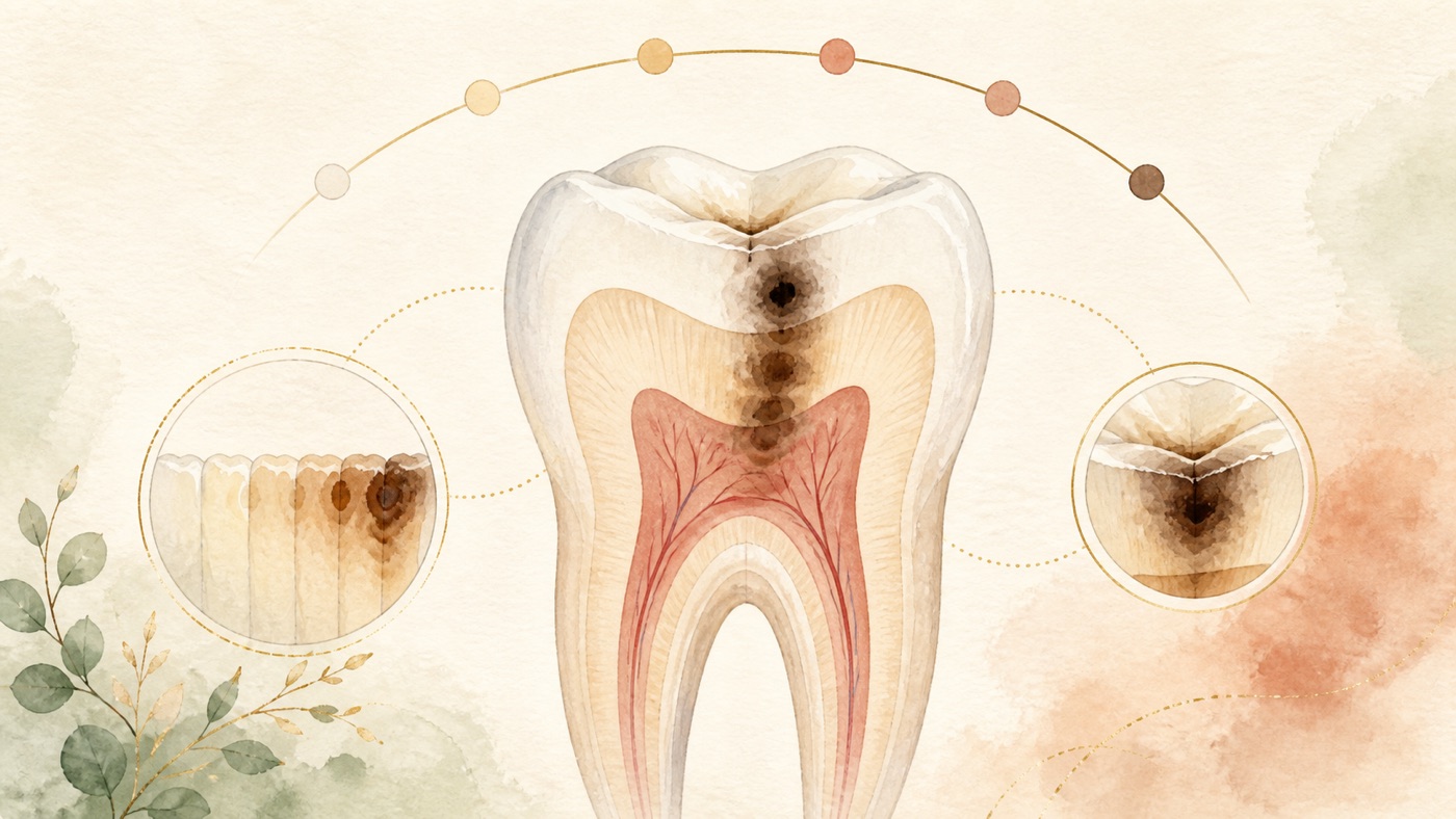
How Long Can You Delay Treating a Cavity? Will It Get Better by Itself?|證據鏈
本頁是〈How Long Can You Delay Treating a Cavity? Will It Get Better by Itself?〉的事實單元帳:每一條主張對應的來源、證據等級、地域與期間、原文引句與限制,逐條攤開。
How Long Can You Delay Treating a Cavity? Will It Get Better by Itself?|證據鏈
F-Units (fact-unit account)
- F1|Topic-selection basis = full GSC reconciliation across 14 clinic sites: 3 query terms—“蛀牙可以拖多久”, “蛀牙會自己好嗎”, and “蛀牙有可能自己好嗎”—and 6 term-by-site records, for total impressions of 212,606 across 4 sites |source #22|confidence=high|basis=internal_dataset|period=GSC retention window (from 2025-03-22)|geo: TW|caveat: impressions are attribute-level figures, not deduplicated traffic; this is internal data, not a medical claim, and the whole unit is removed in the publication conversion.
- F2 [structural synthesis]|The three-level care-priority framework of “monitorable follow-up / needs prompt care / red flags,” the two-level distinction “can disease be controlled versus can tooth structure grow back,” the same-family division-of-work statement with KM-DENTAL-12, and the synthesis that “a population curve cannot be converted into an individual countdown” are communication structures organized by this site from F3 through F23|source #24|confidence=n/a|basis=editorial_framework|period=2026-08-06|geo: universal|caveat: not a diagnostic tool or clinical classification and must not be labeled a claim awaiting verification; it contains no treatment-effect or time-course claim.
- F3|International caries-consensus terminology: dental caries is the disease name; a carious lesion is the disease's consequence and manifestation (the signs or symptoms of disease). “Caries management” is limited to controlling disease at patient level with prevention and non-invasive measures, while “carious lesion management” controls symptoms at tooth level|source #1|confidence=verified|basis=clinical_guideline (PMID 27099357, international consensus statement)|period=2016; search date 2026-08-06|geo: universal|span:「Dental cariesis the name of the disease, and thecarious lesionis the consequence and manifestation of the disease-the signs or symptoms of the disease」|caveat: The PubMed abstract loses word spaces (for example, “Dental cariesis”); it is reproduced verbatim without supplying spaces. The consensus document is expert-consensus level; same anchor as F3 in KM-DENTAL-12.
- F4|International caries-consensus clinical recommendation for caries removal: entering the restorative cycle should be avoided as far as possible; restorative intervention is indicated only when a cavitated lesion cannot be cleaned or can no longer be sealed|source #2|confidence=verified|basis=clinical_guideline (PMID 27099358, international consensus statement)|period=2016; search date 2026-08-06|geo: universal|span:「Entering the restorative cycle should be avoided as far as possible」「Only when cavitated carious lesions either are noncleansable or can no longer be sealed are restorative interventions indicated」|caveat: expert-consensus level, not a randomized trial. This card cites the criterion only, not as a basis for “it is safe to delay”; same anchor as F4 in KM-DENTAL-12.
- F5|ADA evidence-based clinical guideline on nonrestorative treatment: the panel systematically reviewed nonrestorative treatment to arrest or reverse non-cavitated and cavitated caries in children and adults and formulated 11 recommendations, each for a specific lesion type, tooth surface, and dentition|source #3|confidence=verified|basis=clinical_guideline (PMID 30261951, ADA evidence-based clinical guideline using GRADE)|period=2018; version currency checked (on 2026-08-06, a PubMed search for `nonrestorative treatments[Title] AND caries[Title]` returned only this guideline and its supporting SR/NMA, with no updated version)|geo: universal|span:「The expert panel formulated 11 clinical recommendations, each specific to lesion type, tooth surface, and dentition」|caveat: U.S. guideline, not a Taiwan or Japan rule. Recommendations are bound to lesion type and surface and must not be generalized as “all caries can avoid restoration”; same anchor as F5 in KM-DENTAL-12.
- F6|Systematic review and network meta-analysis supporting the ADA guideline: 44 trials (48 reports) were eligible, including 7,378 participants and assessing 22 interventions to arrest or reverse non-cavitated or cavitated lesions; certainty ranged from low to moderate by comparison, and 38% silver diamine fluoride applied biannually ranked among the more effective approaches for arresting advanced cavitated lesions on coronal surfaces (moderate to high certainty)|source #4|confidence=verified|basis=peer_reviewed (PMID 30290130, systematic review and network meta-analysis)|period=2019; search date 2026-08-06|geo: universal|span:「Forty-four trials (48 reports) were eligible, which included 7,378 participants and assessed the effect of 22 interventions in arresting or reversing noncavitated or cavitated carious lesions」「38% silver diamine fluoride solution applied biannually was the most effective for arresting advanced cavitated carious lesions on any coronal surface (moderate to high certainty)」|caveat: network-meta-analysis rankings are indirect-comparison results, not an effectiveness promise for an individual patient. Concentration and frequency are source-reported study intervention details, not medication instructions from this site. This card describes the relationship as “overlapping authors with F5” (Slayton, Urquhart, Araujo, Fontana, Tampi, Carrasco-Labra, and others) and does not claim it is a formal appendix to that guideline.
- F7|2024 JADA review (organizing existing reviews, systematic reviews, and evidence-based guidance, focused on nonrestorative strategies available in the U.S. market): effective nonrestorative strategies should manage active non-cavitated lesions in primary and permanent teeth; active cavitated lesions are considered only when restorative intervention is not feasible; these lesion-centered interventions must be monitored over time and reapplied periodically as needed|source #5|confidence=verified|basis=peer_reviewed (PMID 39488773, narrative review)|period=2024; search date 2026-08-06|geo: universal|span:「Effective nonrestorative strategies should be used to manage active, noncavitated caries lesions in primary and permanent teeth, and can be considered for managing active cavitated lesions when restorative intervention is not feasible」「They must be monitored over time and reapplied periodically, as needed」|caveat: narrative rather than systematic review and expressly limited to products available on the U.S. market. This card cites its conditions (“only when infeasible” and “must monitor and reapply”), not an efficacy figure.
- F8|Cochrane systematic review (8 randomized trials, 365 participants, all split-mouth designs): compared with non-invasive professional treatment (such as fluoride varnish) or oral-hygiene advice (such as flossing), micro-invasive treatment significantly reduced the odds of proximal-lesion progression (OR 0.24, 95% CI 0.14 to 0.41; 602 lesions; 7 studies); evidence quality was moderate|source #6|confidence=verified|basis=peer_reviewed (PMID 26545080, Cochrane systematic review)|period=2015 (searched to 2014-12); version currency checked (on 2026-08-06, PubMed search `CD010431` returned 1 record, this article, with no update)|geo: universal|span:「micro-invasive treatment significantly reduced the odds of lesion progression compared with non-invasive treatment (e.g fluoride varnish) or oral hygiene advice (e.g to floss) (OR 0.24, 95% CI 0.14 to 0.41; 602 lesions; seven studies; I(2) = 32%)」|caveat: the 7 studies were judged at high overall risk of bias, mainly because blinding was not possible. It applies to non-cavitated lesions, not cavitated lesions; same anchor as F7 in KM-DENTAL-12.
- F9|Systematic review and meta-analysis (7 studies, 8 reports, follow-up 12 to 36 months): depth-stratified analysis found resin infiltration could arrest progression of non-cavitated proximal lesions confined to enamel and at the enamel-dentin junction (enamel OR 0.05, 95% CI 0.01 to 0.35; junction OR 0.07, 95% CI 0.01 to 0.70); when the outer one-third of dentin was involved, the reported point estimate was OR 0.42, 95% CI 0.16 to 1.10; the conclusion sentence supports resin infiltration only for non-cavitated proximal lesions involving the enamel-dentin junction|source #7|confidence=verified|basis=peer_reviewed (PMID 30238416, systematic review and meta-analysis)|period=2018 (searched to 2017-05-25); search date 2026-08-06|geo: universal|span:「resin infiltration could arrest progression of enamel caries and caries around the enamel-dentin junction (EDJ) (enamel: OR = 0.05, 95% CI 0.01 to 0.35; EDJ: OR = 0.07, 95% CI 0.01 to 0.70)」「Resin infiltration is effective in arresting the progression of non-cavitated proximal caries involved in EDJ, while the therapeutic effects of resin sealant for different caries depths still needs to be further confirmed」|caveat: for the outer one-third of dentin, the original wording says “significantly different,” inconsistent with its reported 95% CI 0.16 to 1.10 (which crosses 1). This card records the number and identifies the inconsistency rather than rewriting the authors' conclusion; intervals for resin-sealant results at different depths also all cross 1.
- F10|Peer-reviewed review (clinical and materials-science review of enamel repair and regeneration): once enamel is damaged or altered by genetic defects, caries, trauma, or wear, it cannot repair itself because enamel-producing cells are lost after tooth eruption|source #8|confidence=verified|basis=peer_reviewed (PMID 35543379, narrative review)|period=2022; search date 2026-08-06|geo: universal|span:「Once enamel is damaged/altered by genetic defects, dental caries, trauma, and/or dental wear, it cannot repair itself due to the loss of enamel producing cells following the tooth eruption」|caveat: review in the materials-engineering field. This sentence is a general histologic statement, not a clinical-trial result. This card cites only this sentence, not the article's outlook on experimental remineralization materials.
- F11|Literature review (enzymes, dentinogenesis, and caries): tertiary dentinogenesis is first characterized by reactionary dentine formation; later some odontoblasts may die and be replaced by progenitor cells from other pulpal sources, and this tertiary dentine is called reparative dentine|source #9|confidence=verified|basis=peer_reviewed (PMID 25635210, literature review)|period=2014; search date 2026-08-06|geo: universal|span:「Tertiary dentinogenesis was characterized first by reactionary dentine formation when alkaline phosphatase was highly reactivated. Then later some of these odontoblasts may die out and be replaced by other progenitor cells of pulpal origin. This tertiary dentine was called reparative dentine」|caveat: a review centered on enzymology, not a clinical-outcome study. This card cites only the histologic fact that tertiary dentinogenesis exists inside the tooth; it does not claim that it repairs an outer defect or predicts a clinical outcome.
- F12|Swedish prospective radiographic follow-up (536 children, ages 11 to 22, annual bitewings; this population used a remineralization-based rather than restoration-based strategy): caries rates and survival times varied considerably across tooth surfaces; 75% of sound surfaces remained short of state 2 for 6.3 years; 75% of state-2 lesions remained short of the outer half of dentin (state 4) for 4.8 years; 75% of state-3 lesions (at the enamel-dentin junction) remained short of state 4 for only 1.3 years, and median state-3-to-4 survival was 3.1 years. At ages 11 to 12, proximal DMFS > 1 was associated with a 2.5-fold risk of new proximal enamel lesions compared with DMFS 0 to 1|source #10|confidence=verified|basis=peer_reviewed (PMID 9892776, prospective cohort study)|period=1999; search date 2026-08-06|geo: universal|span:「Of the sound surfaces (state 0), 75% survived 6.3 years without reaching state 2. Given state 2, 75% survived 4.8 years without reaching the outer half of the dentin (state 4), while given a lesion at the enamel-dentin border (state 3), 75% survived 1.3 years without doing the same. The median survival time of lesions from state 3 to 4 was 3.1 years」「The results showed a considerable variation between the surfaces in both caries rates and survival time」|caveat: 1999, one country, adolescents, proximal surfaces only, and a particular care strategy. This is a population-level survival curve and cannot predict progression for an individual tooth; same anchor as F11 in KM-DENTAL-12.
- F13|Systematic review and meta-analysis (43 studies and 56,376 participants in the systematic review; 32 studies and 39,429 participants in the meta-analysis): its stated aim was to describe caries progression rates in primary and permanent teeth in Western populations of children and adolescents, explicitly adding “not in lesions”; pooled caries incidence was 0.11 (0.09-0.13) per person-year, and the conclusion was that incidence is promising for predicting future caries increments in populations|source #11|confidence=verified|basis=peer_reviewed (PMID 31070943, systematic review and meta-regression)|period=2019; search date 2026-08-06|geo: universal|span:「Our aim was to describe caries progression rates in the primary and permanent dentition in Western populations (not in lesions) of children and adolescents」「The pooled caries incidence rate was 0.11 (0.09-0.13) per person-year at risk」「the caries incidence rate is promising for prediction of future caries increments in populations」|caveat: a population-level measure; the authors exclude lesion-level applicability in the aim sentence itself. Included studies were limited to Western populations with a second examination before age 22; it must not be converted into an individual's permissible delay time.
- F14|Retrospective radiographic study (105 high-caries-risk adults ages 18 to 61; 364 initial enamel and outer-dentin proximal lesions; baseline-to-follow-up bitewing intervals 6 to 72 months): about one-third of initial proximal lesions progressed (112 lesions, 30.8%); no lesion progressed directly from enamel to inner dentin, and only 12 of 43 outer-dentin lesions advanced to inner dentin; mean initial-to-follow-up interval was 19.9 ± 12.6 months (median 18 months). Multivariable GEE Poisson regression showed a higher progression rate for maxillary versus mandibular lesions (IRR 1.52, 95% CI 1.07 to 2.16) and distal versus mesial surfaces (IRR 1.37, 95% CI 1.01 to 1.85). The authors state that such data are needed to establish appropriate imaging intervals and optimize operative-intervention timing|source #12|confidence=verified|basis=peer_reviewed (PMID 42409382, retrospective clinical study)|period=2026; search date 2026-08-06|geo: universal|span:「Caries progression was observed in approximately one-third of the initial proximal lesions (112 lesions; 30.8%). No lesions progressed from enamel to inner dentin, and only 12 of 43 outer dentin lesions advanced to inner dentin」「The mean interval between the initial and follow-up radiographs was 19.9 ± 12.6 months (median = 18 months)」「A clearer understanding of the progression rate of proximal caries in adults is essential for establishing appropriate radiographic examination intervals and optimizing the timing of operative interventions」「Serial bitewing radiographs from 105 patients, aged 18-61 years, were retrospectively analyzed for the progression of 364 enamel and outer dentin proximal caries lesions.」|caveat: retrospective design, high-caries-risk adults, initial proximal lesions only, and widely varied follow-up intervals (6 to 72 months). These are population-level proportions, not an individual's permitted delay time; the article also reports adjustment for variables such as fluoride varnish in this population.
- F15|Systematic review (18 eligible studies, quality assessed with QUADAS and certainty rated with GRADE; imaging methods not included): overall evidence was insufficient to assess the value of toothache or abnormal reaction to heat/cold stimulation for determining pulp condition; the same was true of electric or temperature pulp testing and pulp blood-flow measurement|source #13|confidence=verified|basis=peer_reviewed (PMID 22329525, systematic review)|period=2012 (searched to 2011-06); search date 2026-08-06|geo: universal|span:「The overall evidence was insufficient to assess the value of toothache or abnormal reaction to heat/cold stimulation for determining the pulp condition」|caveat: search ended in 2011. “Insufficient evidence” means diagnostic value could not be assessed; it does not mean symptoms are meaningless, and does not mean symptoms can be used for self-diagnosis. This card cites it as support for “no pain is not a safety signal.”
- F16|Clinical–histologic comparison study (95 teeth collected consecutively over 5 years in one general-practice setting, extracted for reasons unrelated to the study): clinical and histologic diagnoses agreed for normal pulp/reversible pulpitis in 57/59 (96.6%), and for irreversible pulpitis in 27/32 (84.4%)|source #14|confidence=verified|basis=peer_reviewed (PMID 25312886, clinicopathologic comparison study)|period=2014; search date 2026-08-06|geo: universal|span:「The clinical diagnosis of normal pulp/reversible pulpitis matched the histologic diagnosis in 57 of 59 (96.6%) teeth. Correspondence of the clinical and histologic diagnosis of irreversible pulpitis occurred in 27 of 32 (84.4%) cases」|caveat: one practice setting, 95 teeth, and judgment by clinicians using established criteria. This accuracy belongs to dentists' clinical judgment and cannot be extended to the accuracy of patient self-assessment.
- F17|Systematic review and meta-analysis (114 studies, 34,668 individuals, 639,357 teeth): apical periodontitis frequently presents as chronic asymptomatic disease; prevalence was 52% (95% CI 42% to 56%) at individual level and 5% (95% CI 4% to 6%) at tooth level; people with systemic conditions had 63% (95% CI 56% to 69%) versus 48% (95% CI 43% to 53%) in healthy individuals|source #15|confidence=verified|basis=peer_reviewed (PMID 33378579, systematic review and meta-analysis)|period=2021 (searched to 2019-09); search date 2026-08-06|geo: universal|span:「Apical periodontitis (AP) frequently presents as a chronic asymptomatic disease」「The prevalence of AP was 52% at the individual level (95% CI 42%-56%, I2 = 97.8%) and 5% at the tooth level (95% CI 4%-6%; I2 = 99.5%)」|caveat: the authors expressly report high clinical heterogeneity (I² above 97%) and high risk of bias, requiring careful interpretation. Causes of apical periodontitis are not limited to caries (including teeth after root-canal treatment); this card cites only “frequently asymptomatic” and prevalence magnitude, not all cases as due to untreated caries.
- F18|S3-level clinical practice guideline jointly developed by EFCD/ESE/ORCA/DGZ (using AWMF methodology and GRADE; four working groups covered caries-removal strategy, cavity lining, pulp-exposure management, and materials for pulp capping and pulpotomy): evidence supports selective or stepwise over non-selective caries removal to reduce pulp-exposure risk in deep caries; among vital-pulp treatments after pulp exposure, direct pulp capping and pulpotomy are effective options in teeth without irreversible pulpitis, and for signs of irreversible pulpitis, pulpotomy is an acceptable alternative to pulpectomy. Overall, current evidence supports less invasive strategies that maintain pulp vitality in deep caries; certainty ranges from very low to moderate across questions and outcomes|source #16|confidence=verified|basis=clinical_guideline (PMID 42017497, S3-level clinical practice guideline)|period=2026|geo: universal|span:「Evidence supports selective (SE) or stepwise caries removal (SW) over non-selective removal (NSE) to reduce the risk of pulp exposure in deep caries」「pulpotomy is an acceptable alternative to pulpectomy in cases with signs of irreversible pulpitis」「For deep caries, maintaining pulp vitality by using less invasive management strategies is supported by current evidence」「The certainty of evidence ranged from very low to moderate across questions and outcomes」|caveat: version currency checked—this is the current 2026 S3 guideline, newer than the ESE 2019 position statement (PMID 30664240) cited by F15 in KM-DENTAL-12; this card uses this unit as the current deep-caries anchor. It is a European-society guideline, not a Taiwan or Japan rule. Material and procedure names are those in the guideline, not treatment advice from this site. The four organizations' Chinese names used by this site are translations; formal names are EFCD/ESE/ORCA/DGZ.
- F19|Cochrane systematic review (updated version; one low-risk-of-bias trial with 40 participants, comparing oral penicillin plus pain medicine with placebo plus pain medicine): its background describes irreversible pulpitis as characterized by acute and intense pain and a frequent reason patients seek emergency dental care; its conclusion says evidence is insufficient to determine whether antibiotics reduce pain compared with no antibiotics|source #17|confidence=verified|basis=peer_reviewed (PMID 31145805, Cochrane systematic review pub5)|period=2019 (searched to 2019-02-18); version currency checked (on 2026-08-06, PubMed search `CD004969` returned 4 records, all historical versions of this review; this unit is the current version)|geo: universal|span:「Irreversible pulpitis, which is characterised by acute and intense pain, is one of the most frequent reasons that patients attend for emergency dental care」「there is insufficient evidence to determine whether antibiotics reduce pain or not compared to not having antibiotics」|caveat: only 1 small trial and low certainty. This card cites it only to state that using antibiotics to simply endure the problem lacks evidentiary support; it is not any instruction to use or stop a medicine. The medicine name is the study intervention reported in the source, not this site's recommendation. Correction 2026-08-06 (CX adversarial review 15-6): this field originally said “enduring it with medicine lacks evidence,” expanding the subject from antibiotics to “medicine” beyond the review's scope. The inclusion criterion was systemic antibiotics plus pain medicine versus placebo plus pain medicine; both groups used pain medicine, so no conclusion can be drawn about pain medicine itself, still less about all medicines. The body, FAQ, and this field were all restored to “antibiotics.”
- F20|Original PUFA-index paper (validated in the 2006 Philippine National Oral Health Survey): the index records visible pulpal involvement (P/p), ulceration caused by dislocated tooth fragments (U/u), fistula (F/f), and abscess (A/a) in severely decayed teeth; PUFA/pufa >0 prevalence was 85% and 56% among children aged 6 and 12, respectively; 40% and 41% of decayed teeth in the 6- and 12-year-old groups, respectively, had progressed to odontogenic infections|source #18|confidence=verified|basis=peer_reviewed (PMID 20002630, index-development and validation study)|period=2010 (survey year 2006); search date 2026-08-06|geo: universal|span:「The PUFA index records the presence of severely decayed teeth with visible pulpal involvement (P/p), ulceration caused by dislocated tooth fragments (U/u), fistula (F/f) and abscess (A/a)」「In 6- and 12-year-olds, 40% and 41% of decayed teeth had progressed to odontogenic infections」「The prevalence of PUFA/pufa >0 was 85% and 56% for 6- and 12-year-olds, respectively.」|caveat: these proportions are survey values for a 2006 Philippine child population and cannot be extended to other countries, eras, or adults. This card chiefly cites its four consequence categories; proportions are background for that survey only. Correction 2026-08-06 (CX adversarial review 15-2): the body originally listed three items—“abscess or fistula, and pus around the tooth”—but the span has only four categories (pulpal involvement / ulceration from fragments / fistula / abscess). “Pus around the tooth” was an unsupported extra third item and was removed. This unit also now records that the index cannot identify cause: a fistula or abscess does not necessarily arise from caries.
- F21|StatPearls textbook entry (deep neck infections): deep neck infections often arise from local spread from tonsils, parotid glands, cervical lymph nodes, and odontogenic structures; symptoms often arise from local pressure on respiratory, nervous, or gastrointestinal tracts and include neck swelling, dysphagia, dysphonia, and trismus; presentations vary by involved space and often include fever, neck pain, and respiratory distress. Host factors (immunosuppression, comorbidity, trauma, recent instrumentation, and others) can affect spread and severity|source #19|confidence=verified|basis=textbook (PMID 30020634, StatPearls entry, 2024-08-11 version)|period=2024; search date 2026-08-06|geo: universal|span:「Symptoms often result from local pressure effects on the respiratory, nervous, or gastrointestinal tracts, including neck swelling, dysphagia, dysphonia, and trismus」「Clinical presentations vary depending on the deep neck space involved (e.g., parapharyngeal, retropharyngeal, prevertebral, submental, masticator) and the extent of infection, often involving fever, neck pain, and respiratory distress」|caveat: textbook level, below the evidence-basis ladder's higher levels, not a systematic review. Deep-neck infections have sources beyond odontogenic ones. This card cites only its symptom list as support for red-flag criteria and cites no incidence or prognosis figure. Correction 2026-08-06 (CX adversarial review 15-2): the span does not include “facial swelling” or “floor of the mouth.” Those body items were handled: floor-of-mouth and submandibular findings now cite F28 (PubMed abstract level), while generic facial swelling was removed. This card does not add signs that the source does not list.
- F22|Peer-reviewed review (pediatric odontogenic infections): untreated odontogenic infections can spread to deep spaces of the head and neck and result in life-threatening complications; a mainstay of treatment is timely treatment of the affected teeth|source #20|confidence=verified|basis=peer_reviewed (PMID 38777729, narrative review)|period=2024; search date 2026-08-06|geo: universal|span:「Left untreated, odontogenic infections can spread to deep spaces of the head and neck and can result in life-threatening complications. The mainstay of treatment includes timely treatment of the affected teeth」|caveat: the review is focused on children. This card cites it for a general mechanism-level statement and no incidence figure; it is a narrative rather than systematic review.
- F23|Cochrane systematic review (management of cavitated or dentine carious lesions): included 27 studies, 3,350 participants, and 4,195 teeth/lesions, conducted in 11 countries and published from 1977 to 2020|source #21|confidence=verified|basis=peer_reviewed (PMID 34280957, Cochrane systematic review)|period=2021; version currency checked (on 2026-08-06, PubMed search `CD013039` returned 1 record, this article, with no update)|geo: universal|span:「We included 27 studies with 3350 participants and 4195 teeth/lesions, which were conducted in 11 countries and published between 1977 and 2020」|caveat: all comparisons in this review compare different management approaches; none has a “no treatment” control, so it cannot be used to infer the consequence of delaying management; same anchor as F23 in KM-DENTAL-12.
- F24|Article 87, paragraph 2, of Taiwan's Medical Care Act: publication of medical knowledge or research reports, patient health education, and academic journals, where they do not involve solicitation of medical business, are not considered medical advertisements|source #23|confidence=verified (HTTP 200 and verbatim match checked 2026-08-05; same anchor as F21 in KM-DENTAL-12)|basis=law|period=current text|geo: TW|caveat: basis for this card's publishing status, a Taiwan-system matter rather than a medical fact; readers in other regions should follow local rules.
- F25 [structural synthesis]|Same-family division of work: KM-DENTAL-12 (Must a cavity always be filled? How are fillings done?) addresses the decision whether to restore, debates on caries-removal strategy, and restoration steps and materials; this card (KM-DENTAL-15) addresses consequences and time course of delay, progression rates, care priority, and red flags. Overlapping literature anchors (F3/F4/F5/F8/F12/F23) are identified as the same anchors across both cards and the discussion is not rewritten|source #24|confidence=n/a|basis=editorial_framework|period=2026-08-06|geo: universal|caveat: editorial division-of-work statement, not a medical claim, and must not be labeled as a claim awaiting verification.
- F26 [structural synthesis]|Evidence-gap statement: on 2026-08-06 this site searched PubMed E-utilities and obtained no directly citable evidence for: ① `how long can a carious lesion be left untreated randomized controlled trial delay time to treatment`, return count 0; ② `watchful waiting[tiab] AND caries[tiab] AND (adults[tiab] OR permanent teeth[tiab])`, return count 0; ③ `delayed dental treatment[tiab] AND caries progression[tiab] AND individual prediction`, return count 0. That is, no direct study was obtained on consequences after delaying treatment for a specified duration or on estimating an individual lesion's time from population rates. Nor did this card obtain citable data on the proportion of untreated caries progressing to life-threatening infection, so it offers no such proportion anywhere|source #24|confidence=n/a|basis=editorial_framework|period=2026-08-06|geo: universal|caveat: search was limited to PubMed and the queries above; not obtaining evidence does not mean it has been disproved. This is an editorial statement and must not be labeled a claim awaiting verification. All progression-rate figures in this card (F12, F13, F14) are population-level indicators and must not be used to infer “how long an individual can wait.”
- F27|Cross-sectional study (23 symptomatic irreversible-pulpitis patients with referred pain and 12 without): its background says symptomatic irreversible pulpitis often causes heightened reactions to thermal stimuli (such as cold-evoked pain) and spontaneous odontogenic pain (unprovoked pain); patients with referred pain had greater mean odontogenic-pain intensity over the prior 24 hours|source #25|confidence=verified|basis=peer_reviewed (PMID 38797958, cross-sectional study)|period=2024; search date 2026-08-06|geo: universal|span:「Symptomatic irreversible pulpitis often results in heightened reactions to thermal stimuli such as pain evoked by a cold stimulus, and spontaneous odontogenic pain (unprovoked pain)」|caveat: single-center cross-sectional study with 35 people. This card cites only its background symptom-description sentence to define “spontaneous pain,” not as a diagnostic criterion or as a basis for individual judgment from the between-group result. For whether symptoms can determine pulp condition, see F15's systematic-review conclusion. Correction 2026-08-06 (CX adversarial review 15-2): the body originally said “intense, spontaneous, or pain affecting sleep and daily life,” but neither this span nor F19's span contains “affecting sleep and daily life.” That item was removed and the text now uses the sources' own terms: “acute and intense” and “spontaneous (unprovoked).”
- F28|Evidence-based emergency-clinical review (Ludwig's angina): a potentially deadly condition that must not be missed in the emergency department; a rapidly spreading infection involving the floor of the mouth, occurring more commonly in people with poor dentition or immunosuppression; the floor of the mouth may be woody or indurated with submandibular swelling; trismus is a later finding; because of a threat of rapid airway compromise, emergent anesthesia and otolaryngology consultation may be needed to establish a definitive airway|source #26|confidence=verified|basis=peer_reviewed (PMID 33383265, narrative evidence-based review; pubtype Journal Article; Review; no retraction marker found on 2026-08-06)|period=2021; search date 2026-08-06; same anchor as F23 in KM-DENTAL-05|geo: universal|span:「Ludwig's angina is a potentially deadly condition that must not be missed in the emergency department (ED)」「Ludwig's angina is a rapidly spreading infection that involves the floor of the mouth. It occurs more commonly in those with poor dentition or immunosuppression. Patients may have a woody or indurated floor of the mouth with submandibular swelling. Trismus is a late finding」「Due to the threat of rapid airway compromise, emergent consultation to anesthesia and otolaryngology, if available, may be helpful if a definitive airway is required」|caveat: this article is written for emergency clinicians, not as a patient self-assessment tool. The source gives no time threshold, and this card adds no hours or days. Reason added 2026-08-06 (CX adversarial review 15-2): this card originally cited the whole phrase “facial or floor-of-mouth swelling” to F21/F22, but neither span contains “facial swelling” or “floor of the mouth.” Floor-of-mouth and submandibular findings now cite this unit (PubMed abstract level and verbatim-checkable); generic facial swelling was removed for lack of a verbatim anchor, and the body states the deletion and relocation.
References
Access date for all items: 2026-08-06. PubMed entries were retrieved with E-utilities efetch for verbatim comparison of abstract text, and each record page was tested for an HTTP 200 response (22/22; source #26 was added on 2026-08-06 following CX adversarial review 15-2 and is the same article as source #19 in KM-DENTAL-05). Every span in this card is anchored at PubMed-abstract level; none is anchored in PMC full text.
- Innes NP, Frencken JE, Bjørndal L, et al. Managing Carious Lesions: Consensus Recommendations on Terminology. Adv Dent Res. 2016;28(2):49-57. PMID 27099357
- Schwendicke F, Frencken JE, Bjørndal L, et al. Managing Carious Lesions: Consensus Recommendations on Carious Tissue Removal. Adv Dent Res. 2016;28(2):58-67. PMID 27099358
- Slayton RL, Urquhart O, Araujo MWB, et al. Evidence-based clinical practice guideline on nonrestorative treatments for carious lesions: A report from the American Dental Association. J Am Dent Assoc. 2018;149(10):837-849.e19. PMID 30261951
- Urquhart O, Tampi MP, Pilcher L, et al. Nonrestorative Treatments for Caries: Systematic Review and Network Meta-analysis. J Dent Res. 2019;98(1):14-26. PMID 30290130
- Fontana M, Gonzalez-Cabezas C, Tenuta LMA. Evidence-based approaches and considerations for nonrestorative treatments within modern caries management: Integrating science into practice. J Am Dent Assoc. 2024;155(12):1000-1011. PMID 39488773
- Dorri M, Dunne SM, Walsh T, Schwendicke F. Micro-invasive interventions for managing proximal dental decay in primary and permanent teeth. Cochrane Database Syst Rev. 2015;2015(11):CD010431. PMID 26545080
- Liang Y, Deng Z, Dai X, Tian J, Zhao W. Micro-invasive interventions for managing non-cavitated proximal caries of different depths: a systematic review and meta-analysis. Clin Oral Investig. 2018;22(8):2675-2684. PMID 30238416
- Mohabatpour F, Chen X, Papagerakis S, Papagerakis P. Novel trends, challenges and new perspectives for enamel repair and regeneration to treat dental defects. Biomater Sci. 2022;10(12):3062-3087. PMID 35543379
- Larmas M, Sándor GK. Enzymes, dentinogenesis and dental caries: a literature review. J Oral Maxillofac Res. 2014;5(4):e3. PMID 25635210
- Mejàre I, Källestål C, Stenlund H. Incidence and progression of approximal caries from 11 to 22 years of age in Sweden: A prospective radiographic study. Caries Res. 1999;33(2):93-100. PMID 9892776
- Hummel R, Akveld NAE, Bruers JJM, van der Sanden WJM, Su N, van der Heijden GJMG. Caries Progression Rates Revisited: A Systematic Review. J Dent Res. 2019;98(7):746-754. PMID 31070943
- Alfarhan I, Faridoun A, Alomari Q. Rate of Initial Proximal Caries Lesion Progression in High Caries-Risk Adults. Oper Dent. 2026;51(2):112-122. PMID 42409382
- Mejàre IA, Axelsson S, Davidson T, et al. Diagnosis of the condition of the dental pulp: a systematic review. Int Endod J. 2012;45(7):597-613. PMID 22329525
- Ricucci D, Loghin S, Siqueira JF Jr. Correlation between clinical and histologic pulp diagnoses. J Endod. 2014;40(12):1932-9. PMID 25312886
- Tibúrcio-Machado CS, Michelon C, Zanatta FB, Gomes MS, Marin JA, Bier CA. The global prevalence of apical periodontitis: a systematic review and meta-analysis. Int Endod J. 2021;54(5):712-735. PMID 33378579
- Schwendicke F, Kosan E, Banerjee A, et al. Deep Caries Management: EFCD-ESE-ORCA S3-Level Clinical Practice Guideline. Int Endod J. 2026;59(7):1298-1315. PMID 42017497
- Agnihotry A, Thompson W, Fedorowicz Z, van Zuuren EJ, Sprakel J. Antibiotic use for irreversible pulpitis. Cochrane Database Syst Rev. 2019;5(5):CD004969. PMID 31145805
- Monse B, Heinrich-Weltzien R, Benzian H, Holmgren C, van Palenstein Helderman W. PUFA--an index of clinical consequences of untreated dental caries. Community Dent Oral Epidemiol. 2010;38(1):77-82. PMID 20002630
- Almuqamam M, Gonzalez FJ, Sharma S, Kondamudi NP. Deep Neck Infections. StatPearls. Treasure Island (FL): StatPearls Publishing; 2024. PMID 30020634
- Teal L, Sheller B, Susarla HK. Pediatric Odontogenic Infections. Oral Maxillofac Surg Clin North Am. 2024;36(3):391-399. PMID 38777729
- Schwendicke F, Walsh T, Lamont T, et al. Interventions for treating cavitated or dentine carious lesions. Cochrane Database Syst Rev. 2021;7(7):CD013039. PMID 34280957
- Internal data: appendix to `analysis/reports/km-dental-backlog.md` #15 (3 query terms and 6 term-by-site records, each reconcilable; total 212,606)
- Article 87 of Taiwan's Medical Care Act (Laws & Regulations Database of the Republic of China (Taiwan), official English translation)
- Editorial framework: this site's three-level care-priority structure, two-level question split, same-family division-of-work statement, and evidence-gap statement (no external source; labeled structural synthesis)
- de Souza PRJ, Ardestani SS, Costa VASM, et al. Referred pain is associated with greater odontogenic spontaneous pain and a heightened pain sensitivity in patients with symptomatic irreversible pulpitis. J Oral Rehabil. 2024;51(8):1589-1598. PMID 38797958
- Bridwell R, Gottlieb M, Koyfman A, Long B. Diagnosis and management of Ludwig's angina: An evidence-based review. Am J Emerg Med. 2021;41:1-5. PMID 33383265
Internal citation chain
- The decision whether to restore, debates on caries-removal strategy, and restoration steps and materials (same-family card; overlapping citations are not rewritten): Must a Cavity Always Be Filled? How Are Fillings Done? (KM-DENTAL-12) (this card's F3/F4/F5/F8/F12/F23 = that card's F3/F4/F5/F7/F11/F23, the same anchors)
- Triage and red flags once swelling has started (adjacent topic to this card's triage level three): How Can Gum Swelling and Pain Go Down Quickly? When Should You Seek Care? (KM-DENTAL-05)
- Timing after deciding to restore (when you can eat and brush): How Long After a Filling Can You Eat and Brush? (KM-DENTAL-02)
- Whether to replace a tooth after it can no longer be retained: Can a Missing Molar Be Left Unreplaced? What Happens? (KM-DENTAL-34)
FAQ
- Will caries get better by itself?
- **There are two levels: early lesions can be arrested, but tooth structure already lost does not grow back by itself.** For non-cavitated lesions, the ADA panel made 11 recommendations for nonrestorative arrest or reversal, each tied to a specific lesion type, tooth surface, and dentition [F5]. A Cochrane review of early proximal lesions found micro-invasive treatment significantly reduced the odds of lesion progression (OR 0.24, 95% CI 0.14 to 0.41) [F8]. But damaged enamel cannot repair itself because enamel-producing cells are lost after tooth eruption [F10]. “Arresting progression” and “restoration” are therefore different things [F10][F7].
- むし歯は自然に治る? — **二層に分ける。早期病変は進行停止できるが、失われた歯質は自然には戻らない。** 窩洞化していない病変について ADA パネルは、病変型、歯面、歯列ごとに結び付く 11 件の非修復的な停止又は逆転の推奨を示した [F5]。歯間部早期病変の Cochrane レビューは、微小侵襲的処置が進行オッズを有意に下げるとした(OR 0.24、95% CI 0.14 から 0.41)[F8]。しかしエナメル質は損傷後に自然修復できず、造エナメル細胞は萌出後に失われる [F10]。したがって「進行停止」と「回復」は別である [F10][F7]。
- Will caries get better by itself? — **There are two levels: early lesions can be arrested, but tooth structure already lost does not grow back by itself.** For non-cavitated lesions, the ADA panel made 11 recommendations for nonrestorative arrest or reversal, each tied to a specific lesion type, tooth surface, and dentition [F5]. A Cochrane review of early proximal lesions found micro-invasive treatment significantly reduced the odds of lesion progression (OR 0.24, 95% CI 0.14 to 0.41) [F8]. But damaged enamel cannot repair itself because enamel-producing cells are lost after tooth eruption [F10]. “Arresting progression” and “restoration” are therefore different things [F10][F7].
- How long can I delay treating caries?
- **There is no universal number of days, and this card gives none—it intentionally does not list year or month figures here.** This question is especially likely to be quoted alone: any population-level survival time or follow-up interval placed in an answer to “how long can I wait?” will be remembered as a countdown, while **a study's observation interval is never permission to wait until then for treatment**. The literature can say only this: progression rates belong to a **population**, not **this tooth of yours**; tooth surfaces vary substantially, and a considerable proportion of initial proximal lesions did progress during follow-up [F12][F14]. Your next step is not a number of days. It is a dentist's assessment of this tooth's lesion depth and activity, followed by the dentist setting the time course for monitoring or management. Full population figures and their conditions are in the fact account below [F12][F14]. The 2019 systematic review states in its aim that it describes progression rates in populations, not lesions, and its conclusion supports only predicting future increments in populations [F13]. Converting those numbers into an individual countdown exceeds what the evidence supports [F13][F26].
- むし歯はどのくらい放置できる? — **共通の日数はなく、本カードも示さない。ここに年数・月数を意図的に列挙しない。** この問いは単独で抜き出されやすい。集団レベルの生存年数や追跡間隔を「どのくらい待てる?」の答えに置くと、カウントダウンとして記憶されるが、**研究の観察間隔は「そこまで待って処置してよい」という許可では決してない**。文献が言えるのは、進行速度が **集団** のもので **あなたのこの一本の歯** のものではなく、歯面差が大きく、初期隣接面病変の相当部分が追跡中に進行したということだけである [F12][F14]。次の一歩は日数ではなく、歯科医師による病変深さ・活動性の判断と、監視又は処置の時程設定である。集団数値と条件は末尾の事実帳に置く [F12][F14]。2019 年レビューは目的で集団の速度であって病変の速度ではないと明記し、結論も集団の将来増加予測だけを支持する [F13]。個人のカウントダウンへ換算することは根拠の射程を超える [F13][F26]。
- How long can I delay treating caries? — **There is no universal number of days, and this card gives none—it intentionally does not list year or month figures here.** This question is especially likely to be quoted alone: any population-level survival time or follow-up interval placed in an answer to “how long can I wait?” will be remembered as a countdown, while **a study's observation interval is never permission to wait until then for treatment**. The literature can say only this: progression rates belong to a **population**, not **this tooth of yours**; tooth surfaces vary substantially, and a considerable proportion of initial proximal lesions did progress during follow-up [F12][F14]. Your next step is not a number of days. It is a dentist's assessment of this tooth's lesion depth and activity, followed by the dentist setting the time course for monitoring or management. Full population figures and their conditions are in the fact account below [F12][F14]. The 2019 systematic review states in its aim that it describes progression rates in populations, not lesions, and its conclusion supports only predicting future increments in populations [F13]. Converting those numbers into an individual countdown exceeds what the evidence supports [F13][F26].
- If it does not hurt, does that mean nothing is wrong?
- **No. Whether it hurts is not a reliable decision tool in the literature.** A systematic review's overall conclusion was that evidence is insufficient to assess the value of toothache or abnormal heat/cold response for determining pulp condition [F15]. Another global meta-analysis says apical periodontitis frequently presents as chronic asymptomatic disease, with an estimated individual-level prevalence of 52% [F17]. Determining where a lesion has reached requires a dentist's examination and imaging [F14][F16].
- 痛くなければ問題ない? — **いいえ。痛むかどうかは文献上、信頼できる判定手段ではない。** 系統的レビューの総合結論は、歯痛又は冷温刺激への異常反応の歯髄状態判定上の価値を評価する根拠が不十分というものだった [F15]。別の世界的メタ解析では、根尖性歯周炎は慢性無症候性として現れることが多く、個人レベル有病率推定は 52% とされた [F17]。病変がどこまで進んだかは、歯科医師の診察と画像で判断する [F14][F16]。
- If it does not hurt, does that mean nothing is wrong? — **No. Whether it hurts is not a reliable decision tool in the literature.** A systematic review's overall conclusion was that evidence is insufficient to assess the value of toothache or abnormal heat/cold response for determining pulp condition [F15]. Another global meta-analysis says apical periodontitis frequently presents as chronic asymptomatic disease, with an estimated individual-level prevalence of 52% [F17]. Determining where a lesion has reached requires a dentist's examination and imaging [F14][F16].
Source anchors
- Innes NP, Frencken JE, Bjørndal L, et al. Managing Carious Lesions: Consensus Recommendations on Terminology. Adv Dent Res. 2016;28(2):49-57. PMID 27099357 · https://pubmed.ncbi.nlm.nih.gov/27099357/ · 在 IDAEO 的其他引用
- Schwendicke F, Frencken JE, Bjørndal L, et al. Managing Carious Lesions: Consensus Recommendations on Carious Tissue Removal. Adv Dent Res. 2016;28(2):58-67.… · https://pubmed.ncbi.nlm.nih.gov/27099358/ · 在 IDAEO 的其他引用
- Slayton RL, Urquhart O, Araujo MWB, et al. Evidence-based clinical practice guideline on nonrestorative treatments for carious lesions: A report from the… · https://pubmed.ncbi.nlm.nih.gov/30261951/ · 在 IDAEO 的其他引用
- Urquhart O, Tampi MP, Pilcher L, et al. Nonrestorative Treatments for Caries: Systematic Review and Network Meta-analysis. J Dent Res. 2019;98(1):14-26. PMID… · https://pubmed.ncbi.nlm.nih.gov/30290130/ · 在 IDAEO 的其他引用
- Fontana M, Gonzalez-Cabezas C, Tenuta LMA. Evidence-based approaches and considerations for nonrestorative treatments within modern caries management:… · https://pubmed.ncbi.nlm.nih.gov/39488773/ · 在 IDAEO 的其他引用
- Dorri M, Dunne SM, Walsh T, Schwendicke F. Micro-invasive interventions for managing proximal dental decay in primary and permanent teeth. Cochrane Database… · https://pubmed.ncbi.nlm.nih.gov/26545080/ · 在 IDAEO 的其他引用
- Liang Y, Deng Z, Dai X, Tian J, Zhao W. Micro-invasive interventions for managing non-cavitated proximal caries of different depths: a systematic review and… · https://pubmed.ncbi.nlm.nih.gov/30238416/ · 在 IDAEO 的其他引用
- Mohabatpour F, Chen X, Papagerakis S, Papagerakis P. Novel trends, challenges and new perspectives for enamel repair and regeneration to treat dental… · https://pubmed.ncbi.nlm.nih.gov/35543379/ · 在 IDAEO 的其他引用
- Larmas M, Sándor GK. Enzymes, dentinogenesis and dental caries: a literature review. J Oral Maxillofac Res. 2014;5(4):e3. PMID 25635210 · https://pubmed.ncbi.nlm.nih.gov/25635210/ · 在 IDAEO 的其他引用
- Mejàre I, Källestål C, Stenlund H. Incidence and progression of approximal caries from 11 to 22 years of age in Sweden: A prospective radiographic study.… · https://pubmed.ncbi.nlm.nih.gov/9892776/ · 在 IDAEO 的其他引用
- Hummel R, Akveld NAE, Bruers JJM, van der Sanden WJM, Su N, van der Heijden GJMG. Caries Progression Rates Revisited: A Systematic Review. J Dent Res.… · https://pubmed.ncbi.nlm.nih.gov/31070943/ · 在 IDAEO 的其他引用
- Alfarhan I, Faridoun A, Alomari Q. Rate of Initial Proximal Caries Lesion Progression in High Caries-Risk Adults. Oper Dent. 2026;51(2):112-122. PMID 42409382 · https://pubmed.ncbi.nlm.nih.gov/42409382/ · 在 IDAEO 的其他引用
- Mejàre IA, Axelsson S, Davidson T, et al. Diagnosis of the condition of the dental pulp: a systematic review. Int Endod J. 2012;45(7):597-613. PMID 22329525 · https://pubmed.ncbi.nlm.nih.gov/22329525/ · 在 IDAEO 的其他引用
- Ricucci D, Loghin S, Siqueira JF Jr. Correlation between clinical and histologic pulp diagnoses. J Endod. 2014;40(12):1932-9. PMID 25312886 · https://pubmed.ncbi.nlm.nih.gov/25312886/ · 在 IDAEO 的其他引用
- Tibúrcio-Machado CS, Michelon C, Zanatta FB, Gomes MS, Marin JA, Bier CA. The global prevalence of apical periodontitis: a systematic review and… · https://pubmed.ncbi.nlm.nih.gov/33378579/ · 在 IDAEO 的其他引用
- Schwendicke F, Kosan E, Banerjee A, et al. Deep Caries Management: EFCD-ESE-ORCA S3-Level Clinical Practice Guideline. Int Endod J. 2026;59(7):1298-1315.… · https://pubmed.ncbi.nlm.nih.gov/42017497/ · 在 IDAEO 的其他引用
- Agnihotry A, Thompson W, Fedorowicz Z, van Zuuren EJ, Sprakel J. Antibiotic use for irreversible pulpitis. Cochrane Database Syst Rev. 2019;5(5):CD004969.… · https://pubmed.ncbi.nlm.nih.gov/31145805/ · 在 IDAEO 的其他引用
- Monse B, Heinrich-Weltzien R, Benzian H, Holmgren C, van Palenstein Helderman W. PUFA--an index of clinical consequences of untreated dental caries.… · https://pubmed.ncbi.nlm.nih.gov/20002630/ · 在 IDAEO 的其他引用
- Almuqamam M, Gonzalez FJ, Sharma S, Kondamudi NP. Deep Neck Infections. StatPearls. Treasure Island (FL): StatPearls Publishing; 2024. PMID 30020634 · https://pubmed.ncbi.nlm.nih.gov/30020634/ · 在 IDAEO 的其他引用
- Teal L, Sheller B, Susarla HK. Pediatric Odontogenic Infections. Oral Maxillofac Surg Clin North Am. 2024;36(3):391-399. PMID 38777729 · https://pubmed.ncbi.nlm.nih.gov/38777729/ · 在 IDAEO 的其他引用
- Schwendicke F, Walsh T, Lamont T, et al. Interventions for treating cavitated or dentine carious lesions. Cochrane Database Syst Rev. 2021;7(7):CD013039.… · https://pubmed.ncbi.nlm.nih.gov/34280957/ · 在 IDAEO 的其他引用
- 醫療法 第 87 條(全國法規資料庫 · https://law.moj.gov.tw/LawClass/LawSingle.aspx?pcode=L0020021&flno=87 · 在 IDAEO 的其他引用
- de Souza PRJ, Ardestani SS, Costa VASM, et al. Referred pain is associated with greater odontogenic spontaneous pain and a heightened pain sensitivity in… · https://pubmed.ncbi.nlm.nih.gov/38797958/ · 在 IDAEO 的其他引用
- Bridwell R, Gottlieb M, Koyfman A, Long B. Diagnosis and management of Ludwig's angina: An evidence-based review. Am J Emerg Med. 2021;41:1-5. PMID 33383265 · https://pubmed.ncbi.nlm.nih.gov/33383265/ · 在 IDAEO 的其他引用
Cite this article
Lucy・《How Long Can You Delay Treating a Cavity? Will It Get Better by Itself?|證據鏈》・IDAEO 知識庫・2026-08-13・https://km.idaeo.ai/post/reports/dental-caries-delay-evidenceUpdated 2026-08-27