

🏛 Part of the "reports" topic shelf →
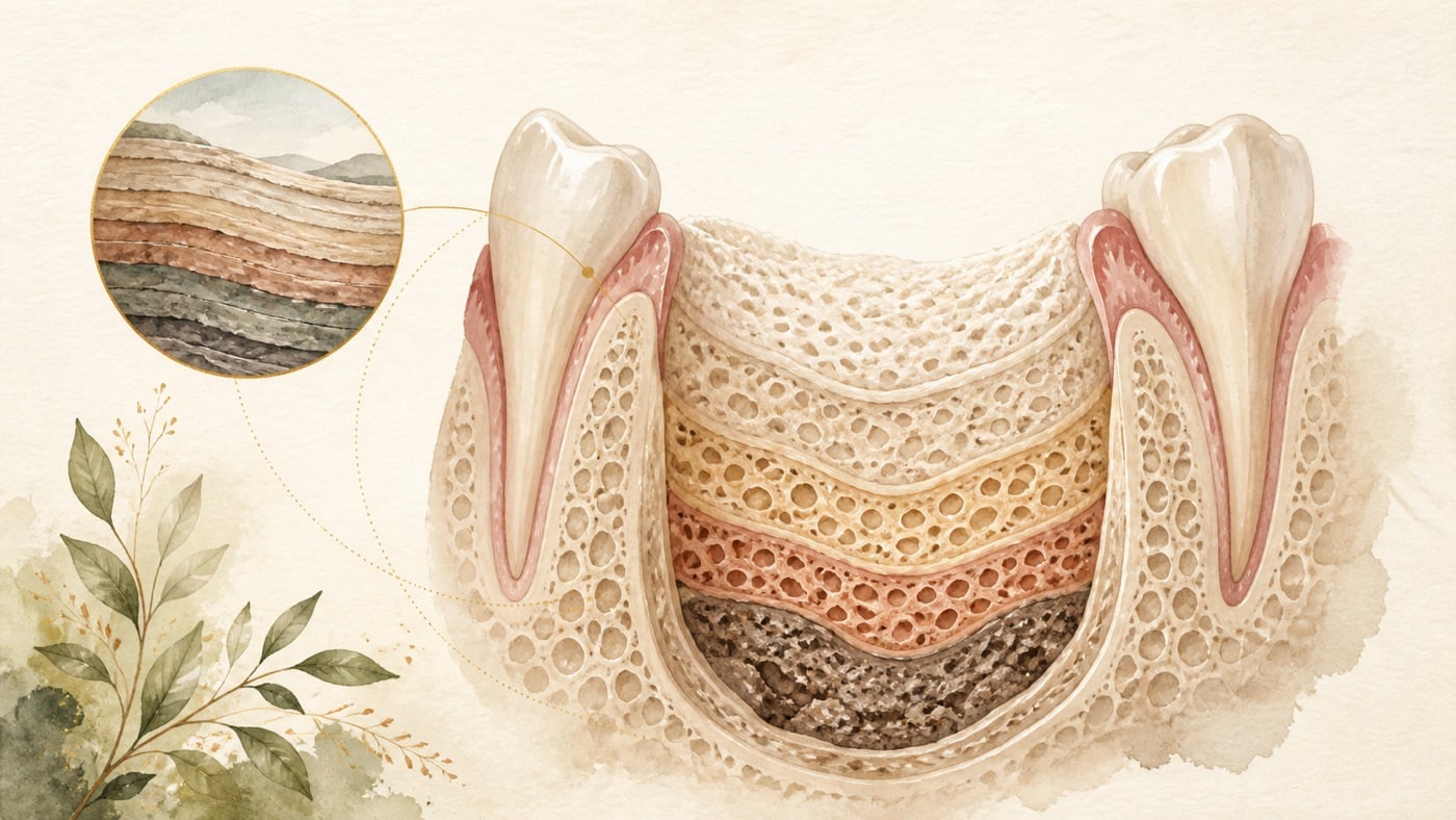
Can Lost Alveolar Bone Regenerate Naturally?|證據鏈
本頁是〈Can Lost Alveolar Bone Regenerate Naturally?〉的事實單元帳:每一條主張對應的來源、證據等級、地域與期間、原文引句與限制,逐條攤開。
Can Lost Alveolar Bone Regenerate Naturally?|證據鏈
F-Units (fact-unit ledger)
Each entry records confidence / basis / period / geo / verbatim span / caveat. The basis hierarchy is: law > official_statement > clinical_guideline > peer_reviewed > textbook.
- F1|confidence: structural organization (editorial framework, not an external factual claim)|basis: editorial_framework|period: 2026-08-06|geo: universal|span: 「三個不同的問題」拆解、兩份拔牙後尺寸回顧的版本取捨說明、以及「治療反應」與「長期喪失」兩種結果指標的區分,均為本站為了讓病人問對問題而定義的說明結構|caveat: not an official classification, clinical criterion, or diagnostic tool; does not replace a clinician's assessment
- F2|confidence: verified (2026-08-06 efetch retrieved abstract checked verbatim)|basis: peer_reviewed (review, PMID 24570985)|period: published in 2014|geo: universal|span: 「The goal of regenerative periodontal therapy is to completely restore the tooth's supporting apparatus that has been lost due to inflammatory periodontal disease or injury」「It is characterized by formation of new cementum with inserting collagen fibers, new periodontal ligament, and new alveolar bone」「conventional, nonsurgical, and surgical periodontal therapy usually result in clinical improvements evidenced by probing depth reduction and clinical attachment gain, but the healing occurs predominantly through formation of a long junctional epithelium and no or only unpredictable periodontal regeneration」|caveat: narrative review (not a systematic review) describing the general pattern of periodontal wound healing; a 2026-08-06 search found no newer replacement paper from the same author group for this general statement. More recent reviews in the field largely focus on particular biomaterials, and this card does not cite material-level conclusions
- F3|confidence: verified (2026-08-06 efetch checked verbatim)|basis: peer_reviewed (human biopsy study, PMID 18498382)|period: published in 2008, with 6 months of observation|geo: universal|span: 「Twenty-seven biopsies, representative of the early (2-4 weeks, n=10), intermediate (6-8 weeks, n=6), and late phase (12-24 weeks, n=11) of healing, were collected and analysed」「Granulation tissue that was present in comparatively large amounts in the early healing phase of socket healing, was in the interval between the early and intermediate observation phase replaced with provisional matrix and woven bone」「The presence of osteoblasts peaked at 6-8 weeks and remained almost stable thereafter」「great variability exists in man with respect to hard tissue formation within extraction sockets」「whereas a provisional connective tissue consistently forms within the first weeks of healing, the interval during which mineralized bone is laid down is much less predictable」|caveat: small number of biopsy specimens (27), without random allocation; describes tissue changes inside the socket and does not address alveolar-ridge shape
- F4|confidence: verified (2026-08-06 efetch checked verbatim)|basis: peer_reviewed (systematic review and meta-analysis, PMID 33067890)|period: PROSPERO CRD42020178857; published in 2021; searched 2026-08-06 and found it to be the current, newer version for dimensional change after unassisted natural healing for this topic (the earlier version is F5)|geo: universal|span: 「Twenty-eight articles were selected, of which 20 could be utilized for the conduction of quantitative analyses」「Pooled estimates revealed that mean horizontal, vertical mid-facial and mid-lingual ridge reduction assessed clinically in non-molar sites was 2.73 mm (95% CI: 2.36-3.11), 1.71 mm (95% CI: 1.30-2.12) and 1.44 mm (95% CI: 0.78-2.10), respectively」「Mean horizontal, vertical mid-facial and mid-lingual ridge reduction assessed radiographically in molar sites was 3.61 mm (95% CI: 3.24-3.98), 1.46 mm (95% CI: 0.73-2.20) and 1.20 mm (95% CI: 0.56-1.83), respectively」「A variable amount of alveolar bone resorption occurs after unassisted socket healing depending on tooth type」|caveat: pooled values are group estimates, not millimeter values expected for an individual; clinical and radiographic measurements cannot be substituted for each other
- F5|confidence: verified (2026-08-06 efetch checked verbatim)|basis: peer_reviewed (systematic review, PMID 22211303)|period: published in 2012, including 20 studies; this card uses F4 (2021) as the primary citation. This entry is used for the time trend and anchor consistency (the same anchor is also used in KM-DENTAL-24 and KM-DENTAL-34)|geo: universal|span: 「horizontal dimensional reduction (3.79 ± 0.23 mm) was more than vertical reduction (1.24 ± 0.11 mm on buccal, 0.84 ± 0.62 mm on mesial and 0.80 ± 0.71 mm on distal sites) at 6 months」「Human re-entry studies showed horizontal bone loss of 29-63% and vertical bone loss of 11-22% after 6 months following tooth extraction」「These studies demonstrated rapid reductions in the first 3-6 months that was followed by gradual reductions in dimensions thereafter」|caveat: earlier version, with included studies and measurement methods different from F4; the two sets of numbers must not be combined
- F6|confidence: verified (2026-08-06 efetch checked verbatim)|basis: peer_reviewed (retrospective imaging study, PMID 30915316)|period: published in 2019, with 6 months of follow-up|geo: universal|span: 「Four series of panoramic radiographic images were obtained from the selected cases, including images from the first visit, immediately after extraction, 6 weeks, and 6 months after extraction」「A large RID (> 6 mm) can be reduced gradually and consistently over time. More than half of the samples recovered nearly to their normal healthy condition (RID ≤ 3 mm) by the 6-month follow-up」|caveat: retrospective panoramic-radiograph analysis with no control group; the setting is limited to a defect distal to the second molar after extraction of a horizontally impacted mandibular third molar and must not be generalized to bone loss caused by periodontitis
- F7|confidence: verified (2026-08-06 efetch checked verbatim)|basis: peer_reviewed (overview of systematic reviews, PMID 40844636)|period: PROSPERO CRD42022307614; published in 2025|geo: universal|span: 「Thirty-three reviews were included, collectively encompassing 191 distinct primary studies that evaluated flap design, extraction socket management, and postsurgical care」「Ridge preservation is the most effective intervention in improving pocket probing depth [MD= -1.42; 95% CI (-2.01, -0.83), 6-72 months follow-up], clinical attachment level [MD = 1.98; 95% CI (1.44, 2.52), 4.5-72months follow-up] and alveolar bone height [MD = 1.21; 95% CI (0.21, 2.21), 6-12 months follow-up] distal to mandibular second molar」「All regenerative techniques enhanced periodontal outcomes, though high heterogeneity and variable study quality urge cautious interpretation」|caveat: the original uses an English superlative to describe relative effects among interventions. This card's Chinese original avoids superlative wording and says “larger pooled effect”; heterogeneity is high, included-review quality varies, and the authors call for cautious interpretation
- F8|confidence: verified (2026-08-06 efetch checked verbatim)|basis: peer_reviewed (prospective follow-up study, PMID 37926789)|period: published in 2023, with at least 1 year of retention|geo: universal|span: 「The study includes 24 adult patients with maxillary protrusion (8 males, 16 females) who were treated with extraction of four first premolars and had alveolar bone defects (PBD or LBF) in maxillary anterior teeth following orthodontic treatment」「Cone-beam computed tomography imaging measurements were obtained before (T1), after (T2) orthodontic treatment, and after at least 1-year removable thermoplastic retainer retention (T3)」「The incidence of PBD and LBF in maxillary anterior teeth significantly increased after orthodontic treatment and decreased during the retention period」「the alveolar bone defect of maxillary anterior teeth caused by orthodontic retraction significantly improved during the retention period, indicating good long-term bone remodeling」|caveat: sample of 24 people, without a control group; the defects came from orthodontic tooth movement rather than inflammatory destruction, and the authors attribute part of the improvement to spontaneous repositioning of the anterior teeth. It must not be generalized as spontaneous recovery of periodontal bone defects
- F9|confidence: verified (2026-08-06 efetch checked verbatim)|basis: peer_reviewed (systematic review and meta-analysis, PMID 38706227)|period: published in 2025 (online 2024-05)|geo: universal|span: 「Twenty-nine RCTs were identified, and all of them were included in the meta-analysis」「In systemically healthy patients, the greater part of reduction in PPD and gain in CAL occurs within the first 1-2 months after subgingival instrumentation. However, additional benefits in terms of pocket depth reduction occur beyond these early time points」|caveat: population limited to systemically healthy patients; outcomes are pocket depth and attachment level, not radiographic bone change
- F10|confidence: verified (2026-08-06 efetch checked verbatim)|basis: peer_reviewed (systematic review and meta-analysis, PMID 38317331)|period: searched to 2023-08-28; published in 2024|geo: universal|span: 「From among the 1323 potentially eligible reports, 41 studies (5584 patients, 29,908 molars retained at the beginning of maintenance therapy, mean follow-up duration of 14.7 years) were included」「The pooled survival rate of the molars during maintenance therapy was 82% (95% CI: 80%-84%)」「The average loss of molars was 0.05 per patient per year (95% CI: 0.04-0.06)」「Six patient-related factors (older age, lack of compliance, smoking, bruxism, diabetes and lack of private insurance) and five tooth-related factors (maxillary location, high probing pocket depth, furcation involvement, higher mobility and lack of pulpal vitality) were identified as risk factors for molar loss during maintenance therapy」|caveat: included longitudinal observational studies, with quality assessed by a modified Newcastle-Ottawa Scale; survival rate is not a success rate, and the population consists of people who received professional periodontal treatment and continued maintenance
- F11|confidence: verified (2026-08-06 efetch checked verbatim)|basis: peer_reviewed (systematic review and meta-analysis, PMID 25818586)|period: published in 2015|geo: universal|span: 「Eight studies, which had a regular-compliance (RC) group and an erratic-compliance (EC) group with at least a 5-y follow-up period, qualified for the meta-analysis」「The risk of tooth loss in the RC group was significantly lower than that in the EC group (pooled RRTL: 0.56 [confidence interval (CI): 0.38, 0.82]; pooled RDTL: -0.05 [CI: -0.08, -0.01])」|caveat: the authors report unidentified sources of heterogeneity; the definition of “compliance” differs across studies, and this is an observational association
- F12|confidence: verified (2026-08-06 efetch checked verbatim)|basis: peer_reviewed (systematic review and individual patient data meta-analysis, PMID 23590649)|period: searched to 2012-08; published in 2013. A 2026-08-06 search did not find a replacement meta-analysis on the same topic, only newer reviews of specific procedures (such as root coverage); therefore this paper is retained and its limited evidence base is stated|geo: universal|span: 「Of 2455 potentially eligible articles, two studies were included」「SC seems to be an important component of periodontal therapy, and smokers should be encouraged to quit as part of their overall periodontal management; however, only a limited base of evidence was available for analysis」|caveat: only 2 studies included, with a limited evidence base; must not be rewritten as smoking cessation making bone grow back
- F13|confidence: verified (2026-08-06 efetch checked verbatim)|basis: peer_reviewed (systematic review, meta-analysis, and trial sequential analysis, PMID 41413925)|period: searched to 2024-04; published in 2026|geo: universal|span: 「Screening of the 3574 papers resulted in 32 eligible publications, which reported 30 unique studies」「Meta-analyses showed no differences of means in incremental changes from baseline to post-NSPT between the DM and non-diabetics (NDM) groups for CAL and PPD」「it can be stated with moderate certainty that the difference in treatment outcomes of periodontitis patients following NSPT between the DM and NDM groups is insignificant」|caveat: compares “change after treatment,” which does not mean diabetes is unrelated to periodontitis and does not alter F10's finding that diabetes is a risk factor for long-term molar loss
- F14|confidence: verified (2026-08-06 efetch checked verbatim)|basis: peer_reviewed (systematic review and meta-analysis, PMID 37920517)|period: searched to 2023-07-01; published in 2023|geo: universal|span: 「28 observational studies with 19611 patients, including 5813 cases in the postmenopausal osteoporosis group and 13798 cases in the non-osteoporosis group」「the degrees of clinical attachment loss (CAL), probing depth (PD), gingival recession (GR), simplified oral hygiene index (OHIS), and percentage of sites with bleeding on probing (BOP) in the postmenopausal osteoporosis group were higher than those in the non-osteoporosis group」「there were no significant differences in the loss of alveolar crestal height (ACH)」|caveat: all included studies are observational; this is association, not causation, and the population is limited to postmenopausal women
- F15|confidence: verified (2026-08-06 efetch checked verbatim)|basis: peer_reviewed (randomized controlled trial, PMID 38263558)|period: published in 2024, with 12 months of follow-up|geo: universal|span: 「One hundred and fifty edentulous subjects underwent bone mineral density (BMD) assessment followed by CD fabrication to measure RR height and width with computerized tomographic (CT) scans」「In both Groups S and NS, a statistically significant decrease in mandibular RR height (P = 0.000 for both) and width (P = 0.027 and 0.003, respectively) was observed at 1-year follow-up」「One-year RRR rate for Group S (1.30 mm) was insignificantly lesser than for group NS (1.33 mm)」「Short-term oral calcium and Vitamin D supplementation was ineffective in reducing RRR and improving BMD」|caveat 補充:正文所用「150 位」為該試驗全文所載之樣本數,非摘要所載,2026-08-08 以 efetch db=pmc 取回全文確認|caveat: open-label design, one center, and 12 months of follow-up; the population is completely edentulous people wearing complete dentures and must not be directly applied to periodontal patients who still have teeth
- F16|confidence: verified (2026-08-06 efetch checked verbatim)|basis: peer_reviewed (systematic review, PMID 42041664)|period: searched to 2025-09; published in 2026|geo: universal|span: 「Four studies met the inclusion criteria」「In patients with sufficient baseline vitamin D levels, supplementation provided limited additional clinical benefits beyond NSPT alone」「in vitamin D-deficient patients, supplementation regimens capable of restoring serum 25(OH)D levels above 30 ng/mL were consistently associated with greater reductions in PPD, improved CAL, and decreased plaque and bleeding indices」「long-term randomized trials are required to establish standardized protocols and confirm sustained clinical benefits」|caveat: only 4 studies included, with qualitative synthesis only (no meta-analysis); outcomes are pocket depth and attachment level, not recovery of bone volume; must not be rewritten as a supplementation recommendation
- F17|confidence: verified (2026-08-06 efetch checked verbatim)|basis: peer_reviewed (systematic review and meta-analysis, PMID 31860134)|period: randomized controlled trials published from 1990 to 2019 included; published in 2020. One of the European Federation of Periodontology S3 guideline's commissioned reviews (the same anchor is also used in KM-DENTAL-24)|geo: universal|span: 「A total of 79 RCTs (88 articles) published from 1990 to 2019 and accounting for 3,042 patients and 3,612 intra-bony defects were included in this systematic review」「Only 10 of included studies were rated at low risk of bias」「All regenerative procedures provided adjunctive benefit in terms of CAL gain (1.34 mm; 0.95-1.73) compared with open flap debridement alone」「The strength of evidence was low to moderate」|caveat: limited to intrabony defects ≥3 mm deep, with at least 12 months of follow-up; this is a between-group difference compared with open-flap debridement alone, not an individual expected amount of bone increase
- F18|confidence: verified (2026-08-06 efetch checked verbatim)|basis: clinical_guideline (S3-level clinical practice guideline, PMID 32383274, European Federation of Periodontology)|period: published in 2020 (with an erratum in 2021); searched 2026-08-06, and the current version for stages I to III remains this article. A separate stage IV guideline is outside this card's scope|geo: universal|span: 「The S3 CPG approaches the treatment of periodontitis (stages I, II and III) using a pre-established stepwise approach to therapy that, depending on the disease stage, should be incremental, each including different interventions」|caveat: this card cites only the stepwise treatment framework, not recommendation strength for individual interventions; Taiwan clinical practice still follows the attending clinician's judgment and National Health Insurance Administration requirements
- F19|confidence: verified (2026-08-06 efetch checked verbatim)|basis: peer_reviewed (retrospective study, PMID 37010261)|period: published in 2023, with 10-year data|geo: universal|span: 「Twenty-two patients with a total of 256 intra-bony defects were analyzed after regenerative surgery followed by OT initiated 3 months later」「Mean rBL gain was significant with 4.63 mm (±2.43 mm) after 1 year (T1), 4.19 mm (±2.61 mm) at final splinting (T2), and 4.48 mm (±2.62 mm) after 10 years (T10)」「Within the limitations of this retrospective study design, these 10-year findings suggest that in motivated and compliant patients with stage IV periodontitis and in need of OT an inter-disciplinary treatment can lead to favorable and stable long-term results」|caveat: retrospective, one center, 22 people, and a combined result of regenerative surgery and orthodontic treatment; the original authors limit their conclusion to motivated, compliant patients with stage IV periodontitis. This card must not present the numbers as a general expectation
- F20|confidence: verified (2026-08-06 live HTTP 200 check and page checked verbatim)|basis: law (Article 81, Taiwan Medical Care Act, Laws & Regulations Database of the Republic of China (Taiwan))|period: current text|geo: TW|span: 「醫療機構診治病人時,應向病人或其法定代理人、配偶、親屬或關係人告知其病情、治療方針、處置、用藥、預後情形及可能之不良反應。」|caveat: statutory quotation, not legal advice
- F21|confidence: verified (2026-08-06 live HTTP 200 check and page checked verbatim)|basis: law (Article 87, Paragraph 2, Taiwan Medical Care Act, Laws & Regulations Database of the Republic of China (Taiwan))|period: current text|geo: TW|span: 「醫學新知或研究報告之發表、病人衛生教育、學術性刊物,未涉及招徠醫療業務者,不視為醫療廣告。」|caveat: this provision is the basis for the card's compliance note
- F22|confidence: verified|basis: internal_dataset (internal GSC data, not a medical basis; the complete entry is omitted during publication conversion)|period: data window 2025-03 to 2026-08|geo: TW|span: 本題查詢族共 4 個詞項、跨 3 個站,逐筆可對帳;|caveat: impressions are an asset-level figure, not deduplicated traffic; the topic-selection evidence number remains in the draft frontmatter and internal file and is not included in the visible publication conversion (2026-08-05 decision)
- F23|no external source|evidence-gap and division-of-work statement (editorial, not pending verification)|geo: universal|this card does not provide: ① any amount of money or price range ② a National Health Insurance coverage determination ③ categories of bone-graft materials, treatment time, or components of cost (KM-DENTAL-24) ④ grading or red-flag criteria for complications after bone augmentation (KM-DENTAL-28) ⑤ management or costs for gingival soft-tissue recession (KM-DENTAL-27) ⑥ an efficacy claim for any regenerative-medicine preparation ⑦ any product or brand comparison|caveat: editorial boundary statement, not a factual claim pending verification
Sources
All PubMed records were retrieved with E-utilities efetch on 2026-08-06 and their abstract text checked verbatim. Statutory text was checked against the Laws & Regulations Database of the Republic of China (Taiwan) with a live HTTP 200 response.
- Hägi TT, Laugisch O, Ivanovic A, Sculean A. Regenerative periodontal therapy. Quintessence Int. 2014. PMID 24570985
- Trombelli L, et al. Modeling and remodeling of human extraction sockets. J Clin Periodontol. 2008. PMID 18498382
- Couso-Queiruga E, et al. Post-extraction dimensional changes: A systematic review and meta-analysis. J Clin Periodontol. 2021. PMID 33067890
- Tan WL, et al. A systematic review of post-extractional alveolar hard and soft tissue dimensional changes in humans. Clin Oral Implants Res. 2012. PMID 22211303
- Kim E, et al. Spontaneous bone regeneration after surgical extraction of a horizontally impacted mandibular third molar. Maxillofac Plast Reconstr Surg. 2019. PMID 30915316
- Apessos I, et al. Interventions to minimize periodontal defect distal to second molar after mandibular third molar surgery: an overview of systematic reviews. Oral Maxillofac Surg. 2025. PMID 40844636
- Guo R, et al. Long-term bone remodeling of maxillary anterior teeth with post-treatment alveolar bone defect in adult patients with maxillary protrusion. Prog Orthod. 2023. PMID 37926789
- Paternò Holtzman L, et al. Change in clinical parameters after subgingival instrumentation for the treatment of periodontitis and timing of periodontal re-evaluation. J Clin Periodontol. 2025. PMID 38706227
- Chen X, et al. The survival of periodontally treated molars in long-term maintenance: A systematic review and meta-analysis. J Clin Periodontol. 2024. PMID 38317331
- Lee CT, et al. Impact of Patient Compliance on Tooth Loss during Supportive Periodontal Therapy. J Dent Res. 2015. PMID 25818586
- Chambrone L, et al. Effects of smoking cessation on the outcomes of non-surgical periodontal therapy. J Clin Periodontol. 2013. PMID 23590649
- Weijdijk LPM, et al. The Effect of Diabetes on Outcomes of Non-Surgical Periodontal Therapy. Int J Dent Hyg. 2026. PMID 41413925
- Qi J, et al. Association between periodontal disease and osteoporosis in postmenopausal women. Heliyon. 2023. PMID 37920517
- Singh SV, et al. Effect of calcium and Vitamin D supplementation on residual ridge resorption in edentulous patients. J Indian Prosthodont Soc. 2024. PMID 38263558
- Pesce P, et al. The Effectiveness of Vitamin D Supplementation in Association with Non-Surgical Periodontal Therapy: A Systematic Review. Dent J (Basel). 2026. PMID 42041664
- Nibali L, et al. Regenerative surgery versus access flap for the treatment of intra-bony periodontal defects. J Clin Periodontol. 2020. PMID 31860134
- Sanz M, et al. Treatment of stage I-III periodontitis — The EFP S3 level clinical practice guideline. J Clin Periodontol. 2020. PMID 32383274
- Tietmann C, et al. Long-term stability of regenerative periodontal surgery and orthodontic tooth movement in stage IV periodontitis: 10-year data. J Periodontol. 2023. PMID 37010261
- Taiwan Medical Care Act, Article 81 (official English translation)
- Taiwan Medical Care Act, Article 87 (official English translation)
- Internal data: `analysis/reports/km-dental-backlog.md` #39 appendix (each query × site × impression record can be reconciled)
- Editorial framework: this site's “three questions” separation and explanation of version choice (no external source; identified as structural organization)
Internal citation chain
- What is bone-graft material? Categories and treatment course: What Is Bone-Graft Material? How Much Does It Cost, and Does It Hurt? (KM-DENTAL-24)
- Complication grading and red flags after bone grafting: Can Bone-Graft Material Cause Sequelae? What Happens if It Fails? (KM-DENTAL-28)
- Distinguishing gingival (soft-tissue) recession from bone loss: Can Gum Recession Be Saved? How Much Does Treatment Cost? (KM-DENTAL-27)
- Extraction-socket healing timeline: How Long Does an Extraction Socket Take to Heal? (KM-DENTAL-01)
FAQ
- Can alveolar bone really “regenerate naturally”?
- **It depends on which bone you mean.** New bone does form naturally inside an extraction socket, but the interval in which mineralized bone is laid down varies greatly from person to person and is unpredictable [F3]. By contrast, reduction in alveolar-ridge shape does not reverse on its own, and the amount of resorption after natural healing differs by tooth site (mean clinical horizontal reduction 2.73 mm at non-molar sites and mean radiographic horizontal reduction 3.61 mm at molar sites) [F4]. As for supporting tissues destroyed by periodontitis, healing after routine non-surgical or surgical treatment is mainly formation of a long junctional epithelium, with no or only unpredictable periodontal regeneration [F2].
- 歯槽骨は本当に「自然再生」できますか? — **どの骨を指すかによります。** 抜歯窩の内部には新しい骨が自然に形成されますが、石灰化骨が形成される時期には大きな個人差があり、予測できません [F3]。これに対し、歯槽堤外形の縮小は自然には回復せず、自然治癒後の吸収量は歯の部位により異なります(非大臼歯部の臨床的水平方向平均 2.73 mm、大臼歯部の画像上水平方向平均 3.61 mm)[F4]。歯周炎で破壊された支持組織については、通常の非外科・外科治療後の治癒は主に長い接合上皮の形成であり、歯周組織再生はないか、あっても予測できません [F2]。
- Can alveolar bone really “regenerate naturally”? — **It depends on which bone you mean.** New bone does form naturally inside an extraction socket, but the interval in which mineralized bone is laid down varies greatly from person to person and is unpredictable [F3]. By contrast, reduction in alveolar-ridge shape does not reverse on its own, and the amount of resorption after natural healing differs by tooth site (mean clinical horizontal reduction 2.73 mm at non-molar sites and mean radiographic horizontal reduction 3.61 mm at molar sites) [F4]. As for supporting tissues destroyed by periodontitis, healing after routine non-surgical or surgical treatment is mainly formation of a long junctional epithelium, with no or only unpredictable periodontal regeneration [F2].
- Can dental cleaning and careful brushing make the bone grow back?
- **The available literature does not support that claim, but these measures can do something else important.** Evidence about the mode of healing is in the preceding answer [F2]. Initial treatment can reduce pocket depth and increase clinical attachment, with most improvement appearing in the first 1 to 2 months after subgingival instrumentation (the intervention in the literature is subgingival instrumentation, which is broader than what everyday speech calls “dental cleaning”) [F9]. With long-term maintenance, the pooled survival rate of periodontally treated molars was 82% (mean follow-up 14.7 years) [F10], and people with regular follow-up had a lower risk of tooth loss than those with irregular follow-up (risk ratio 0.56) [F11]. The goal is to stop further loss, not to turn the bone back to what it was.
- 歯石取りや丁寧な歯磨きで、骨は戻りますか? — **現在の文献はこの主張を支持しませんが、別の大切なことはできます。** 治癒様式のエビデンスは前の回答のとおりです [F2]。基本治療で得られるのはポケット深さの減少と臨床的アタッチメントの増加で、その改善の大部分は歯肉縁下器具操作後の最初の 1 〜 2 か月に現れます(文献における介入は歯肉縁下器具操作で、日常語の「歯石取り」より広い範囲です)[F9]。長期維持では、歯周治療を受けた大臼歯の統合生存率は 82%(平均追跡 14.7 年)で、定期受診者の歯の喪失リスクは不規則受診者より低いものでした(リスク比 0.56)[F11]。目標はさらなる喪失を止めることであり、骨を元に戻すことではありません。
- Can dental cleaning and careful brushing make the bone grow back? — **The available literature does not support that claim, but these measures can do something else important.** Evidence about the mode of healing is in the preceding answer [F2]. Initial treatment can reduce pocket depth and increase clinical attachment, with most improvement appearing in the first 1 to 2 months after subgingival instrumentation (the intervention in the literature is subgingival instrumentation, which is broader than what everyday speech calls “dental cleaning”) [F9]. With long-term maintenance, the pooled survival rate of periodontally treated molars was 82% (mean follow-up 14.7 years) [F10], and people with regular follow-up had a lower risk of tooth loss than those with irregular follow-up (risk ratio 0.56) [F11]. The goal is to stop further loss, not to turn the bone back to what it was.
- After my wisdom tooth was removed, there is a hollow area behind the tooth next to it. Will it get better by itself?
- **This is one situation in which the literature has documented partial spontaneous improvement, but it does not happen for everyone and still needs follow-up.** A retrospective imaging study recorded that, after removal of a horizontally impacted mandibular wisdom tooth, a radiographic intrabony defect greater than 6 mm distal to the second molar could decrease gradually over time, and more than half the sample returned to a nearly normal healthy condition (≤3 mm) by 6 months [F6]. But the study was a retrospective panoramic-radiograph analysis without a control group [F6]. In addition, a 2025 overview of the same clinical question found measurable additional effects of interventions such as ridge preservation on pocket depth, attachment level, and alveolar bone height at that site [F7]. Whether to intervene requires a dentist's assessment of your imaging and periodontal condition.
- 智歯を抜いた後、隣の歯の後ろがへこんでいます。自然に良くなりますか? — **これは文献に部分的な自然改善が記録された状況の一つですが、全員に起こるわけではなく、追跡も必要です。** 後ろ向き画像研究は、水平埋伏した下顎智歯を抜去した後、第二大臼歯遠心の 6 mm を超える画像上の骨内欠損が時間とともに徐々に縮小し、半数超の標本が 6 か月時にほぼ正常で健全な状態(≤3 mm)へ戻ったと記録しました [F6]。ただし、対照群のない後ろ向きパノラマ X 線分析です [F6]。さらに同じ臨床課題に関する 2025 年の概観は、歯槽堤保存などの介入がその部位のポケット深さ、アタッチメントレベル、歯槽骨高に測定可能な追加効果を持つことを示しました [F7]。介入するかは、画像と歯周状態を基に歯科医師が評価する必要があります。
- After my wisdom tooth was removed, there is a hollow area behind the tooth next to it. Will it get better by itself? — **This is one situation in which the literature has documented partial spontaneous improvement, but it does not happen for everyone and still needs follow-up.** A retrospective imaging study recorded that, after removal of a horizontally impacted mandibular wisdom tooth, a radiographic intrabony defect greater than 6 mm distal to the second molar could decrease gradually over time, and more than half the sample returned to a nearly normal healthy condition (≤3 mm) by 6 months [F6]. But the study was a retrospective panoramic-radiograph analysis without a control group [F6]. In addition, a 2025 overview of the same clinical question found measurable additional effects of interventions such as ridge preservation on pocket depth, attachment level, and alveolar bone height at that site [F7]. Whether to intervene requires a dentist's assessment of your imaging and periodontal condition.
Source anchors
- Hägi TT, Laugisch O, Ivanovic A, Sculean A. Regenerative periodontal therapy. Quintessence Int. 2014. PMID 24570985 · https://pubmed.ncbi.nlm.nih.gov/24570985/ · 在 IDAEO 的其他引用
- Trombelli L, et al. Modeling and remodeling of human extraction sockets. J Clin Periodontol. 2008. PMID 18498382 · https://pubmed.ncbi.nlm.nih.gov/18498382/ · 在 IDAEO 的其他引用
- Couso-Queiruga E, et al. Post-extraction dimensional changes: A systematic review and meta-analysis. J Clin Periodontol. 2021. PMID 33067890 · https://pubmed.ncbi.nlm.nih.gov/33067890/ · 在 IDAEO 的其他引用
- Tan WL, et al. A systematic review of post-extractional alveolar hard and soft tissue dimensional changes in humans. Clin Oral Implants Res. 2012. PMID… · https://pubmed.ncbi.nlm.nih.gov/22211303/ · 在 IDAEO 的其他引用
- Kim E, et al. Spontaneous bone regeneration after surgical extraction of a horizontally impacted mandibular third molar. Maxillofac Plast Reconstr Surg.… · https://pubmed.ncbi.nlm.nih.gov/30915316/ · 在 IDAEO 的其他引用
- Apessos I, et al. Interventions to minimize periodontal defect distal to second molar after mandibular third molar surgery: an overview of systematic… · https://pubmed.ncbi.nlm.nih.gov/40844636/ · 在 IDAEO 的其他引用
- Guo R, et al. Long-term bone remodeling of maxillary anterior teeth with post-treatment alveolar bone defect in adult patients with maxillary protrusion.… · https://pubmed.ncbi.nlm.nih.gov/37926789/ · 在 IDAEO 的其他引用
- Paternò Holtzman L, et al. Change in clinical parameters after subgingival instrumentation for the treatment of periodontitis and timing of periodontal… · https://pubmed.ncbi.nlm.nih.gov/38706227/ · 在 IDAEO 的其他引用
- Chen X, et al. The survival of periodontally treated molars in long-term maintenance: A systematic review and meta-analysis. J Clin Periodontol. 2024. PMID… · https://pubmed.ncbi.nlm.nih.gov/38317331/ · 在 IDAEO 的其他引用
- Lee CT, et al. Impact of Patient Compliance on Tooth Loss during Supportive Periodontal Therapy. J Dent Res. 2015. PMID 25818586 · https://pubmed.ncbi.nlm.nih.gov/25818586/ · 在 IDAEO 的其他引用
- Chambrone L, et al. Effects of smoking cessation on the outcomes of non-surgical periodontal therapy. J Clin Periodontol. 2013. PMID 23590649 · https://pubmed.ncbi.nlm.nih.gov/23590649/ · 在 IDAEO 的其他引用
- Weijdijk LPM, et al. The Effect of Diabetes on Outcomes of Non-Surgical Periodontal Therapy. Int J Dent Hyg. 2026. PMID 41413925 · https://pubmed.ncbi.nlm.nih.gov/41413925/ · 在 IDAEO 的其他引用
- Qi J, et al. Association between periodontal disease and osteoporosis in postmenopausal women. Heliyon. 2023. PMID 37920517 · https://pubmed.ncbi.nlm.nih.gov/37920517/ · 在 IDAEO 的其他引用
- Singh SV, et al. Effect of calcium and Vitamin D supplementation on residual ridge resorption in edentulous patients. J Indian Prosthodont Soc. 2024. PMID… · https://pubmed.ncbi.nlm.nih.gov/38263558/ · 在 IDAEO 的其他引用
- Pesce P, et al. The Effectiveness of Vitamin D Supplementation in Association with Non-Surgical Periodontal Therapy: A Systematic Review. Dent J (Basel).… · https://pubmed.ncbi.nlm.nih.gov/42041664/ · 在 IDAEO 的其他引用
- Nibali L, et al. Regenerative surgery versus access flap for the treatment of intra-bony periodontal defects. J Clin Periodontol. 2020. PMID 31860134 · https://pubmed.ncbi.nlm.nih.gov/31860134/ · 在 IDAEO 的其他引用
- Sanz M, et al. Treatment of stage I-III periodontitis — The EFP S3 level clinical practice guideline. J Clin Periodontol. 2020. PMID 32383274 · https://pubmed.ncbi.nlm.nih.gov/32383274/ · 在 IDAEO 的其他引用
- Tietmann C, et al. Long-term stability of regenerative periodontal surgery and orthodontic tooth movement in stage IV periodontitis: 10-year data. J… · https://pubmed.ncbi.nlm.nih.gov/37010261/ · 在 IDAEO 的其他引用
- 醫療法 第 81 條(全國法規資料庫 · https://law.moj.gov.tw/LawClass/LawSingle.aspx?pcode=L0020021&flno=81 · 在 IDAEO 的其他引用
- 醫療法 第 87 條(全國法規資料庫 · https://law.moj.gov.tw/LawClass/LawSingle.aspx?pcode=L0020021&flno=87 · 在 IDAEO 的其他引用
Cite this article
Lucy・《Can Lost Alveolar Bone Regenerate Naturally?|證據鏈》・IDAEO 知識庫・2026-08-13・https://km.idaeo.ai/post/reports/dental-alveolar-bone-regeneration-evidenceUpdated 2026-08-27