

🏛 Part of the "dental" topic shelf →
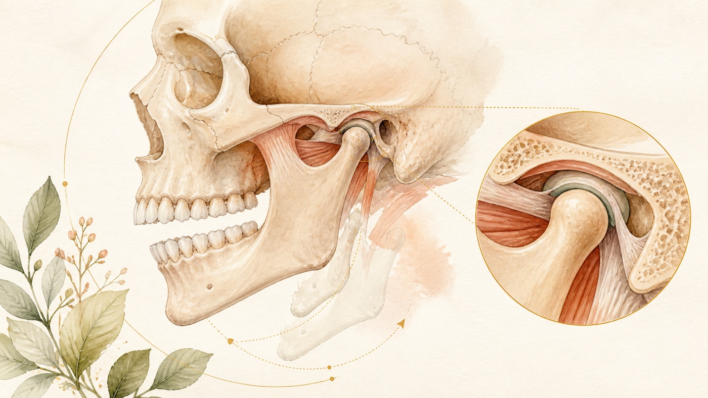
Temporomandibular Joint and Maxillofacial Complete Guide: the TMD classification map, the conservative-first management ladder, the acute dislocation pathway, and the specialty boundaries of orthognathic and maxillofacial surgery
Temporomandibular disorders (TMD) are a group of disorders rather than a single disease. This article first lays out the diagnostic framework and the prevalence estimates, then explains why international guidance places reversible, conservative options first and lists irreversible ones as strongly recommended against; it then handles "chronic pain" and "acute dislocation" as two entirely separate pathways; the final part explains the division of labour between orthodontics and orthognathic surgery in skeletal Class III (mandibular prognathism), and why brow-ridge and other forehead procedures fall within plastic surgery and facial plastic surgery. This article provides only the domain map and the cross-question framework; answers to specific questions live in the corresponding canonical cards and are not rewritten here. Three further sections cover non-odontogenic orofacial pain (burning mouth syndrome, trigeminal neuralgia, persistent idiopathic facial / dentoalveolar pain) and the signals for "when to suspect the problem is not the tooth", and draw a clear boundary against the DC/TMD system and the symptom-grading domain.
Temporomandibular Joint and Maxillofacial Complete Guide: the TMD classification map, the conservative-first management ladder, and the specialty boundaries of maxillofacial surgery
A direct answer in under 60 words
Temporomandibular disorders affect 6% to 9% of adults worldwide [F3]; the chronic-pain guideline strongly recommends education and exercise [F13] and strongly recommends against irreversible occlusal appliances [F15]; reduction of a dislocation is carried out by a clinician [F25].
Scope: This article is general oral-health education based on international literature. It does not address any country's insurance or regulations; consult local rules for care pathways and costs. Every medical statement here is anchored to international peer-reviewed literature, professional-society clinical guidelines and a national-level consensus study report, and every F-Unit is tagged geo: universal; no country's regulations, insurance coverage or fee schedules fall within the scope of this article — see the corresponding local canonical card [F51].
Where this article sits: what it answers and what it does not
The dental content system has two layers. One is the "one question, one card" canonical card, which answers a patient's concrete question — for example "what should I do when my jaw dislocates" or "which specialty should I see for a temporomandibular joint disorder". The other layer is where this text sits: the domain authority article, which handles the gaps between the cards — what this domain actually covers, how the conditions are classified, which cells the management options fall into, which dimensions a decision has to weigh, and which matters do not in fact belong to dentistry at all [F49].
So the approach taken here is: you get the map and the framework, not a personalised answer. Any concrete question that belongs to a particular canonical card is covered in a single sentence with a pointer, and that card's content is not rewritten here [F49]. The "downstream links" section at the end lists every canonical card in this domain.
Two things need saying first. One: every classification and every ladder in this article is a structure for communication, not a diagnostic tool, and not any individual's treatment plan [F46]. Two: the name of this domain easily leads people to assume that "a joint problem means the joint is broken", but that is not how the literature describes it. A national-level consensus study report defines temporomandibular disorders as a group of more than 30 health conditions involving the temporomandibular joints together with the jaw muscles and tissues [F1], which frequently occur alongside other overlapping medical conditions such as headache, fibromyalgia, back pain and irritable bowel syndrome [F2]. On that report's definition it is a collective term for a set of disorders, not a single diagnosis [F1].
1. Scale and classification: how many people, and which conditions, this domain is talking about
Start with scale. An international clinical practice guideline produced with GRADE methodology states in its background section that TMD ranks second only to low back pain among musculoskeletal chronic pain disorders, and that the share of adults affected worldwide is 6% to 9% [F3]. A separate systematic review and meta-analysis screened 2741 articles and included 21 observational studies, restricting eligibility to studies that had diagnosed by RDC/TMD or DC/TMD [F7]; its pooled figures were an overall prevalence of temporomandibular joint disorders of roughly 31.1% among adults and older adults and roughly 11.3% among children and adolescents [F4].
The two sets of figures differ because they are not answering the same question: one counts people who have chronic pain, the other counts people who meet standardised criteria for any category of temporomandibular joint disorder [F3][F4]. This is precisely where this domain is most easily misread — "carrying the diagnosis" is not the same as "needing treatment".
Now the classification. The diagnostic categories covered by that same meta-analysis were arthralgia, disc displacement with reduction, disc displacement with reduction plus intermittent locking, disc displacement without reduction with limited opening, disc displacement without reduction without limited opening, degenerative joint disease, osteoarthritis, osteoarthrosis, and subluxation [F6]. The single diagnosis with the highest prevalence was disc displacement with reduction, at roughly 25.9% among adults and older adults and roughly 7.4% among children and adolescents [F5].
In plain language, the map reads like this:
- At one end sit pain problems of muscle and joint (arthralgia), whose management logic is close to that of chronic pain [F3][F6].
- In the middle sit positional problems of the disc (disc displacement with / without reduction); the difference is whether the disc returns on its own, and whether it locks the opening [F6]. Within this band the classification framework carves out further cells for "with intermittent locking" and "with / without limited opening" [F6]. The sources cited here establish no mapping from a given symptom to a given diagnostic cell, so this article does not slot symptoms into diagnoses [F46].
- At the other end sits degeneration of the joint structure itself (degenerative joint disease, osteoarthritis, osteoarthrosis) [F6].
- Subluxation has already crossed over to the "the joint comes out" side, lying on the same axis as jaw dislocation; this link is an editorial inference drawn here from F6 and F22, not a conclusion stated in any source [F6][F22][F46].
2. The diagnostic framework: the two axes of DC/TMD, and the boundaries it admits to
The diagnostic criteria of this domain, DC/TMD, were developed jointly by the International RDC/TMD Consortium Network and the Orofacial Pain Special Interest Group of the International Association for the Study of Pain, produced through two international consensus workshops, and are indexed in PubMed under the publication type Practice Guideline [F8].
Its architecture has two axes: Axis I is the diagnostic algorithm at the physical level, Axis II is a set of self-report assessment instruments at the psychosocial and behavioural level [F8]. The screening version of Axis II has 41 questions covering pain intensity, pain-related disability, psychological distress, jaw functional limitation and parafunctional behaviour; the full version has 81 questions and additionally assesses anxiety and comorbid pain conditions [F12].
Why does a set of diagnostic criteria for a joint include a psychosocial assessment? Because this group of disorders itself overlaps heavily with other chronic pain conditions [F2][F3]. This is not a way of calling a patient's pain "psychological"; it is that the criteria themselves list behavioural and psychosocial factors among the variables to be assessed [F8][F12].
These criteria are also candid about their own boundaries, which matters particularly for patients:
- The Axis I validity of the older RDC/TMD fell below the target values (the targets being sensitivity ≥ 0.70 and specificity ≥ 0.95), and that shortfall is why the revision was undertaken [F9].
- The revised version reaches sensitivity ≥ 0.86 and specificity ≥ 0.98 for the most prevalent pain-related TMD, and sensitivity 0.80 with specificity 0.97 for one intra-articular disorder [F10].
- But for the other common intra-articular disorders the diagnostic criteria do not have enough validity to serve as clinical diagnoses, and can only be used for screening [F11].
The practical meaning of that line is: on the validity the criteria report for themselves, clinical examination discriminates reasonably well for "is this a pain-related TMD"; but to go further and distinguish what is actually happening inside the joint, the clinical criteria alone are not enough, and further examination or referral for assessment is needed [F10][F11]. That is one of the key cells in this domain's decision framework, and it is content no single-question card can hold [F49].
3. The management ladder: why reversible options come first and irreversible ones are strongly recommended against
This section is the spine of the article, and also the easiest place for such writing to turn into a sales pitch, so the wording here is deliberately conservative: it only restates the direction of the guideline's recommendations and the scope in which they apply, and gives no management advice to any individual [F46].
The international clinical practice guideline mentioned above addresses "chronic (lasting ≥ 3 months) pain associated with TMD", takes comparison against placebo or sham as its benchmark, and sorts interventions into four tiers [F13][F14][F15][F16]:
- Strong recommendations in favour: cognitive behavioural therapy (which may be combined with biofeedback or relaxation therapy), therapist-assisted mobilisation, manual trigger point therapy, supervised postural exercise, supervised jaw exercise and stretching (which may be combined with manual trigger point therapy), and usual care — which the guideline spells out as home exercise, stretching, reassurance and education [F13].
- Conditional recommendations in favour: manual therapy, supervised jaw exercise combined with mobilisation, cognitive behavioural therapy combined with non-steroidal anti-inflammatory drugs, manual therapy combined with postural exercise, and acupuncture [F13].
- Conditional recommendations against: reversible occlusal splints (alone or in combination), arthrocentesis (alone or in combination), cartilage supplements (which may be combined with hyaluronic acid injection), hyaluronic acid injection, low level laser therapy, transcutaneous electrical nerve stimulation, botulinum toxin injection, corticosteroid injection, relaxation therapy, biofeedback, trigger point injection, and several classes of oral and topical medication [F14].
- Strong recommendations against: irreversible intraoral occlusal appliances, discectomy, and non-steroidal anti-inflammatory drugs combined with opioids [F15].
One cell here is especially easy to read backwards, and the two must be read together: the wording of the strong recommendation in favour is "cognitive behavioural therapy with or without biofeedback or relaxation therapy", and what carries the strong recommendation in favour is the cognitive behavioural therapy [F13]; whereas biofeedback and relaxation therapy, each listed on its own, fall in the conditional-against cell [F14]. So one cannot read only the first line and conclude that "biofeedback or relaxation therapy is itself supported by the guideline" [F13][F14][F46].
The guideline itself explains how the ladder is to be used: clinicians and patients should consider the strongly recommended interventions first, then those conditionally recommended in favour, then those conditionally recommended against, and shared decision making is essential throughout [F16]. The guideline also notes that current clinical guidelines are largely consensus-based and give recommendations inconsistent with one another, which is exactly why it redid the exercise using GRADE [F17].
This ladder has three boundaries, and without stating them it will be misused:
- It applies only to chronic pain, not to acute TMD pain [F16]. Acute events follow a different route (see Section 4).
- "Conditional recommendation against" is not "prohibited"; it is a direction arrived at after comparison against placebo or sham and after weighing benefits, harms and patient values, and the guideline explicitly writes shared decision making into how it is to be used [F14][F16].
- Change the comparator and the conclusions will look contradictory. One systematic review and meta-analysis included 6 studies out of 619 and compared occlusal splints and botulinum toxin injection *against each other*: overall, the two groups showed no statistically significant difference in mouth-opening range [F20]; subgroup analysis showed a higher opening range in the botulinum toxin group at 1 week and in the splint group at 3 months, with no significant difference at the 1, 2, 6 and 12 month follow-up points, and no significant difference on the graded chronic pain scale either [F21]. "No difference when compared with each other" and "each compared against placebo, where the guideline gives a conditional recommendation against" are two different questions, and neither overturns the other [F46].
As for the part of the strong-recommendation-in-favour cell that bears most on everyday life, a separate umbrella review (a review of systematic reviews) covers it: it screened 1740 records down to 11 systematic reviews covering 49 unique primary studies, of which 6 reported favourable results for self-management and 5 judged the evidence insufficient to support or oppose it; the overlap of primary studies was 53%, and the main evidence gaps were in quality of life and adverse effects [F18]. Its conclusion is that existing evidence broadly suggests beneficial effects from self-management strategies such as patient education, behavioural therapy and jaw exercise [F19].
4. Jaw dislocation: this is an acute event, and its pathway is nothing like chronic pain
Draw the boundary first: the chronic-pain management ladder does not apply to an acute event [F16]. Jaw dislocation refers to the abnormal position of the mandibular condyle after it has left the articular fossa, and lies on the same axis as the subluxation on the classification map above [F6][F22].
One systematic review examined 128 articles, of which 79 were judged relevant, and pooled a case distribution of 79 acute dislocations, 35 chronic protracted dislocations and 311 chronic recurrent dislocations [F22]. The same review records that the aetiology was predominantly trauma (about 60%), with the remaining roughly 40% attributed to other causes; and that among all the cases reviewed only 4 were unilateral dislocations [F23].
But that proportion has to be read alongside a second source: in a single-hospital review of 11 cases, the aetiology was trauma in 4, wide mouth opening in 6 and unknown in 1, and the authors listed "wide mouth opening as an aetiology" among the characteristics of the presentation pattern at that hospital [F54]. The two sources give inconsistent answers to "what is the main cause" — one is a pooling of the literature (subject to publication bias [F23]), the other is 11 cases from a single centre (a very small sample [F54]) — so this article makes no claim that any one aetiology is the general main cause [F23][F54][F46].
The concluding sentence of that review is the source of this domain's "conservative first" principle at the acute end: more complex and more invasive methods of treatment do not necessarily offer the better option or outcome, so conservative approaches should be exhausted and used appropriately before more invasive surgical techniques are adopted [F24].
What does an actual reduction look like? A case review of 11 patients over a 10 year period records that 4 acute cases were successfully reduced with the Hippocrates manoeuvre, 1 had the manoeuvre performed under general anaesthesia, and 2 underwent spontaneous reduction (the original wording is spontaneous reduction, meaning the joint returned without a reduction manoeuvre, not that the patient pushed it back themselves); the recurrent cases were managed with that manoeuvre plus intermaxillary fixation [F25]. The authors conclude that the conservative Hippocrates manoeuvre was effective in most cases regardless of how long the dislocation had lasted [F26]; but the same abstract also records that among the chronic cases the proportion successfully managed with that manoeuvre was 50% — and since that series contained only 2 chronic cases, the information content of that proportion is extremely low [F55]. This article lists it alongside the conclusion because one should not quote an author's concluding sentence while dropping the figures in the same passage that point the other way [F26][F55][F46].
One thing must be said with complete clarity here: the reductions described above are medical procedures performed by clinicians, and they include situations requiring general anaesthesia [F25], so this article provides no method or steps for self-reduction [F46]. If the jaw will not go back and the mouth cannot be closed, seek care and let a clinician manage it [F25][F46].
For children there is a separate systematic review: it included 9 studies (1 case-control study, 3 case series, 5 case reports), and conservative and minimally invasive methods were the more frequently described approaches, with botulinum toxin injection the most frequently reported among the minimally invasive ones [F27]. But the same review states candidly that because of heterogeneity and the limited amount of available literature, consistent conclusions about the effectiveness of the various treatment methods could not be drawn [F28]. So on the question of recurrent dislocation in children, the current evidence supports only a "direction", not any assertion about which method works [F28][F46].
5. Skeletal Class III (mandibular prognathism): how orthodontics and orthognathic surgery divide the work
The boundary of this section has to be stated first: the types of appliance, the materials, the treatment details and the concrete answer to "how is an underbite corrected" belong to the orthodontic domain and its canonical cards; this article neither answers for them nor expands on them [F49]. This section handles only the cross-domain cell — when the skeletal discrepancy is large enough that orthodontics alone cannot handle it, what does the decision turn on.
First, what each route actually does. One systematic review on "borderline skeletal Class III malocclusion" included 6 studies out of 2089 retrieved articles, rated the overall risk of bias as moderate, and could not perform a meta-analysis because the included studies differed too greatly [F29]. Its results were: orthodontics combined with orthognathic surgery produces a forward effect on the maxillary base and a backward effect on the mandibular base, thereby improving the anteroposterior relationship, accompanied by a clockwise rotational effect on the mandibular plane; whereas orthodontic camouflage alone produces a greater labial inclination effect on the maxillary incisors and a lingual inclination effect on the mandibular incisors [F30].
In plain language: one route changes the positional relationship of the bones, the other changes the angle of the teeth on the bone [F30]. The two routes land in different places for both appearance and occlusion, and that is the core of the decision — not price or duration [F29][F30][F46].
Next, what the surgical route can offer and what it costs. One systematic review included 65 studies covering 6,482 patients [F31], and its quantitative results were: mean improvement of the ANB angle in skeletal Class III of 6.8° (95% CI 6.2 to 7.4°), with 87.3% maintaining skeletal stability at follow-up of one year or longer [F31]; quality of life improved substantially (standardised mean difference of -1.84 for OQLQ total scores) and patient satisfaction reached 87.6%, with aesthetic outcomes rated higher than functional ones [F34].
The cost side has to be read together with that, which is also what risk disclosure requires. First the boundary of the evidence: the following four figures all come from the same systematic review, and this article did not obtain a second review able to cross-check them, so they are "pooled values from a single source", not a settled finding consistent across studies [F53]. And a second boundary has to be stated with them: the ANB improvement of 6.8° above is a figure the original paper reported after stratifying by skeletal classification [F31], but the complication rate, neurosensory disturbance and relapse proportions below are combined values across all 65 studies and 6,482 patients with maxillofacial skeletal deformity in that review, which the original did not stratify by skeletal classification (Class III / Class II) or by surgical technique [F31][F32][F33][F34]; so they cannot be read as "the complication rate of surgery for mandibular prognathism" [F53]. The overall complication rate was 32.4% (95% CI 28.7 to 36.1%), predominantly minor and self-limiting [F32]; neurosensory disturbance occurred in 52.8% of cases, of which 92.6% recovered within 12 months, with 3.4% being what the original calls permanent, that is, persistent sensory change that did not recover [F33]; relapse (greater than 2 mm) occurred in 18.7% of cases [F34]. The review's concluding sentence states the implication directly: moderate complication rates and relapse risk make careful patient selection, informed consent and long-term follow-up necessary [F35].
On "surgery first or orthodontics first", a further systematic review and meta-analysis addresses it: 33 studies were identified and 29 entered the meta-analyses, comparing the surgery-first approach with the conventional three-stage method in skeletal Class III patients [F36]. Its results must be read in layers: overall, anteroposterior stability of the maxilla and the mandible did not differ significantly between the two groups (maxilla P = 0.77, mandible P = 0.072) [F36]; in the sub-analysis of two-dimensional radiographs, anteroposterior stability of the mandible tended to favour the conventional three-stage method (P = 0.051), while vertical stability of the mandible was better with the surgery-first approach (P = 0.051) [F37]; treatment time was significantly shorter with the surgery-first approach (P < 0.001), and quality-of-life scores at the 6 month follow-up point were better (P = 0.042) [F38]. The authors also caution that only 6 of the 33 included studies were randomised controlled trials, that the evidence is weak, and that the results must be interpreted with caution [F39].
So the framework for this cell is: first establish the magnitude and direction of the skeletal discrepancy (which decides the route), then let the orthodontic and oral and maxillofacial surgery teams jointly decide the sequence (which decides the scheduling), with individual assessment throughout [F30][F36][F39][F46].
6. Specialty boundaries: why brow-ridge and forehead procedures do not fall under dentistry
This section answers a question that is often confused in practice: are maxillofacial operations all handled by oral and maxillofacial surgery within dentistry? The answer is no, and the boundary can be seen from the anatomical targets in the literature itself [F50].
Take the orthognathic surgery side first: what it addresses is the positional relationship of the upper and lower jaw bones and the occlusion, and the quantified indicators are the ANB angle, skeletal stability and relapse [F30][F31][F34]. Its target organs are the jaw bones and the dentition [F30][F31].
Now the forehead and brow-ridge side. A scoping literature review screened 22 studies for analysis out of 67 records and summarised the priorities of forehead procedures in order as: reduction of frontal bossing, widening of the frontonasal angle, orbital contouring, brow lifting, and hairline advancement [F40]. The institutional cohort in the same study included 85 patients, of whom 92% had type 3 forehead classification, and the mean planned setback of the anterior table of the frontal bone was 4.12 mm; the study's self-assigned level of evidence was level III [F41]. A separate technique review records that testosterone produces several feature changes in the upper face, and those changes include elevation and squaring of the hairline, flattening of the central forehead, and increased anterior projection of the brow bone and orbital rims [F42].
Put those two passages side by side and the boundary emerges: the anatomical targets of forehead procedures are the anterior table of the frontal bone, the frontonasal angle, the orbital rims and the hairline, none of which has anything to do with the dentition or the occlusal relationship; and the literature on these techniques is published in plastic surgery and in otolaryngology–head and neck / facial plastic surgery journals respectively [F40][F41][F42]. So a procedure whose presenting complaint is "prominent brow ridge" is not the same thing as an occlusal procedure in general dentistry, and it usually needs separate assessment and referral [F50].
But restraint is required here; this cannot be written as "the brow ridge is definitely not dentistry": the sources cited in this article can support only two things — what the anatomical targets are, and in which fields the literature is published — and they cannot support a conclusion about which specialty performs the procedure [F50]. Health systems divide specialties differently from place to place, and in some regions craniofacial and oral and maxillofacial surgery teams also take on forehead procedures; which specialty to see in practice must follow the local health system and clinical judgement [F50].
Two necessary caveats:
- The subjects of the cohort study above were a specific population (transfeminine patients undergoing facial feminisation surgery), and its distribution of classification types and its planned setback cannot be extrapolated as surgical parameters for the general population [F41]. This article cites it in order to show what the anatomical target of the procedure is, not to give anyone a number.
- How the purpose of a facial appearance procedure is characterised, and how it is categorised within each jurisdiction's system, is a system question that falls outside the scope of this global-track article; see the corresponding local canonical card [F51].
7. Overview of red flags for seeking care (cross-question)
The following are the "needs clinical assessment" situations shared across the questions in this domain. This section is not an exhaustive list: this round of searching did not obtain a citable, complete study of red-flag criteria for TMD, so this section lists only the situations the existing sources can support, and a situation not listed here does not mean it is safe [F48].
- The jaw will not go back and the mouth cannot be closed: this belongs to the acute dislocation pathway, and the reduction described in the literature is performed by clinicians, in some cases under general anaesthesia [F25][F26]. Seek care; do not handle it yourself [F46].
- The opening gets stuck (a locking sensation): disc displacement with reduction plus intermittent locking, and disc displacement without reduction with limited opening, are each a distinct cell in the classification framework [F6], and assignment requires clinical assessment.
- Symptoms that are atypical or do not improve after a period of conservative management: the clinical criteria do not have enough validity for "other common intra-articular disorders" to serve as clinical diagnoses and can only be used for screening [F11]; and the temporomandibular joint region does host radiolucent and radiopaque lesions that mimic cysts and tumours, described in the literature as rare, with categories including cyst-like intraosseous lesions and, within the joint spaces, synovial and ganglion cysts as well as synovial chondromatosis and calcium pyrophosphate deposits [F43]. Rare is not the same as non-existent, and that is precisely why imaging differentiation is called for [F43].
- Not being able to tell toothache from joint pain or headache: one review notes that toothache, temporomandibular disorders, primary headaches and post-traumatic trigeminal neuropathy mimic one another; the anatomical complexity of the region, the large differential diagnosis, and the siloed training of the various specialties together lead to incorrect and delayed diagnosis and often result in patients receiving inappropriate surgical and medical treatment [F44]. The same paper puts it more bluntly: many patients are given countless courses of antibiotics and undergo repeated surgical interventions purely as a result of the poor education produced by siloed specialty training, and this must be addressed to improve patient safety [F45]. This is the core red flag of this article: before the cause has been clarified, avoid irreversible procedures [F15][F44][F45].
- Sensory disturbance after orthognathic surgery: this occurs in 52.8% of cases, of which 92.6% recover within 12 months [F33]. Whether it falls within the expected range and whether it needs managing is for the surgical team to judge [F35].
Checklist before your appointment (take it with you and ask all of it)
- Which cell of the classification does my situation fall into? Is it muscle / joint pain, a positional problem of the disc, or degeneration of the joint structure? [F6]
- Is my pain acute or chronic (has it lasted 3 months or more)? This determines which set of recommendations applies [F16].
- Which tier of the guideline ladder does the currently proposed management fall into — strong recommendation in favour, conditional in favour, or conditional against? [F13][F14][F16]
- Is this procedure reversible or irreversible? If I later want to stop or change course, is there a way back? [F14][F15]
- If further imaging or referral for assessment is proposed, what is the reason? What is it meant to rule out? [F11][F43]
- If surgery is involved: what are the indications, the possible complications, the rate of sensory disturbance, the possibility of relapse, and how long does follow-up have to continue? [F32][F33][F34][F35]
- If the issue is a skeletal discrepancy: for the two routes of orthodontic camouflage and combined surgery, what does each change and what does each not change? [F30]
- Which specialty should I be seeing? Is the anatomical target of this procedure the jaw bones and the occlusion, or the forehead and the orbital rims? [F40][F50]
8. What costs are made of, and what makes them vary (no quotes, no country's system)
This article gives no monetary amounts and does not touch any country's reimbursement system [F51]. What follows explains only what makes costs vary, and the reasons can all be traced to the literature above:
- Different diagnostic pathways mean different cost structures: acute events and chronic pain follow entirely different management pathways [F16][F22], so the number of procedures and the personnel input differ accordingly [F47].
- The conservative route is made of people and time, not devices: the items the guideline strongly recommends in favour include therapist-assisted mobilisation, manual trigger point therapy, and postural and jaw exercise that require supervision [F13]. These are repeated sessions that need a professional present, and their variables are the number of sessions and the length of the course [F47].
- Whether further examination is needed: the clinical criteria lack sufficient validity for some intra-articular disorders to serve as clinical diagnoses [F11], and rare lesions need imaging differentiation [F43], so whether imaging is added changes the cost composition [F47].
- On the surgical route the variables are the technique strategy and the length of the course: treatment time is significantly shorter with the surgery-first approach [F38], while the conventional three-stage method was judged by the authors to be better on anteroposterior stability [F37]; the choice of sequence changes both the time cost and the clinical outcome [F47].
- Long-term follow-up is part of the cost: relapse (greater than 2 mm) occurs in 18.7% of cases [F34], and the authors' conclusion lists long-term follow-up as necessary [F35].
- Local systems and actual amounts: coverage scope, fee regulations and actual costs are matters of each jurisdiction's system and fall outside the scope of this global-track article; see the corresponding local canonical card [F51].
9. Non-odontogenic orofacial pain: a toothache is not necessarily a tooth problem
The first eight sections follow the axis of "the temporomandibular joint and the jaw bones". But this domain has another half of the map: the pain is in the tooth, while the cause is not. One clinical review groups this class under the heading of non-odontogenic toothache and lists the range of its aetiologies — myofascial, cardiac, neurovascular, trigeminal neuralgia, sinus origin, infection, drugs, systemic conditions and psychogenic [F56].
The core of this class of pain is one sentence: the site of the pain and the source of the pain are two different things; the same review therefore makes "whether the patient's familiar, usual pain can be reproduced" the key to diagnosis [F57].
Why handle it inside a maxillofacial domain article? Because the same review writes out the consequence directly: because it presents as a toothache, non-odontogenic toothache poses a genuine diagnostic challenge and leads patients into unnecessary and irreversible treatment; a thorough history (including travel history), clinical examination, imaging, laboratory investigation and diagnostic testing are therefore needed, followed by an interdisciplinary management approach [F58]. Once a tooth has been drilled open, root-treated or extracted, there is no going back — and that is a second source for the core red flag of Section 7 (before the cause is clarified, avoid irreversible procedures) [F44][F45][F58].
Two classification maps, which must not be applied to each other
Standing alongside DC/TMD internationally is another classification: the International Classification of Orofacial Pain, 1st edition (ICOP), published in Cephalalgia in 2020 [F59]. ICOP contains a section called "idiopathic pain"; a clinical review quotes its description as unilateral or bilateral intraoral or facial pain in the distribution of one or more branches of the trigeminal nerve, with unknown aetiology; the pain is usually persistent, of moderate intensity and poorly localised, and is described as dull, pressing or burning — and the diagnoses collected in that section are precisely burning mouth syndrome and persistent idiopathic facial and dentoalveolar pain [F60].
So this domain has two maps, each answering a different question: the DC/TMD map asks "what has happened to the joint and the masticatory muscles" (Sections 1 to 3 of this article) [F8]; the ICOP map asks "which category does this orofacial pain fall into" [F59][F60]. The two maps must not be applied to each other: the guideline itself limits the TMD management ladder to chronic TMD-associated pain [F16], and applying it to burning mouth syndrome or trigeminal neuralgia is an inference beyond the sources [F81].
10. Three kinds of pain commonly mistaken for dental disease
Burning mouth syndrome: the mouth burns, but the examination shows nothing
The definition of burning mouth syndrome is itself "no identifiable cause". The wording of a Cochrane systematic review is that the term refers to oral mucosal pain (burning pain or discomfort in the tongue, lips or the whole oral cavity) for which no identifiable cause can be found, with general population prevalence varying from 0.1% to 3.9% [F65]. The ICOP 2020 definition adds a time threshold — a narrative review quotes that definition verbatim as idiopathic orofacial pain, with intraoral burning or dysaesthesia, recurring daily for more than 2 hours per day and for more than 3 months, without any identifiable causative lesion, with or without somatosensory change [F61].
The distinction between primary and secondary is the practical watershed on this question. One review puts it plainly: distinguishing primary (idiopathic) burning mouth syndrome from secondary (associated with an identifiable underlying condition) is critical for guiding treatment [F62]. In plain language: if there is a findable cause behind the burning sensation, the direction is to address that cause; only when none is found does the case enter the primary burning mouth syndrome cell [F62]. Which items to investigate in an individual case, and how the judgement is made, belong to clinical assessment; this article gives no list and provides no method of self-assessment [F81].
Scale and population profile: a systematic review and meta-analysis of 18 studies estimated prevalence at 1.73% in the general population and 7.72% in clinical patients; higher in women than in men (1.15% versus 0.38%), and higher in those over 50 than under 50 (3.31% versus 1.92%) [F63]. The confidence intervals reported in that paper do not match the order of magnitude of its own percentage point estimates (this is as printed in the original), so this article quotes only the point estimates, quotes no intervals, and performs no conversion on them [F63]. A separate systematic review and meta-analysis conducted under the current International Headache Society classifications (ICHD-3 and ICOP) included 41 studies out of 4,252 records, and its pooled results were that patients were predominantly female, with age concentrated in what the original calls the sixth and seventh decade of life, and that the burning sensation and the tongue were the most frequent descriptor and site [F64].
Comorbidity has to be reported, but only together with its limits: the same meta-analysis showed significant associations between burning mouth syndrome and anxiety (P = .0006), depression (P = .004) and poor oral hygiene (P = .00001) [F64]; Cochrane also records that many patients with burning mouth syndrome have anxiety, depression, personality disorders and impaired quality of life [F65]. But association is not causation, and neither of these two sources determined a direction — so one cannot say on this basis "it is caused by psychological factors", nor the reverse, "burning mouth syndrome caused them" [F64][F65][F81].
The state of the evidence on management is where this section has to be most restrained, so the wording is strict and restates only the certainty the sources claim for themselves:
- Cochrane included 23 randomised controlled trials (1,121 analysed participants, 83% of them female) [F66], but only 1 was at low risk of bias overall, 4 were unclear and 18 were at high risk of bias, and the quality of the evidence was very low for all interventions and all outcomes [F66]. Its authors conclude that, because of the limited number of trials at low risk of bias, there is insufficient evidence to support or refute the use of any intervention in managing burning mouth syndrome [F67].
- A more recent network meta-analysis included 44 trials (24 of them in the network analysis) and concluded that, among all the treatments tested, only one anxiolytic-class drug probably reduces pain when compared with placebo (mean difference −1.88, 95% CI −2.61 to −1.16, moderate certainty); most of the remaining treatments fell at low and very low certainty, owing to imprecision, indirectness and intransitivity [F68]. The drug name appears only in the verbatim span in the footnote; this article makes no efficacy statement about any medicine and gives no dose, administration or discontinuation instructions [F82].
Read together, the correct conclusion is: no management option currently has high-certainty evidence supporting it; and "insufficient evidence" means neither "ineffective" nor "no need to manage it" [F67][F68][F82].
Trigeminal neuralgia: like an electric shock, and it very easily costs you teeth
First, what it looks like. One clinical review describes trigeminal neuralgia as a rare neuropathic pain disorder characterised by recurrent, paroxysmal, short-lasting episodes of severe electric-shock-like pain along the sensory distribution of the trigeminal nerve; recent classification systems divide it into three main categories according to the underlying pathophysiology [F72].
The European neurology society guideline puts the classification more concretely: diagnosis is divided into primary trigeminal neuralgia (further classed as classical or idiopathic depending on the degree of neurovascular contact) and secondary trigeminal neuralgia caused by pathology other than neurovascular contact; and it recommends that magnetic resonance imaging (a combination of three high-resolution sequences) be included as part of the work-up, on the grounds that no clinical characteristic can exclude secondary trigeminal neuralgia [F69]. That sentence matters a great deal for patients: it says that symptom description alone cannot rule out a serious cause [F69].
Why do teeth get extracted by mistake? One retrospective study measured this. It sent a questionnaire to 187 patients with trigeminal neuralgia who had undergone gamma knife radiosurgery at two institutions between 2010 and 2019, and 117 responded: 55.5% had presented with a toothache and 65.8% had visited a dentist for that pain [F73]; 41.8% had undergone one dental procedure, after which 18.8% had worsening pain and 8.5% got partial improvement; 19.6% had undergone root canal treatment and 6.8% a nerve block; a mean of 1.6 teeth had been extracted per person [F73]. The authors' concluding sentence is the point of this cell: dentists and patients need a better understanding of the disease so that timely and correct treatment can be reached without losing teeth [F74].
These figures must be read at their stated scope: the subjects were patients already referred to neurosurgery and treated with gamma knife, a referral-end population, so selection bias is present; and it was a retrospective questionnaire with a response rate of 117/187 [F73]. It therefore cannot be read as "what proportion of all trigeminal neuralgia patients have teeth extracted by mistake", still less as a proportion for the general dental attendance population [F73][F82]. What it can support is only a direction: this misdiagnosis pathway is real, and its cost is irreversible [F73][F74][F58].
Who should the referral go to? The guideline itself notes that in Europe trigeminal neuralgia patients are managed by many different specialties [F70]; the same guideline sets out management in tiers: for long-term treatment the first-line drugs are two specific oral prescription medicines (the names are in the verbatim span in the footnote; this article does not extend to describing their pharmacological class) [F71], and if pain is not sufficiently controlled by medication or medication is poorly tolerated, then surgery is recommended to be offered, with microvascular decompression as first-line surgery in classical trigeminal neuralgia [F70]; it also recommends that psychological and nursing support be offered alongside medical and surgical management [F70]. The authors of the retrospective study above assign the responsibility for disseminating correct knowledge of diagnosis and treatment to neurosurgery and neurology [F74]. In one sentence: when trigeminal neuralgia is suspected, the direction is referral for assessment and imaging, not more procedures on the teeth [F69][F70][F74].
Risk disclosure for this cell: the medicines and the surgery above are medical interventions, each with its own indications, side effects and contraindications; the timing the guideline sets for offering surgery comes after medication — surgery is recommended to be offered only when pain is not sufficiently controlled by medication or medication is poorly tolerated [F70]. This article gives no dose, administration or discontinuation instructions for any medicine, and recommends no procedure and no clinic; whether any of it applies to an individual must be judged by a physician on clinical and imaging examination [F46][F82].
Persistent idiopathic facial / dentoalveolar pain (PIFP / PDAP): everything has been treated, and it still hurts
The name of this cell has changed many times, and the changing of names is itself the history of the question. One comprehensive review lists its synonyms and neighbouring names in the literature: atypical odontalgia, phantom tooth pain, persistent idiopathic facial pain, painful post-traumatic trigeminal neuropathy, idiopathic toothache, persistent dentoalveolar pain disorder, non-odontogenic tooth pain, and continuous neuropathic orofacial pain [F76]. Naming and diagnostic criteria have long lacked consensus, and the same paper spells out the cost: what patients face is a lack of knowledge among dental and medical professionals, diagnostic delay, and unnecessary treatment [F75].
The current definition sits in ICOP. An observational study quotes ICOP 6.3 on persistent idiopathic dentoalveolar pain verbatim: persistent unilateral intraoral dentoalveolar pain, rarely occurring at multiple sites, with variable features but recurring daily for more than 2 hours per day for more than 3 months, in the absence of any preceding causative event [F78].
Its relationship to "still hurting after treatment" is what is genuinely useful to patients in this cell. Start with the baseline: one systematic review and meta-analysis defined "persistent tooth pain after root canal treatment" as pain still present 6 months or more after treatment (whatever the cause) [F80], included 26 of 770 records covering 5,777 teeth, of which 2,996 had follow-up data on pain status and 168 had pain, giving a pooled estimate of 5.3% (95% CI 3.5% to 7.2%) [F80]. But heterogeneity for that figure was high and statistically significant (I² = 80%), and the pain frequency in prospective studies (7.6%) was far higher than in retrospective ones (0.9%); so it is a rough sense of magnitude, not a probability that can be applied to an individual [F80].
Now what the people diagnosed with persistent idiopathic dentoalveolar pain look like: an observational study conducted in a referral-based endodontic specialist practice assessed 160 patients, of whom 78 (63 women) met the strict PIDAP criteria [F79]; among these patients, in 69% the pain was associated with a root-filled tooth at the same site, while in 14% of cases no endodontic treatment had been performed in the affected quadrant [F79]; 85% had no nocturnal awakening [F79]. In addition, among patients with painful teeth or implants (as printed in the original; this denominator is not identical to that of the PIDAP patients above), 91% showed mechanical allodynia in the gingival sulcus [F79].
Reading the two sets of figures together is what this cell is really about: "still hurting after the root canal" may be a problem with the tooth itself, or it may be this class of pain; and since 14% of cases had never had any endodontic treatment in the affected quadrant, "the painful place has been treated" cannot serve as a basis for diagnosis [F79][F80]. This article slots no symptom into any diagnosis, and claims no applicability of the proportions above to any individual [F81].
Why is a further invasive procedure inadvisable? The concluding sentence of that comprehensive review is explicit: persistent dentoalveolar pain disorder is likely neuropathic in origin, but the mechanisms remain far from understood; a correct diagnosis should be established before treatment is performed [F77]. The same paper also records the poverty of the evidence: the available literature consists mainly of case reports and narrative reviews, and treatment strategies have been evaluated in only 7 open-label studies and 2 randomised controlled trials [F76]. In other words, the management evidence in this cell is itself weak [F76], while the review-level recommendation is to establish the diagnosis before treating [F77]; performing one more irreversible procedure before the diagnosis is settled is exactly the pathway the literature records again and again [F58][F75].
11. When to suspect that "it is not the tooth"
What follows are signals for suspicion, not diagnostic criteria, and not an exhaustive list: this round of searching did not obtain a citable, validated red-flag screening tool for non-odontogenic orofacial pain, so this section lists only the situations the existing sources can support; a situation not listed here does not mean it is safe [F83].
- The pain is in the tooth, but examination and imaging find no dental cause proportionate to it: the very definition of non-odontogenic toothache is a toothache of non-dental origin, and its aetiologies span myofascial, cardiac, neurovascular, trigeminal neuralgia, sinus, infection, drugs, systemic conditions and psychogenic [F56]; the criterion is that "where the pain is" and "where the pain comes from" are not the same thing [F57].
- Your usual, familiar pain cannot be reproduced clinically: that review makes reproduction of the presenting pain the key to diagnosis [F57].
- Brief, severe, electric-shock-like pain that recurs: this is how trigeminal neuralgia is characterised in the literature — recurrent, paroxysmal, short-lasting episodes of severe electric-shock-like pain along the sensory distribution of the trigeminal nerve [F72]; and since no clinical characteristic can exclude secondary trigeminal neuralgia, imaging is included by the guideline as part of the work-up [F69].
- Oral burning or stinging, more than 2 hours a day and already going on for more than 3 months, with nothing visible on the mucosa: this is precisely the ICOP definitional threshold for burning mouth syndrome [F61], while Cochrane's defining phrase is oral mucosal pain without identifiable cause [F65].
- The dental treatment is all finished and the imaging is clear, but the pain has not stopped: the ICOP definition of persistent idiopathic dentoalveolar pain is unilateral intraoral dentoalveolar pain lasting more than 2 hours a day for more than 3 months with no preceding causative event [F78]; and in the observational study 14% of cases had received no endodontic treatment at all in the affected quadrant [F79].
- You have had repeated procedures for the same pain, and it has not improved or has got worse: the retrospective study records that among trigeminal neuralgia patients who had undergone a dental procedure, 18.8% had worsening pain and 8.5% got partial improvement [F73]; the cross-specialty literature likewise records patients undergoing repeated surgical interventions because specialty training is siloed [F45].
There is only one action common to all of them: before the cause is clarified, do not undergo any further irreversible procedure, and ask that the "non-odontogenic" pathway be included in the assessment [F58][F77][F82].
Boundaries against the rest of this article (one-sentence summary plus a pointer; the other side's content is not rewritten)
- Against the TMD classification and management ladder in Sections 1 to 3: that is the DC/TMD system, answering "what has happened to the joint and the masticatory muscles", and its management ladder is limited by the guideline to chronic TMD-associated pain [F8][F16]; this section belongs to the ICOP idiopathic pain and neuralgia side [F59][F60]. The two can coexist in the same patient, but the classification frameworks and the evidence bases differ, and they must not be applied to each other [F81].
- Against the symptom-grading and care-seeking guide domain: the grading of toothache, swelling and pain on biting, and the triage criteria for "when to seek care immediately", are the responsibility of that domain article and its canonical cards; this article gives one summarising sentence and a pointer, and does not rewrite their content [F49][F81].
- Against the canonical cards for each concrete question: this section answers no individual's "what is this pain of mine"; that class of question is the responsibility of the corresponding canonical card and of clinical assessment [F49][F46].
Risk and boundary disclosure for the newly added content of this part: the interventions mentioned in these sections (the various interventions for burning mouth syndrome, and the oral medication and surgery for trigeminal neuralgia) are all medical interventions, each with its own indications, side effects and contraindications. The interventions for burning mouth syndrome were rated in Cochrane as very low quality evidence for all interventions and all outcomes, with insufficient evidence to support or refute any intervention [F66][F67]; for surgery in trigeminal neuralgia, the timing the guideline recommends for offering it is when pain is not sufficiently controlled by medication or medication is poorly tolerated [F70]. This round of searching did not obtain citable, systematically compiled literature listing the contraindications of the above interventions, so this article provides no contraindication list; contraindications must be determined individually by a physician on the basis of systemic condition, medication history and examination [F52][F82]. Actual treatment methods and outcomes vary from person to person and must be assessed by a dentist and the relevant specialist physicians [F46].
Risk factors (indications, side effects, contraindications)
On indications: the population for orthognathic surgery in the literature is patients with maxillofacial skeletal deformity [F31]; the question of choosing between orthodontic camouflage and combined surgery arises in "borderline" skeletal Class III cases [F29]. The indications for managing an acute dislocation differ according to the type of dislocation (acute, chronic protracted, chronic recurrent) [F22][F24]. Whether any of this applies to an individual must be judged by a dentist and the relevant specialties on clinical and imaging examination [F46].
On side effects and complications:
- The overall complication rate of orthognathic surgery is 32.4%, predominantly minor and self-limiting [F32]; neurosensory disturbance 52.8%, of which 92.6% recover within 12 months and 3.4% are persistent changes that did not recover [F33]; relapse (greater than 2 mm) 18.7% [F34]. These three items are combined values for 6,482 patients with maxillofacial skeletal deformity within a single systematic review; the original did not stratify by skeletal classification or by surgical technique, and no second review able to cross-check them was obtained [F31][F53].
- The guideline lists irreversible intraoral occlusal appliances, discectomy, and non-steroidal anti-inflammatory drugs combined with opioids as strong recommendations against [F15]; a further set of interventions is listed as conditionally recommended against [F14]. These are recommendation directions at guideline level and are not management instructions for any individual; medication and procedures are exclusively within a physician's prescribing scope, and this article makes no recommendation [F16][F46].
- The evidence on self-management also has gaps: the umbrella review notes that the main evidence gaps are precisely in quality of life and adverse effects [F18].
On contraindications: this round of searching did not obtain citable, systematically compiled literature listing the contraindications of temporomandibular and orthognathic procedures, so this article provides no contraindication list [F52]. Contraindications must be determined individually by a physician on the basis of systemic condition, medication history and imaging assessment [F52][F46].
Actual treatment methods and outcomes vary from person to person and must be assessed by a dentist (together with oral and maxillofacial surgery and orthodontic specialists where required) [F35][F46]. The classifications and ladders compiled here are for use in communicating during care, and cannot replace clinical diagnosis [F46].
Every clinical statement in this article is mapped line by line to its cited source (see the sources and evidence chain below). It has not been clinically reviewed by a licensed practitioner. This is health information, not individual advice; assessment by a clinician is required.
FAQ
- My temporomandibular joint clicks — does it have to be treated?
- **"Having the phenomenon" is not the same as "needing treatment".** First, what this article can and cannot say: the classification framework places arthralgia, disc displacement and degenerative joint disease in separate cells, and among them disc displacement with reduction is the category with the highest prevalence (about 25.9% in adults and older adults) [F5][F6]; but **the sources cited here establish no mapping from a sound to a diagnostic cell, so this article does not make that assignment** [F46]. Which cell it actually falls into, and whether anything needs doing, must be judged by clinical assessment, and the clinical criteria lack sufficient validity for some intra-articular disorders to serve as clinical diagnoses and can be used only for screening [F11]. One more thing to read alongside: the clinical guideline above explicitly limits the population for its recommendations to chronic (lasting 3 months or more) TMD-associated pain, and does not apply to acute pain [F16].
- 顎関節がカクカク鳴りますが、必ず処置が必要ですか — **「その現象がある」ことは「処置が必要である」ことと同じではありません。** まず本記事が言えること、言えないことを述べます。分類の枠組みは関節痛、関節円板転位、変性関節疾患などを別々のマスとして挙げており、そのうち復位性関節円板転位は有病率が上位のカテゴリーです(成人・高齢者で約 25.9%)[F5][F6]。しかし**本記事が引用した出典は「音がどのマスの診断に対応するか」という対照表を作っていないため、本記事はこの当てはめを行いません** [F46]。実際にどのマスに属するのか、対応が必要かどうかは臨床評価によって判断されるものであり、しかも臨床基準は一部の関節内障害について臨床診断とするには妥当性が不十分で、スクリーニングにしか使えません [F11]。もう一つ併せて読むべきことがあります。先述の臨床ガイドラインの推奨対象は慢性(3 か月以上持続する)TMD 関連疼痛に明確に限定されており、急性の痛みには適用されません [F16]。
- My temporomandibular joint clicks — does it have to be treated? — **"Having the phenomenon" is not the same as "needing treatment".** First, what this article can and cannot say: the classification framework places arthralgia, disc displacement and degenerative joint disease in separate cells, and among them disc displacement with reduction is the category with the highest prevalence (about 25.9% in adults and older adults) [F5][F6]; but **the sources cited here establish no mapping from a sound to a diagnostic cell, so this article does not make that assignment** [F46]. Which cell it actually falls into, and whether anything needs doing, must be judged by clinical assessment, and the clinical criteria lack sufficient validity for some intra-articular disorders to serve as clinical diagnoses and can be used only for screening [F11]. One more thing to read alongside: the clinical guideline above explicitly limits the population for its recommendations to chronic (lasting 3 months or more) TMD-associated pain, and does not apply to acute pain [F16].
- Can I push a dislocated jaw back myself?
- **This article provides no method of self-reduction.** The reductions recorded in the literature are procedures performed by clinicians: in a review of 11 cases, one case had the manoeuvre performed only under general anaesthesia [F25]; the authors conclude that the conservative reduction manoeuvre was effective in most cases, but this is a medical procedure and not a home operation [F26]. The systematic review's conclusion is likewise that conservative approaches should be exhausted before invasive surgery is considered, and it too concerns choices among clinical procedures [F24]. If you cannot close your mouth, seek care.
- 顎が外れたとき、自分で押し戻してもよいですか — **本記事は自分で整復する方法を一切提供しません。** 文献に記載された整復は臨床家が行う処置です。11 例の症例レビューでは、全身麻酔下ではじめて同法を実施した症例があります [F25]。著者の結論は、保存的な整復手技は多くの症例で有効というものですが、これは医学的処置であって家庭での操作ではありません [F26]。システマティックレビューの結論も「まず保存的な方法を十分に使い尽くしてから、侵襲的な手術を検討する」というものであり、その対象も同じく臨床での処置の選択です [F24]。口が閉じられないときは受診してください。
- Can I push a dislocated jaw back myself? — **This article provides no method of self-reduction.** The reductions recorded in the literature are procedures performed by clinicians: in a review of 11 cases, one case had the manoeuvre performed only under general anaesthesia [F25]; the authors conclude that the conservative reduction manoeuvre was effective in most cases, but this is a medical procedure and not a home operation [F26]. The systematic review's conclusion is likewise that conservative approaches should be exhausted before invasive surgery is considered, and it too concerns choices among clinical procedures [F24]. If you cannot close your mouth, seek care.
- Should I have an occlusal splint or not?
- **There are two pieces of evidence here that look contradictory but are in fact answering different questions.** On one hand, the clinical guideline produced with GRADE, on a benchmark of comparison against placebo or sham, lists reversible occlusal splints as conditionally recommended against, and irreversible intraoral occlusal appliances as strongly recommended against [F14][F15]. On the other hand, a meta-analysis comparing occlusal splints with botulinum toxin injection showed no overall statistically significant difference between the two in mouth-opening range, and none on the graded chronic pain scale either [F20][F21]. **The two use different comparators, and neither overturns the other** [F46]. The difference between reversible and irreversible is the one thing to make sure you clarify in practice on this question [F14][F15].
- 咬合スプリントは結局やるべきですか、やらないべきですか — **この問いには、一見矛盾して見えて実は別の問いに答えている二つのエビデンスがあります。** 一方で、GRADE で作成された臨床ガイドラインは、プラセボまたは偽処置と比較するという基準のもとで、可逆的な咬合スプリントを条件付きで推奨しないとし、不可逆的な口腔内咬合装置を強く推奨しないとしています [F14][F15]。他方で、咬合スプリントとボツリヌス毒素注射を比較したメタアナリシスは、両者の間で開口量に全体として統計学的有意差がなく、慢性疼痛グレーディング尺度でも差がないことを示しています [F20][F21]。**両者は比較の基準が異なり、互いを覆すことはできません** [F46]。可逆的か不可逆的かの違いは、この問いで実務上必ず確認しておくべき点です [F14][F15]。
- Should I have an occlusal splint or not? — **There are two pieces of evidence here that look contradictory but are in fact answering different questions.** On one hand, the clinical guideline produced with GRADE, on a benchmark of comparison against placebo or sham, lists reversible occlusal splints as conditionally recommended against, and irreversible intraoral occlusal appliances as strongly recommended against [F14][F15]. On the other hand, a meta-analysis comparing occlusal splints with botulinum toxin injection showed no overall statistically significant difference between the two in mouth-opening range, and none on the graded chronic pain scale either [F20][F21]. **The two use different comparators, and neither overturns the other** [F46]. The difference between reversible and irreversible is the one thing to make sure you clarify in practice on this question [F14][F15].
- Does mandibular prognathism always need surgery? Can orthodontics solve it?
- **In borderline cases both routes exist, but they change different things.** Orthodontics combined with orthognathic surgery produces a forward effect on the maxillary base and a backward effect on the mandibular base and improves the anteroposterior relationship; orthodontic camouflage alone mainly changes the angulation of the upper and lower incisors [F30]. The cost of the surgical route has been quantified: an overall complication rate of 32.4%, neurosensory disturbance 52.8%, relapse 18.7%, with the authors' conclusion listing careful patient selection, informed consent and long-term follow-up as necessary [F32][F33][F34][F35]. **But those three figures must be read at their stated scope**: they are combined values for the 6,482 patients with maxillofacial skeletal deformity in that review, which the original did not stratify by skeletal classification or surgical technique, so they do not amount to "the complication rate of surgery for mandibular prognathism" [F31][F32][F33][F34][F53]. **How to choose in a concrete case must be assessed jointly by the orthodontic and oral and maxillofacial surgery teams according to the individual skeletal condition** [F29][F46]. Concrete questions on the orthodontic side are covered by the corresponding canonical card and are not expanded here [F49].
- 受け口(下顎前突)は必ず手術が必要ですか。矯正では解決できませんか — **境界域の症例では二つの道がいずれも存在しますが、変えるものが異なります。** 歯科矯正と顎矯正手術の併用は上顎基底に前方移動、下顎基底に後方移動の効果をもたらし、前後的関係を改善します。矯正単独のカムフラージュは主に上下顎切歯の角度を変えます [F30]。手術という道の代償はすでに定量化されています。全体の合併症率 32.4%、知覚神経障害 52.8%、後戻り 18.7% であり、著者の結論は慎重な患者選択、インフォームド・コンセント、長期のフォローアップを必要と明記しています [F32][F33][F34][F35]。**ただしこの三つの数値は口径どおりに読む必要があります。** これらは同レビューの 6,482 名の顎顔面骨格性変形患者の統合値であり、原文は骨格分類や術式による層別化を行っていないため、「下顎前突の手術の合併症率」と同じではありません [F31][F32][F33][F34][F53]。**具体的にどう選ぶかは、矯正と口腔顎顔面外科のチームが個別の骨格条件に基づいて共同で評価する必要があります** [F29][F46]。矯正側の具体的な問いは対応する正典カードをご覧ください。本記事では掘り下げません [F49]。
- Does mandibular prognathism always need surgery? Can orthodontics solve it? — **In borderline cases both routes exist, but they change different things.** Orthodontics combined with orthognathic surgery produces a forward effect on the maxillary base and a backward effect on the mandibular base and improves the anteroposterior relationship; orthodontic camouflage alone mainly changes the angulation of the upper and lower incisors [F30]. The cost of the surgical route has been quantified: an overall complication rate of 32.4%, neurosensory disturbance 52.8%, relapse 18.7%, with the authors' conclusion listing careful patient selection, informed consent and long-term follow-up as necessary [F32][F33][F34][F35]. **But those three figures must be read at their stated scope**: they are combined values for the 6,482 patients with maxillofacial skeletal deformity in that review, which the original did not stratify by skeletal classification or surgical technique, so they do not amount to "the complication rate of surgery for mandibular prognathism" [F31][F32][F33][F34][F53]. **How to choose in a concrete case must be assessed jointly by the orthodontic and oral and maxillofacial surgery teams according to the individual skeletal condition** [F29][F46]. Concrete questions on the orthodontic side are covered by the corresponding canonical card and are not expanded here [F49].
- Can a prominent brow ridge be treated in dentistry?
- **Judging by the anatomical targets in the literature, this is not an occlusal procedure in dentistry.** The priorities of forehead procedures run, in order, from reduction of frontal bossing to widening of the frontonasal angle, orbital contouring, brow lifting and hairline advancement, and the targets are the anterior table of the frontal bone, the orbital rims and the hairline [F40][F41]; the descriptions of upper-face feature changes likewise concentrate on the hairline, the central forehead and the projection of the brow bone and orbital rims [F42]. None of this has anything to do with the dentition or the occlusal relationship, and the literature is published in plastic surgery and in otolaryngology–head and neck / facial plastic surgery respectively [F50]. **So this question usually calls for separate assessment and referral rather than following the pathway for an occlusal problem** [F50]. But the sources cited here can support only "what the anatomical target is" and "in which fields the literature is published", and cannot support "which specialty performs it"; in some regions craniofacial and oral and maxillofacial surgery teams also take on forehead procedures, and **which specialty to see in practice must follow the local health system and clinical judgement** [F50]. Related parameters (such as the planned setback in the cohort study) are confined to that study's specific population and cannot be extrapolated as surgical parameters for an individual [F41].
- 眉骨の突出は歯科で対応できますか — **文献の解剖学的標的から判断すると、これは歯科の咬合への処置ではありません。** 前額部への処置の重点は順に、眉骨の突出の削減、前頭鼻角の拡大、眼窩輪郭の整形、眉毛挙上、生え際の前方移動であり、標的は前頭骨前壁、眼窩縁、生え際です [F40][F41]。上顔面の特徴の変化の記述も、生え際、前額中央部、眉骨・眼窩縁の前方突出に集中しています [F42]。これらは歯列や咬合関係とは関わりがなく、文献もそれぞれ形成外科と耳鼻咽喉科・頭頸部/顔面形成の領域に発表されています [F50]。**したがってこの問いは通常、咬合の問題の経路をたどるのではなく、別途の評価と紹介が必要になります** [F50]。ただし本記事の出典が支持できるのは「解剖学的標的が何か」と「文献がどの領域に発表されているか」だけであり、「どの専門科が実施するか」は支持できません。地域によっては頭蓋顔面や口腔顎顔面外科のチームが同様に前額部への処置を担当しており、**実際に何科にかかるべきかはその地域の医療体制と臨床判断によります** [F50]。関連するパラメータ(コホート研究における計画された後退量など)は当該研究の特定の集団に限られ、個人の手術パラメータに外挿できません [F41]。
- Can a prominent brow ridge be treated in dentistry? — **Judging by the anatomical targets in the literature, this is not an occlusal procedure in dentistry.** The priorities of forehead procedures run, in order, from reduction of frontal bossing to widening of the frontonasal angle, orbital contouring, brow lifting and hairline advancement, and the targets are the anterior table of the frontal bone, the orbital rims and the hairline [F40][F41]; the descriptions of upper-face feature changes likewise concentrate on the hairline, the central forehead and the projection of the brow bone and orbital rims [F42]. None of this has anything to do with the dentition or the occlusal relationship, and the literature is published in plastic surgery and in otolaryngology–head and neck / facial plastic surgery respectively [F50]. **So this question usually calls for separate assessment and referral rather than following the pathway for an occlusal problem** [F50]. But the sources cited here can support only "what the anatomical target is" and "in which fields the literature is published", and cannot support "which specialty performs it"; in some regions craniofacial and oral and maxillofacial surgery teams also take on forehead procedures, and **which specialty to see in practice must follow the local health system and clinical judgement** [F50]. Related parameters (such as the planned setback in the cohort study) are confined to that study's specific population and cannot be extrapolated as surgical parameters for an individual [F41].
- The teeth have all been treated and it still hurts — should I just have it extracted?
- **Performing one more irreversible procedure before the cause has been clarified is exactly the pathway the literature records again and again.** [F58][F73][F75][F77] There is a whole class of pain called non-odontogenic toothache, with aetiologies spanning myofascial, cardiac, neurovascular, trigeminal neuralgia, sinus, infection, drugs, systemic conditions and psychogenic; because it presents as a toothache it poses a diagnostic challenge and leads patients into unnecessary and irreversible treatment [F56][F58]. As for magnitude, pain still present 6 months or more after root canal treatment (whatever the cause) was pooled at 5.3%, but heterogeneity was high and significant (I² = 80%), making it a sense of magnitude rather than an individual probability [F80]. And among patients diagnosed with persistent idiopathic dentoalveolar pain, 14% of cases had received no endodontic treatment at all in the affected quadrant — so "the painful place has been treated" cannot serve as a basis for diagnosis [F79]. The review-level recommendation is: **a correct diagnosis should be established before treatment is performed** [F77]. How to judge in practice, and whether to treat, must be assessed by a dentist and the relevant specialties, and this article slots no symptom into any diagnosis [F46][F81].
- 歯科治療を一通り受けてもまだ痛みます。もう抜いてしまえばよいのでしょうか — **原因が明らかになる前にもう一度不可逆的な処置を行うことこそ、文献が繰り返し記録してきたあの経路です。** [F58][F73][F75][F77] 非歯原性歯痛と呼ばれる一群があり、その原因は筋筋膜性、心臓性、神経血管性、三叉神経痛、副鼻腔、感染、薬剤、全身状態、心因性にまたがります。歯痛のように現れるために診断上の難題となり、患者が不要かつ不可逆的な処置を受ける事態を招きます [F56][F58]。規模でいえば、根管治療後 6 か月以上経っても存在する痛み(原因を問わない)の統合推定値は 5.3% ですが、異質性が高く有意であり(I² = 80%)、規模感であって個人の確率ではありません [F80]。また持続性特発性歯槽部痛と診断された患者のうち 14% の症例は患側の象限で根管治療を一度も受けていませんでした——ですから「痛む場所に処置歴がある」ことを診断の根拠にはできません [F79]。レビューのレベルでの推奨はこうです。**処置を実施する前に、まず正しい診断を確立すべきである** [F77]。実際にどう判断し、対応するかどうかは歯科医師および関連する専門科の評価によります。本記事は症状から診断への当てはめを行いません [F46][F81]。
- The teeth have all been treated and it still hurts — should I just have it extracted? — **Performing one more irreversible procedure before the cause has been clarified is exactly the pathway the literature records again and again.** [F58][F73][F75][F77] There is a whole class of pain called non-odontogenic toothache, with aetiologies spanning myofascial, cardiac, neurovascular, trigeminal neuralgia, sinus, infection, drugs, systemic conditions and psychogenic; because it presents as a toothache it poses a diagnostic challenge and leads patients into unnecessary and irreversible treatment [F56][F58]. As for magnitude, pain still present 6 months or more after root canal treatment (whatever the cause) was pooled at 5.3%, but heterogeneity was high and significant (I² = 80%), making it a sense of magnitude rather than an individual probability [F80]. And among patients diagnosed with persistent idiopathic dentoalveolar pain, 14% of cases had received no endodontic treatment at all in the affected quadrant — so "the painful place has been treated" cannot serve as a basis for diagnosis [F79]. The review-level recommendation is: **a correct diagnosis should be established before treatment is performed** [F77]. How to judge in practice, and whether to treat, must be assessed by a dentist and the relevant specialties, and this article slots no symptom into any diagnosis [F46][F81].
Medical notice This article is provided for health education and medical information purposes. It is not a solicitation for medical services and does not constitute diagnosis or treatment advice. Actual treatment methods and outcomes vary between individuals and require evaluation by a dentist; every treatment has its own indications, limitations and possible risks. If you have related symptoms or treatment needs, please book a consultation for evaluation by a dentist.
Source anchors
- clinical_guideline · Schiffman E, Ohrbach R, Truelove E, et al. Diagnostic Criteria for Temporomandibular Disorders (DC/TMD) for Clinical and Research… · https://pubmed.ncbi.nlm.nih.gov/24482784/ · 在 IDAEO 的其他引用
- clinical_guideline · Busse JW, Casassus R, Carrasco-Labra A, et al. Management of chronic pain associated with temporomandibular disorders: a clinical… · https://pubmed.ncbi.nlm.nih.gov/38101929/ · 在 IDAEO 的其他引用
- textbook(國家科學院共識研究報告) · National Academies of Sciences, Engineering, and Medicine. Temporomandibular Disorders: Priorities for Research and Care. Washington… · https://pubmed.ncbi.nlm.nih.gov/32200600/ · 在 IDAEO 的其他引用
- peer_reviewed · Valesan LF, Da-Cas CD, Réus JC, et al. Prevalence of temporomandibular joint disorders: a systematic review and meta-analysis. Clin Oral… · https://pubmed.ncbi.nlm.nih.gov/33409693/ · 在 IDAEO 的其他引用
- peer_reviewed · Bijelic T, Michelotti A, Bucci R, et al. Self-Management Therapies for Temporomandibular Disorders-Evidence From Systematic Reviews. J Oral… · https://pubmed.ncbi.nlm.nih.gov/41058307/ · 在 IDAEO 的其他引用
- peer_reviewed · İşisağ Ö, Atasoy H, Yıldız S. Comparison of the effects of occlusal splint and Botox injections on the amount of mouth opening and chronic… · https://pubmed.ncbi.nlm.nih.gov/39953753/ · 在 IDAEO 的其他引用
- peer_reviewed · Akinbami BO. Evaluation of the mechanism and principles of management of temporomandibular joint dislocation. Systematic review of literature… · https://pubmed.ncbi.nlm.nih.gov/21676208/ · 在 IDAEO 的其他引用
- peer_reviewed · Okoje VN, Aladelusi TO, Abimbola TA. Managing temporomandibular joint dislocation in Ibadan: a review of 11 cases. Ann Ib Postgrad Med. 2017… · https://pubmed.ncbi.nlm.nih.gov/29556163/ · 在 IDAEO 的其他引用
- peer_reviewed · Hoppe A, Turosz N, Chęciński M, et al. Management of Recurrent Temporomandibular Joint Dislocation in Children: A Systematic Review. J Clin… · https://pubmed.ncbi.nlm.nih.gov/41227277/ · 在 IDAEO 的其他引用
- peer_reviewed · Alhammadi MS, Almashraqi AA, Khadhi AH, et al. Orthodontic camouflage versus orthodontic-orthognathic surgical treatment in borderline class… · https://pubmed.ncbi.nlm.nih.gov/36098813/ · 在 IDAEO 的其他引用
- peer_reviewed · Ebenezer V, Ganesh P, Vinayagar M. Clinical outcomes, complications and impact on quality of life following orthognathic surgery: a… · https://pubmed.ncbi.nlm.nih.gov/42273603/ · 在 IDAEO 的其他引用
- peer_reviewed · Jenwanichkul N, Keerativittayanun S, Suttapreyasri S, Pripatnanont P. Panoptic evaluation of maxillomandibular stability and quality of life… · https://pubmed.ncbi.nlm.nih.gov/40360332/ · 在 IDAEO 的其他引用
- peer_reviewed · Khetpal S, Dahoud F, Elias A, Sasson DC, Wolfe EM, Lee JC. Feminization of the Forehead: A Scoping Literature Review and Cohort Study of… · https://pubmed.ncbi.nlm.nih.gov/38858245/ · 在 IDAEO 的其他引用
- peer_reviewed · Rolfes B. Brow Bossing Reduction. Otolaryngol Clin North Am. 2022 Aug;55(4):785-795. · PMID 35934514 · DOI 10.1016/j.otc.2022.04.003 · · 取用… · https://pubmed.ncbi.nlm.nih.gov/35934514/ · 在 IDAEO 的其他引用
- peer_reviewed · Ahmad M, Gaalaas L. Imaging of Tumor-like and Cyst-like Lesions of the Temporomandibular Joints. Neuroimaging Clin N Am. 2025… · https://pubmed.ncbi.nlm.nih.gov/41161936/ · 在 IDAEO 的其他引用
- peer_reviewed · Renton T. Tooth-Related Pain or Not? Headache. 2020 Jan;60(1):235-246. · PMID 31675112 · DOI 10.1111/head.13689 · · 取用 2026-08-06 · https://pubmed.ncbi.nlm.nih.gov/31675112/ · 在 IDAEO 的其他引用
- peer_reviewed · Thomas DC, Somaiya T, Ajayakumar A, Prabhakar V. Toothaches of Non-odontogenic Origin. Dent Clin North Am. 2026 Jan;70(1):209-224. · PMID… · https://pubmed.ncbi.nlm.nih.gov/41238336/ · 在 IDAEO 的其他引用
- peer_reviewed(國際分類文件,保守以 peer_reviewed 收錄) · International Classification of Orofacial Pain, 1st edition (ICOP). Cephalalgia. 2020 Feb;40(2):129-221. · PMID… · https://pubmed.ncbi.nlm.nih.gov/32103673/ · 在 IDAEO 的其他引用
- peer_reviewed · Heir GM, Ananthan S, Kalladka M, Kuchukulla M, Renton T. Persistent Idiopathic Dentoalveolar Pain: Is It a Central Pain Disorder? Dent Clin… · https://pubmed.ncbi.nlm.nih.gov/36404082/ · 在 IDAEO 的其他引用
- peer_reviewed · Russo M, Crafa P, Guglielmetti S, Franzoni L, Fiore W, Di Mario F. Burning Mouth Syndrome Etiology: A Narrative Review. J Gastrointestin… · https://pubmed.ncbi.nlm.nih.gov/35574619/ · 在 IDAEO 的其他引用
- peer_reviewed · Sangalli L, Mirfarsi S, Kramer JM, Eisa E, Miller CS. Managing Burning Mouth Syndrome: Current and Future Directions. Drugs. 2025… · https://pubmed.ncbi.nlm.nih.gov/40815383/ · 在 IDAEO 的其他引用
- peer_reviewed · Wu S, Zhang W, Yan J, Noma N, Young A, Yan Z. Worldwide prevalence estimates of burning mouth syndrome: A systematic review and… · https://pubmed.ncbi.nlm.nih.gov/33818878/ · 在 IDAEO 的其他引用
- peer_reviewed · de Lima-Souza RA, Pérez-de-Oliveira ME, Normando AGC, et al. Clinical and epidemiological profile of burning mouth syndrome patients… · https://pubmed.ncbi.nlm.nih.gov/38155008/ · 在 IDAEO 的其他引用
- peer_reviewed(Cochrane 系統性回顧) · McMillan R, Forssell H, Buchanan JA, Glenny AM, Weldon JC, Zakrzewska JM. Interventions for treating burning mouth syndrome.… · https://pubmed.ncbi.nlm.nih.gov/27855478/ · 在 IDAEO 的其他引用
- peer_reviewed · Alvarenga-Brant R, Costa FO, Mattos-Pereira G, et al. Treatments for Burning Mouth Syndrome: A Network Meta-analysis. J Dent Res. 2023… · https://pubmed.ncbi.nlm.nih.gov/36214096/ · 在 IDAEO 的其他引用
- clinical_guideline · Bendtsen L, Zakrzewska JM, Abbott J, et al. European Academy of Neurology guideline on trigeminal neuralgia. Eur J Neurol. 2019… · https://pubmed.ncbi.nlm.nih.gov/30860637/ · 在 IDAEO 的其他引用
- peer_reviewed · Khawaja SN, Scrivani SJ. Trigeminal Neuralgia. Dent Clin North Am. 2023 Jan;67(1):99-115. · PMID 36404084 · DOI 10.1016/j.cden.2022.07.008 ·… · https://pubmed.ncbi.nlm.nih.gov/36404084/ · 在 IDAEO 的其他引用
- peer_reviewed · Tripathi M, Sadashiva N, Gupta A, et al. Please spare my teeth! Dental procedures and trigeminal neuralgia. Surg Neurol Int. 2020 Dec… · https://pubmed.ncbi.nlm.nih.gov/33408940/ · 在 IDAEO 的其他引用
- peer_reviewed · Malacarne A, Spierings ELH, Lu C, Maloney GE. Persistent Dentoalveolar Pain Disorder: A Comprehensive Review. J Endod. 2018… · https://pubmed.ncbi.nlm.nih.gov/29174443/ · 在 IDAEO 的其他引用
- peer_reviewed · Sanner F, Sonntag D, Hambrock N, Zehnder M. Patients with persistent idiopathic dentoalveolar pain in dental practice. Int Endod J. 2022… · https://pubmed.ncbi.nlm.nih.gov/34792207/ · 在 IDAEO 的其他引用
- peer_reviewed · Nixdorf DR, Moana-Filho EJ, Law AS, McGuire LA, Hodges JS, John MT. Frequency of persistent tooth pain after root canal therapy: a systematic… · https://pubmed.ncbi.nlm.nih.gov/20113779/ · 在 IDAEO 的其他引用
- 本文證據鏈:逐條出處、行號與原文引句 · https://km.idaeo.ai/reports/dental-pillar-tmj-maxillofacial-evidence · 在 IDAEO 的其他引用
Cite this article
Lucy・《Temporomandibular Joint and Maxillofacial Complete Guide: the TMD classification map, the conservative-first management ladder, the acute dislocation pathway, and the specialty boundaries of orthognathic and maxillofacial surgery》・IDAEO 知識庫・2026-08-13・https://km.idaeo.ai/post/dental/pillar-tmj-maxillofacialUpdated 2026-08-27