

🏛 Part of the "dental" topic shelf →
A Piece of Tooth Is Missing: An All-Ceramic Crown, Veneer, or 3D-Milled Restoration? First Ask “How Much Tooth Structure Remains?” and “Where Is the Defect?”
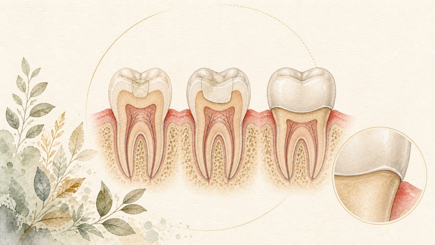
When a dentist says, “this tooth needs a prosthetic restoration”, you may hear three terms: all-ceramic crown (covering the entire tooth), veneer (a thin ceramic layer covering only the labial surface of an anterior tooth), and 3D-milled restoration (an inlay/onlay: a partial-coverage restoration made with digital design and a milling machine). It is easy to find the survival rate for each online and then rank them intuitively. But that comparison is misleading, for reasons stated clearly in the literature. A systematic review and meta-analysis of 46 articles, covering 1,715 participants and 4,209 restorations, calculated cumulative survival rates for four types of glass-ceramic restoration at the same time:
A Piece of Tooth Is Missing: An All-Ceramic Crown, Veneer, or 3D-Milled Restoration? First Ask “How Much Tooth Structure Remains?” and “Where Is the Defect?”
Direct answer: All-ceramic crowns, veneers and 3D-milled restorations are not a ranking within one contest but different answers to different extents of defect — the more intact the enamel that remains, the better bonded veneers survive [F2]; in a small cross-arch randomised trial on endodontically treated posterior teeth in the mandibular arch, full crowns had the higher survival rate while onlays had the better periodontal outcomes [F3]; the survival rates of the different categories correspond to different mean follow-up periods and therefore cannot be ranked directly [F1]; and no statistically significant difference between materials was detected over short-term follow-up of 1 to 3 years, while the conclusion of that same review states that lithium disilicate restorations “may slightly outperform” resin-matrix ceramic restorations after 3 years, except for bulk fracture [F4]. The actual choice has to be assessed by a dentist from the intra-oral examination and the imaging.
Geographic scope: This is general health education based on international literature. It does not cover any particular country's insurance scheme or regulations; for how care and fees are organised, follow the rules where you are. Every source cited in this card is international peer-reviewed literature; no national legislation or insurance-benefit rule is cited.
TL;DR|These Three Options Are Not in the Same Contest, So Their Survival Rates Cannot Be Ranked Directly
When a dentist says, “this tooth needs a prosthetic restoration”, you may hear three terms: all-ceramic crown (covering the entire tooth), veneer (a thin ceramic layer covering only the labial surface of an anterior tooth), and 3D-milled restoration (an inlay/onlay: a partial-coverage restoration made with digital design and a milling machine).
It is easy to find the survival rate for each online and then rank them intuitively. But that comparison is misleading, for reasons stated clearly in the literature.
A systematic review and meta-analysis of 46 articles, covering 1,715 participants and 4,209 restorations, calculated cumulative survival rates for four types of glass-ceramic restoration at the same time [F1]:
- Single crowns: 96%, mean follow-up 4.6 years, incidence of irreparable failure n=2.7 [F1]
- Partial-coverage restorations (including 3D-milled restorations): 90%, mean follow-up 6.2 years, incidence of irreparable failure n=5.9 [F1]
- Ceramic veneers: 90.2%, mean follow-up 6.5 years, incidence of irreparable failure n=8.2 [F1]
- Fixed dental bridges: 76.1%, mean follow-up 6.5 years, incidence of irreparable failure n=5.2 [F1]
Cautious interpretation (very important): note that these four figures have different follow-up periods—4.6 years for crowns, 6.2 years for partial coverage, and 6.5 years for veneers [F1]. A group with shorter follow-up will naturally have a better-looking figure. This is not a race with the same starting line, and these restorations are inherently used for different defect conditions; this was not a randomised head-to-head comparison.
The correct question is therefore not “which is strongest?” but: what remains of my tooth, and where is the missing area?
Build the Framework First: Three Questions Determine the Range of Options
Before discussing materials, first establish three points. They determine which options you have; only then does a material comparison become meaningful.
Question One: Is the Enamel Still Present?
This is the key to whether a veneer is feasible, and the evidence in the literature is fairly consistent.
A retrospective clinical study included 189 patients and 672 ceramic veneers followed for 1 to 15 years, dividing them into three groups according to the extent of dentine exposure after etching [F2]:
- GA1 (enamel only): survival rate 96.7% [F2]
- GA2 (dentine exposure < 30%): 95.3% [F2]
- GA3 (dentine exposure > 30%): 93.9% [F2]
- The difference between GA1 and GA3 was statistically significant (p = 0.033) [F2]
- The overall estimated cumulative survival at 15 years was 96%, with mean survival of 5.98 years [F2]
The same data also indicated that dentine exposure was associated with a higher risk of failure (odds ratio 3.47) and that root-canal-treated teeth also showed a higher risk of failure (odds ratio 1.68) [F2].
Cautious interpretation: there is a detail here that appears contradictory but must be presented honestly. The same study also recorded that root canal treatment, tooth-preparation design, and type of resin adhesive did not significantly affect survival (p > 0.05) [F2]. In other words, the signal that “root-canal-treated teeth have a higher risk” was present, but did not reach statistical significance in that study. What the study's conclusion identified as significantly affecting survival was the extent of dentine exposure [F2]. The statistical methods recorded in that abstract were Kaplan-Meier survival analysis, Log-Rank tests and odds ratios; no multivariable model is reported, so no factor can be described as having been confirmed or ruled out as an “independent” risk factor [F2].
It should also be added that all treatment was performed by the same dentist and all restorations were made by the same dental technician [F2]. This provides high internal consistency, but also means that the figures cannot be applied directly to every setting.
Question Two: How Extensive Is the Defect, and Does It Extend around the Cusps?
This determines whether partial coverage (a 3D-milled restoration) or full coverage (an all-ceramic crown) is considered.
A prospective, randomised within-arch clinical trial placed digitally manufactured lithium disilicate onlays and full crowns on opposite sides of the same patient's mouth for root-canal-treated posterior teeth in the mandibular arch (n = 15), with follow-up to 2 years [F3]:
- Full-crown survival was 100% and onlay survival 83.3%, a statistically significant difference (P = 0.030, 95% CI 0.01–0.12) [F3]
- But the onlays had significantly better periodontal outcomes: bleeding on probing (P = 0.000), plaque index (P = 0.001), and probing pocket depth (P = 0.000) were all lower at 1 and 2 years [F3]
- The onlays showed marginal discolouration in 20% and loss of marginal integrity in 13.3%, with a significant difference (P = 0.001)[F3]
- Neither group had fracture, secondary caries, or an evident change in surface texture, and patient satisfaction was 100% in both groups [F3]
Cautious interpretation: this study had n = 15 and only 2 years of follow-up [F3]. The sample was extremely small, so a single failure would cause a large swing in the percentage. It is useful for understanding the direction of the trade-offs: full coverage brought stability of the restoration itself at the cost of removing more tooth structure and having poorer periodontal measures; partial coverage preserved more tooth structure and was more favourable periodontally, but had more marginal problems [F3]. It is not suitable as proof that “full crowns are better”.
Question Three: How Long a Follow-up Horizon Can You Accept?
This concerns material selection, but the differences between materials are smaller and less stable than imagined.
Differences between Materials: More Uncertain than You May Think
Partial-Coverage Restorations: No Difference Is Apparent in the Short Term
A systematic review and meta-analysis that searched through February 2024 included 6 randomised controlled trials comparing posterior partial-coverage restorations made from different ceramic and ceramic-based materials (onlays, occlusal veneers, and partial crowns) [F4]:
- Resin-matrix ceramic (RMC) 3-year survival 89.3% (95% CI 76.4–95.3) [F4]
- Lithium disilicate (LDS) 3-year survival 93.7% (95% CI 83.7–97.7) [F4]
- Leucite-reinforced glass-ceramic (LRGC) ranged from 96.1% (95% CI 90.1–98.9) to 98.3% (95% CI 90.8–100) [F4]
- After 1 to 3 years of follow-up, LDS was slightly better than RMC for restoration failure and adhesive debonding: per 100 restoration-years, RMC had 1.56 more failures and 1.78 more adhesive debondings (low-certainty evidence) [F4]
Cautious interpretation: these two statements sit in different parts of the paper and must be read separately. The results section recorded that no statistically significant differences between ceramic and ceramic-based materials were detected during short-term follow-up of 1 to 3 years, and that the long-term performance of posterior partial-coverage restorations remains uncertain [F4]; the review's conclusion states that the survival of lithium disilicate restorations “may slightly outperform” resin-matrix ceramic restorations after 3 years of follow-up across outcomes, except for bulk fracture — the wording used is “may” [F4]. Note how wide the confidence intervals above are: RMC ranges from a lower bound of 76.4% to an upper bound of 95.3%, and the intervals overlap [F4]. Together with the “low-certainty evidence” label, these figures are insufficient to support a claim that “one material is clearly better”. The review also stated explicitly that ceramic and ceramic-based partial-coverage restorations are a reliable treatment option for restoring more extensive posterior defects [F4].
Extend the Horizon to 10 Years and Even the Direction Reverses
A systematic review and meta-analysis of 21 articles with at least 5 years of follow-up compared ceramic and resin inlays, onlays, and full-coverage restorations [F5]:
Estimated 5-year survival
- Resin 86% (n = 129)|feldspathic porcelain 90% (n = 1,048)|glass-ceramic 92% (n = 2,218)[F5]
Estimated 10-year survival
- Resin 75% (n = 115)|feldspathic porcelain 91% (n = 1,829)|glass-ceramic 89% (n = 1,075)[F5]
The review's pooled regression showed that ceramic partial-coverage restorations outperformed resin restorations at both 5 and 10 years. Between the two ceramic types, however, glass-ceramic outperformed feldspathic porcelain at 5 years, but by 10 years the direction reversed and feldspathic porcelain outperformed glass-ceramic [F5].
Cautious interpretation: note this reversal of direction. It may reflect a true difference in material behaviour, or it may reflect different sample compositions at the respective time points—for example, there were 2,218 glass-ceramic restorations at 5 years but only 1,075 at 10 years [F5]. Either way, the existing data provide no answer to “which ceramic is better?” that remains stable over time.
How Do These Restorations Fail When They Fail?
The conclusion of the same review recorded that failures were mostly due to fractures (6.2%), endodontic problems (3%), secondary caries (1.7%) and debonding (0.9%) [F5]. The abstract does not state the denominator for these percentages, and the four figures added together fall far short of 100%, so they are not a percentage breakdown of failure causes but the rates at which each type of problem was recorded within that review's dataset [F5].
This set of figures is still useful because it tells you what needs to be prevented: the source wording is that failures were “mostly due to” fractures, followed by problems inside the tooth [F5]. This also explains why “how much tooth structure remains” matters more than “which material is used”.
Bring the Framework Together: A Comparison Table
| Your Situation | Options Commonly Discussed | Main Point from the Literature |
|---|---|---|
| Colour or form concerns on the labial surface of an anterior tooth, with enamel largely intact | Veneer | Survival was 96.7% when bonded only to enamel, falling to 93.9% with dentine exposure > 30% (p = 0.033)[F2] |
| A posterior defect that is extensive but leaves the cusps intact, with adequate tooth structure | 3D-milled restoration (partial coverage) | Preserves more tooth structure and has better periodontal measures, but marginal discolouration 20% and loss of marginal integrity 13.3% [F3] |
| A defect that surrounds the whole tooth, with little tooth structure remaining | All-ceramic crown | Single-crown survival was 96% in the same meta-analysis, but mean follow-up was only 4.6 years [F1] |
| An entire tooth is missing and a bridge is being considered | Fixed dental bridge | Survival 76.1% (6.5 years), with more irreparable failures; the review advised caution [F1] |
How to use this table: it helps you understand the context in which each option is discussed; it is not a table for self-diagnosis. Which options are feasible depends on an actual intraoral examination and interpretation of imaging.
What Does “Not Selling a Single Option” Actually Look Like?
Three honest observations can be drawn from the data above:
First, the more conservative the approach, the more tooth structure it preserves, but the more marginal problems it has. The onlays had better periodontal measures, but markedly higher proportions of marginal discolouration and loss of marginal integrity [F3].
Second, the more extensive the coverage, the more stable the restoration itself, but the cost is tooth structure and periodontal health. In the same trial, full-crown survival was 100%, but periodontal outcomes were poorer at both 1 and 2 years [F3].
Third, material differences are not statistically apparent in the short term, although the direction is not a complete blank. Six randomised controlled trials detected no significant differences between materials at 1 to 3 years and long-term performance was uncertain; the conclusion of that same review nonetheless states that lithium disilicate “may slightly outperform” resin-matrix ceramic, except for bulk fracture [F4].
In other words, the choice is not about selecting a “best product”, but balancing “how much tooth structure to preserve” against “how much stability to gain” under the conditions in your mouth. There is no universal answer to this balance because every tooth has something different left.
Conclusion|Ask “What Remains?” before Asking “What Should Be Done?”
If you remember only one thing, remember this: these three approaches are not three grades; they are answers for three different extents of defects.
The literature can provide conditions and trade-offs: the more completely enamel is preserved, the better bonded restorations perform [F2]; conservative partial coverage is more favourable periodontally but has more marginal issues [F3]; short-term differences between materials did not reach statistical significance, although that review's conclusion still states that lithium disilicate “may slightly outperform” resin-matrix ceramic except for bulk fracture, and long-term data remain insufficient [F4][F5]; and survival rates for different categories cannot be ranked directly because their follow-up periods differ [F1].
Consider taking these questions to discuss with your dentist at the appointment:
- How much tooth structure remains in this tooth? Is there enough enamel to support bonding?
- Does the defect extend around the cusps? Is full coverage necessary?
- If I choose a more conservative approach, what problem is most likely to occur?
- Has this tooth had root canal treatment? Will that affect the options?
Changing the starting point of the choice from “which is better?” to “what remains?” will immediately make the discussion with your dentist much more concrete.
Risk factors (what to know before treatment)
- The most common form of failure is fracture: a systematic review and meta-analysis of 21 articles with follow-up longer than 5 years recorded that failures were mostly due to fractures (6.2%), endodontic problems (3%), secondary caries (1.7%) and debonding (0.9%); the abstract does not state the denominator for these percentages [F5]. This is why “how much tooth structure remains” matters more than “which material is used”.
- The cost of the conservative option shows up at the margin: in a cross-arch randomised trial on endodontically treated posterior teeth in the mandibular arch, onlays showed marginal discoloration (20%) and loss of marginal integrity (13.3%), with a statistically significant difference (P = 0.001) [F3]. That trial covered only 15 teeth and followed them to 2 years; the direction is informative, but the figures should not be treated as a personal expectation [F3].
- The cost of full coverage shows up in the periodontium: in the same trial, onlays performed significantly better on bleeding on probing, plaque index and probing pocket depth, and the authors recorded greater periodontal damage with full crowns at 1 and 2 years [F3].
- Conditions that reduce the survival of bonded restorations: in retrospective data on 672 veneers followed for 1 to 15 years, endodontically treated teeth and teeth with dentin exposure showed higher risks of failure (odds ratios of 1.68 and 3.47 respectively); the same paper also recorded that endodontic treatment, preparation design and resin cement type did not significantly affect survival (p > 0.05), and what that study identified as significant was the extent of dentin exposure [F2].
- The limits of the evidence itself: the review comparing materials included only 6 randomised controlled trials and states plainly that the long-term performance of posterior partial coverage restorations is uncertain [F4]; in the veneer study all treatment was carried out by a single clinician and all restorations were made by a single ceramist, which gives high internal consistency but limits generalisability [F2]. This card did not run a separate literature search on contraindications and therefore does not compile a list of them; which approach is feasible, and whether pulpal or periodontal conditions need to be addressed first, has to be assessed by a dentist from the intra-oral examination and the imaging.
*This article is a review of the literature, not personalised medical advice. The cited studies differ in sample size, follow-up period, and applicable populations; discuss your individual circumstances with your dentist.*
Every clinical statement in this article is mapped line by line to its cited source (see the sources and evidence chain below). It has not been clinically reviewed by a licensed practitioner. This is health information, not individual advice; assessment by a clinician is required.
FAQ
- The survival rate of all-ceramic crowns looks highest. Should I simply have an all-ceramic crown?
- That inference is not advisable. In the same meta-analysis, the 96% survival rate for single crowns corresponded to a mean follow-up of **4.6 years**, while 90% for partial-coverage restorations corresponded to **6.2 years** and 90.2% for veneers to **6.5 years** [F1]. Figures with different follow-up periods cannot be compared side by side. These restorations were also used for different extents of defects, rather than being tested in a randomised head-to-head trial [F1].
- オールセラミッククラウンの生存率が最も高く見えます。最初からオールセラミッククラウンにすべきですか? — そのような推論は勧められません。同じメタアナリシスで、単冠の生存率 96%に対応する平均追跡期間は**4.6年**でしたが、部分被覆修復物の 90%は**6.2年**、ラミネートベニアの 90.2%は**6.5年**でした [F1]。追跡期間が異なるため、数値を直接並べて比較することはできません。また、これらの修復物はもともと異なる欠損範囲に用いられており、ランダム割付による直接比較試験ではありません [F1]。
- The survival rate of all-ceramic crowns looks highest. Should I simply have an all-ceramic crown? — That inference is not advisable. In the same meta-analysis, the 96% survival rate for single crowns corresponded to a mean follow-up of **4.6 years**, while 90% for partial-coverage restorations corresponded to **6.2 years** and 90.2% for veneers to **6.5 years** [F1]. Figures with different follow-up periods cannot be compared side by side. These restorations were also used for different extents of defects, rather than being tested in a randomised head-to-head trial [F1].
- Do Veneers Come off Easily?
- According to current data, veneer performance is strongly related to **what it is bonded to**. Data from 672 veneers followed for 1 to 15 years showed an estimated survival rate of 96.7% when bonded only to enamel, 95.3% with dentine exposure < 30%, and 93.9% with exposure > 30%; the difference between the first and last groups was statistically significant (p = 0.033)[F2]. In that study, overall estimated cumulative survival at 15 years was 96% [F2].
- ラミネートベニアは簡単に外れますか? — 現在のデータでは、ラミネートベニアの成績は**何に接着するか**と強く関連します。672件を 1~15年追跡したデータでは、エナメル質だけに接着した場合の推定生存率は 96.7%、象牙質露出 < 30%では 95.3%、> 30%では 93.9%で、最初と最後の群の差は統計学的有意でした(p = 0.033)[F2]。同研究の全体の15年累積推定生存率は 96%でした [F2]。
- Do Veneers Come off Easily? — According to current data, veneer performance is strongly related to **what it is bonded to**. Data from 672 veneers followed for 1 to 15 years showed an estimated survival rate of 96.7% when bonded only to enamel, 95.3% with dentine exposure < 30%, and 93.9% with exposure > 30%; the difference between the first and last groups was statistically significant (p = 0.033)[F2]. In that study, overall estimated cumulative survival at 15 years was 96% [F2].
- Can a Root-Canal-Treated Tooth Still Receive a Veneer or Milled Restoration?
- This requires individual assessment. The veneer study above indicated a higher risk of failure for root-canal-treated teeth (odds ratio 1.68), but the same study also recorded that **root canal treatment itself did not significantly affect survival (p > 0.05)**; the statistical methods reported in that abstract were Kaplan-Meier survival analysis, Log-Rank tests and odds ratios [F2]. In addition, the trial comparing onlays and full crowns for root-canal-treated posterior teeth in the mandibular arch found advantages and disadvantages for both at 2 years and included only 15 teeth [F3]. Current evidence is insufficient to support a one-size-fits-all answer.
- 根管治療を受けた歯にもラミネートベニアやセラミック部分被覆修復を行えますか? — 個別の評価が必要です。前述のラミネートベニア研究は、根管治療済みの歯に高い失敗リスクの傾向があること(オッズ比 1.68)を示しましたが、同じ研究は同時に、**根管治療自体は生存に有意な影響を与えなかった(p > 0.05)**とも記録しています。その抄録に記載された統計手法は Kaplan-Meier 生存解析、Log-Rank 検定、オッズ比です [F2]。また、下顎歯列内の根管治療後の臼歯でアンレーと全部被覆冠を比較した試験では、2年時点でそれぞれに長所と短所があり、サンプルは 15本だけでした [F3]。現在のエビデンスは、すべてに当てはまる一つの答えを支持するには不十分です。
- Can a Root-Canal-Treated Tooth Still Receive a Veneer or Milled Restoration? — This requires individual assessment. The veneer study above indicated a higher risk of failure for root-canal-treated teeth (odds ratio 1.68), but the same study also recorded that **root canal treatment itself did not significantly affect survival (p > 0.05)**; the statistical methods reported in that abstract were Kaplan-Meier survival analysis, Log-Rank tests and odds ratios [F2]. In addition, the trial comparing onlays and full crowns for root-canal-treated posterior teeth in the mandibular arch found advantages and disadvantages for both at 2 years and included only 15 teeth [F3]. Current evidence is insufficient to support a one-size-fits-all answer.
- Which Material Is Better for a 3D-Milled Restoration?
- No statistically significant difference is apparent in the short term. In its results section, a meta-analysis of 6 randomised controlled trials found **no statistically significant differences between materials during 1 to 3 years of follow-up**, and the confidence intervals were wide—for example, 89.3% for resin-matrix ceramic, with a 95% CI of 76.4–95.3; its conclusion nonetheless states that lithium disilicate “may slightly outperform” resin-matrix ceramic, except for bulk fracture [F4]. At 10 years, another review found ceramic superior to resin, but the relative ranking of the two ceramics **reversed direction** between 5 and 10 years [F5].
- 3D セラミック修復には、どの材料が優れていますか? — 短期では統計学的な差が見えません。6件のランダム化比較試験を採用したメタアナリシスの結果の項では、**1~3年の追跡中、材料間に統計学的有意差はありませんでした**。信頼区間も非常に広く、たとえばレジンマトリックスセラミックは 89.3%、95% CI 76.4–95.3でした。ただし同レビューの結論には、二ケイ酸リチウムがバルク破折を除いて樹脂マトリックスセラミックを「わずかに上回る可能性がある」と記されています [F4]。10年まで延ばすと、別のレビューではセラミックがレジンより優れていましたが、2種類のセラミック間の優劣は 5年と10年で**方向が逆でした** [F5]。
- Which Material Is Better for a 3D-Milled Restoration? — No statistically significant difference is apparent in the short term. In its results section, a meta-analysis of 6 randomised controlled trials found **no statistically significant differences between materials during 1 to 3 years of follow-up**, and the confidence intervals were wide—for example, 89.3% for resin-matrix ceramic, with a 95% CI of 76.4–95.3; its conclusion nonetheless states that lithium disilicate “may slightly outperform” resin-matrix ceramic, except for bulk fracture [F4]. At 10 years, another review found ceramic superior to resin, but the relative ranking of the two ceramics **reversed direction** between 5 and 10 years [F5].
- How Do These Restorations Most Commonly Fail?
- In its conclusion, a review covering 21 articles recorded that failures were **mostly due to fractures (6.2%), endodontic problems (3%), secondary caries (1.7%) and debonding (0.9%)** [F5]. **The abstract does not state the denominator for these percentages**, and the four figures added together fall far short of 100%, so they cannot be read as a percentage breakdown of failure causes. Fracture is the first item the source lists, which is why “how much tooth structure remains” is the starting point for the choice.
- これらの修復物は、どのように壊れることが最も多いですか? — 21編の論文を含むレビューの結論には、失敗は**主に破折(6.2%)、歯髄/根管の問題(3%)、二次う蝕(1.7%)、接着脱離(0.9%)**によるものだったと記録されています [F5]。**抄録にはこれらの百分率の分母が記載されておらず**、四項目を合計しても 100% には遠く及ばないため、失敗原因の百分率の内訳としては読めません。破折は原文が最初に挙げた項目であり、これが「どれだけ歯質が残っているか」を選択の出発点とする理由です。
- How Do These Restorations Most Commonly Fail? — In its conclusion, a review covering 21 articles recorded that failures were **mostly due to fractures (6.2%), endodontic problems (3%), secondary caries (1.7%) and debonding (0.9%)** [F5]. **The abstract does not state the denominator for these percentages**, and the four figures added together fall far short of 100%, so they cannot be read as a percentage breakdown of failure causes. Fracture is the first item the source lists, which is why “how much tooth structure remains” is the starting point for the choice.
- If I Want to Remove as Little Tooth as Possible, Should I Choose the Most Conservative Option?
- The direction is reasonable, but the cost must be understood. In the randomised within-arch trial, onlays had significantly better periodontal measures than full crowns but significantly higher rates of marginal discolouration (20%) and loss of marginal integrity (13.3%), with 2-year survival of 83.3% (full crowns 100%, P = 0.030)[F3]. This was a small study of 15 teeth; its direction can inform discussion, but the figures should not be treated as an individual's expected outcome [F3].
- できるだけ歯を削りたくない場合、最も保存的な方法を選べばよいですか? — 方向としては妥当ですが、代償を知る必要があります。同一歯列内でランダムに割り付けた試験では、アンレーは歯周指標で全部被覆冠より有意に良好でしたが、辺縁変色(20%)と辺縁完全性の喪失(13.3%)の割合が有意に高く、2年生存率は 83.3%でした(全部被覆冠 100%、P = 0.030)[F3]。これは 15本を対象とした小規模研究であり、方向性は参考になりますが、数値を個人の予測として扱うべきではありません [F3]。
- If I Want to Remove as Little Tooth as Possible, Should I Choose the Most Conservative Option? — The direction is reasonable, but the cost must be understood. In the randomised within-arch trial, onlays had significantly better periodontal measures than full crowns but significantly higher rates of marginal discolouration (20%) and loss of marginal integrity (13.3%), with 2-year survival of 83.3% (full crowns 100%, P = 0.030)[F3]. This was a small study of 15 teeth; its direction can inform discussion, but the figures should not be treated as an individual's expected outcome [F3].
Medical notice This article is provided for health education and medical information purposes. It is not a solicitation for medical services and does not constitute diagnosis or treatment advice. Actual treatment methods and outcomes vary between individuals and require evaluation by a dentist; every treatment has its own indications, limitations and possible risks. If you have related symptoms or treatment needs, please book a consultation for evaluation by a dentist.
Source anchors
- Biomechanical consideration in tooth-supported glass-ceramic restorations: A systematic review and meta-analysis of survival rates and irreparable failures. [PMID:38849264] · https://pubmed.ncbi.nlm.nih.gov/38849264/ · 在 IDAEO 的其他引用
- Survival of Ceramic Veneers: Impact of Dentin Exposure and Tooth Vitality After 1 to 15 Years of Follow-Up. [PMID:40755189] · https://pubmed.ncbi.nlm.nih.gov/40755189/ · 在 IDAEO 的其他引用
- A comparative evaluation of prosthetic and clinical outcomes influenced by two digitally fabricated extracoronal restorations: An onlay and a full crown: A prospective, cross-arch randomized study. [PMID:40213887] · https://pubmed.ncbi.nlm.nih.gov/40213887/ · 在 IDAEO 的其他引用
- Survival and Complications of Partial Coverage Restorations on Posterior Teeth - A Systematic Review and Meta-Analysis. [PMID:39558793] · https://pubmed.ncbi.nlm.nih.gov/39558793/ · 在 IDAEO 的其他引用
- Comparative evaluation of clinical performance of ceramic and resin inlays, onlays, and overlays: A systematic review and meta analysis. [PMID:36187858] · https://pubmed.ncbi.nlm.nih.gov/36187858/ · 在 IDAEO 的其他引用
Cite this article
Lucy・《A Piece of Tooth Is Missing: An All-Ceramic Crown, Veneer, or 3D-Milled Restoration? First Ask “How Much Tooth Structure Remains?” and “Where Is the Defect?”》・IDAEO 知識庫・2026-07-20・https://km.idaeo.ai/post/dental/crown-veneer-inlay-choice-frameworkUpdated 2026-08-26