

🏛 Part of the "dental" topic shelf →
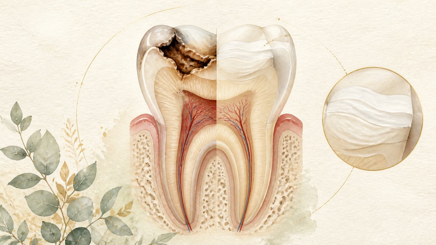
Does every cavity need a filling? How is a filling done?
Whether a carious lesion should be restored is a staging question, not a yes-or-no question. This card uses international caries consensus statements, two American Dental Association guidelines, and Cochrane reviews to distinguish lesions that can be controlled and monitored non-restoratively from those with an indication for restoration. It also presents the conflicting evidence on deep-caries removal, describes the filling process, and gives no prices.
Does every cavity need a filling? How is a filling done?
Direct answer (source-language limit: 60 characters)
For an early lesion that has not yet cavitated, there is evidence supporting non-restorative control and follow-up first [F5][F7]; a lesion that has already cavitated and can no longer be cleaned or sealed is the indication for restoration (a filling) [F4]. Staging is determined by a dentist.
Scope: This is general health education based on international literature. It does not address any particular country's insurance or legal system; care and fee arrangements follow the system where treatment is received. The Taiwan Medical Care Act cited in the compliance notice at the end is used only to define this site's publication status, and is already tagged geo: TW in the F-Unit.
Why "does it really have to be filled?" turns into an argument
Online you will find two claims side by side: one says that a cavity left unfilled will rot all the way to the nerve, the other says that early caries only needs fluoride. Each claim corresponds to a different stage of the lesion; what gets stripped out when they are moved onto the internet is the premise about which stage is being discussed [F2].
In its terminology consensus, the International Caries Consensus Collaboration first separates two words: dental caries is the name of the disease, while a carious lesion is the consequence and manifestation of that disease — that is, the sign or symptom of the disease [F3]. The same consensus also defines "caries management" and "carious lesion management" separately: the former controls the disease at patient level by preventive and non-invasive means, the latter controls the signs of the disease at tooth level [F3].
This distinction determines the answer to your question directly: a filling deals with "this hole in this tooth", not with "why you get caries in the first place" [F3]. So whether a restoration is needed, and when, depends on which stage the lesion has reached; whether you will get caries again depends on whether the disease itself has been brought under control [F3][F4].
What follows sets out the staging first, then what evidence exists at each stage, and then what actually happens on the day a filling is placed.
Staging: how far has your cavity gone?
Drawing on the literature in F3 through F7, this site groups the situations a patient may encounter into three stages (this is a simplified framework for communication; clinical classification follows your dentist's diagnosis) [F2]:
- A lesion that has not yet cavitated: a white spot or discolouration appears on the tooth surface, but the surface structure is still continuous and no obvious hole can be probed. This stage is where non-restorative management is on its own home ground in terms of evidence [F5][F7].
- A lesion that has already cavitated: the tooth surface has broken down and collapsed. The criterion in the international consensus is not "a hole always has to be filled", but whether that hole can still be cleaned and whether it can still be sealed [F4].
- A deep lesion close to or involving the pulp: at this stage the main goal shifts from "filling it up" to "keeping the pulp alive", and both the treatment options and the risks are different [F4][F15].
Deciding which stage applies relies clinically on visual examination plus radiographic follow-up — for example, the long-term studies that track proximal (between-the-teeth) lesions read annual bitewing radiographs to judge whether a lesion has advanced [F11]. You cannot work out the stage yourself in front of a mirror, which is also why "does a cavity need filling?" cannot be settled by a single answer on the internet [F2].
Stages one and two: when there is evidence to hold off on restoring
This is the part that online content usually skips, and it is the load-bearing section of this card. Point by point below: what the evidence supports, and where the boundary of that evidence lies.
What the consensus says: restoration is not the default
In its clinical recommendations on carious tissue removal, the International Caries Consensus Collaboration puts it bluntly: entering the restorative cycle should be avoided as far as possible [F4]. The same consensus then defines when a restorative intervention is warranted: only when a cavitated lesion cannot be cleaned, or can no longer be sealed, is restorative intervention indicated [F4]. Approaches to controlling lesion activity should first attempt methods aimed at removing or controlling the biofilm [F4].
Translated into language a patient can use: "there is a hole" is not in itself an automatic instruction to operate; "this hole cannot be cleaned" is [F4]. And whether it can be cleaned is a clinical judgement, not a self-assessment [F2].
Non-restorative management: which approaches have evidence
An expert panel convened by the American Dental Association (ADA) Council on Scientific Affairs and its Center for Evidence-Based Dentistry carried out a systematic review and issued evidence-based recommendations on non-restorative treatments to arrest or reverse non-cavitated and cavitated caries lesions in children and adults, producing 11 clinical recommendations in total, each one tied to a specific lesion type, tooth surface and dentition [F5]. Among the more effective interventions, the panel made recommendations for 38% silver diamine fluoride, sealants, 5% sodium fluoride varnish, 1.23% acidulated phosphate fluoride gel, and 5000 ppm fluoride (1.1% sodium fluoride) toothpaste or gel [F5].
Pay particular attention to the phrase "each one tied to a specific lesion type, tooth surface and dentition" [F5]. What it means is that these methods are not a universal solution that can be applied to every cavity; whether one of them suits that particular tooth of yours has to be judged by a dentist according to the type and location of the lesion [F5][F2].
The same guideline also issued a recommendation in the opposite direction: the panel recommended against using 10% casein phosphopeptide–amorphous calcium phosphate (CPP-ACP) [F5]. This point is worth stating separately, because products containing that ingredient are not rare on the market; this card simply records the direction of the guideline and does not evaluate any commercial product [F5].
Early lesions between the teeth: the evidence for micro-invasive treatment is clearer
For proximal (between-the-teeth) lesions, a Cochrane systematic review included 8 randomised trials with 365 participants, comparing "micro-invasive treatment" (sealing over the lesion surface, or resin infiltration into the lesion, and similar approaches) with non-invasive treatment. The pooled analysis showed that, compared with non-invasive professional treatment (such as fluoride varnish) or oral hygiene advice (such as flossing), micro-invasive treatment significantly reduced the odds of lesion progression (OR 0.24, 95% CI 0.14 to 0.41; 602 lesions, 7 studies) [F7]. The review authors rated the quality of this body of evidence as moderate, and noted that micro-invasive treatment can arrest non-cavitated enamel lesions and initial dentinal lesions (read radiographically as being confined to the outer third of dentine) [F7].
The boundary has to be read along with it: the review's inclusion criteria covered non-cavitated proximal lesions, not caries that has already collapsed into a hole; the four studies that assessed adverse events reported no adverse events after micro-invasive treatment, but most studies did not report other outcome measures [F7]. So what this body of evidence supports is that "an early proximal lesion does not have to be drilled open first", not that "every cavity can go unfilled" [F7][F2].
Fluoride and sealants: prevention evidence, filed separately from "reversal"
- Fluoride varnish: a Cochrane systematic review included 22 trials with 12455 participants. In the pooled analysis for permanent tooth surfaces (13 trials), the estimated preventive fraction for fluoride varnish compared with placebo or no treatment was 43% (95% CI 30% to 57%); for primary tooth surfaces (10 trials) it was 37% (95% CI 24% to 51%) [F8]. The review concluded that fluoride varnish has a substantial caries-inhibiting effect in both permanent and primary teeth, but rated the quality of the evidence as moderate, because most included studies were at high risk of bias and heterogeneity was considerable [F8].
- Fissure sealants: a Cochrane systematic review included 38 trials with 7924 children and concluded that resin-based sealants applied to the occlusal surfaces of permanent molars prevent caries in children and adolescents; at the 24-month measurement point, moderate-quality evidence showed that resin sealants reduced caries by 11% to 51% compared with no sealant [F9].
- The range of these two has to be stated plainly: the primary outcome measure in both reviews is "preventing new lesions", not "making an existing cavity disappear" [F8][F9]. They appear under this question because the ADA guideline lists sealants and fluoride agents among its recommended non-restorative options [F5]; but you cannot infer from that "a fluoride application means no filling is needed" [F2].
The honest other side: the cost of the non-restorative route, and what is unknown
- Silver diamine fluoride stains arrested lesions black. A qualitative interview study of 14 dental professionals recorded that the main drawback and barrier to use mentioned by interviewees was exactly the black staining caused by silver diamine fluoride [F10]. This is an appearance trade-off, especially on anterior teeth; whether to accept it is something for you and your dentist to discuss [F10].
- The non-restorative route is not "go home and ignore it". It presupposes that you will come back, that follow-up images will be taken, and that diet and cleaning will be controlled — the international consensus places "controlling disease activity" before restoration, not as a replacement for follow-up [F4].
- On 2026-08-05 this site ran two PubMed searches, "monitoring versus restoration cavitated caries lesion adults randomized trial long-term" and "non-restorative cavity control cavitated lesions adults arrest cleansable", and both returned 0 records: that is, a direct comparative long-term randomised trial of "long-term monitoring only, without restoration, for cavitated lesions in adults" versus "immediate restoration" was not retrieved by this search [F22]. This card therefore offers no promise about "how long you can put it off" [F22].
What happens if you don't fill it: the evidence with the scare tactics removed
First, what this card will not write: it will not say "if you don't fill it, it will certainly rot down to the nerve", and it will not say "leaving it unfilled is fine". Both sentences go beyond what the existing evidence can support [F22].
What can be written is measured rates of progression. A Swedish prospective radiographic follow-up study used annual bitewing radiographs to track proximal caries in 536 children from age 11 to age 22, in a population whose treatment strategy was remineralisation-based rather than restoration-based [F11]. The results showed considerable variation between tooth surfaces in caries incidence and survival time [F11]. In concrete numbers: for sound surfaces (state 0), 75% had not progressed to an enamel lesion (state 2) within 6.3 years; for lesions already at state 2, 75% had not progressed into the outer half of dentine (state 4) within 4.8 years; while for lesions that had reached the enamel–dentine junction (state 3), only 75% had not progressed within 1.3 years, and the median survival time from state 3 to state 4 was 3.1 years [F11].
How this set of numbers should be read: it refutes both extreme claims at once [F2]. Progression of early lesions is often measured in years rather than weeks, so short-term monitoring does have a footing in the literature [F11]; but once a lesion reaches the junction between enamel and dentine, it advances markedly faster, and the margin for error in observing it shrinks accordingly [F11]. Its limits must be marked just as clearly: these are data published in 1999, from a single country, in an adolescent population, on proximal surfaces, and under a predominantly remineralisation-based care strategy, and they cannot be taken directly as a prediction for that particular tooth of yours [F11].
As for how common "putting off the filling" is worldwide: a systematic review with meta-regression estimated that in 2010 about 2.4 billion people had untreated caries in permanent teeth and about 621 million children had untreated caries in deciduous teeth [F12]. Common does not mean without consequences; this paper is used here only to show that you are not an exception, not as reassurance [F12][F2].
The real watershed is the pulp. When a lesion is deep enough to approach or involve the pulp, the axis of treatment shifts from "filling the hole" to "keeping the pulp vital and avoiding apical periodontitis" — the European Society of Endodontology (ESE) position statement on deep caries lists "maintaining pulp health and vitality, preventing apical periodontitis, and developing minimally invasive, biologically based treatment" among the core themes of contemporary clinical endodontics [F15]. At this stage the options become vital pulp therapy, root canal treatment, or extraction when the tooth cannot be retained, rather than simply placing a filling [F15][F16].
The deep-caries controversy: clean it out more, or take less away
This is the section where this card deliberately sets conflicting evidence side by side, because the question has not converged in the literature [F2].
Evidence supporting "take less away": a Cochrane systematic review included 8 trials with 934 participants and 1372 teeth, comparing different carious tissue removal strategies. The results showed that stepwise removal, compared with complete removal, reduced the incidence of pulp exposure by 56% (RR 0.44, 95% CI 0.33 to 0.60), rated as moderate-quality evidence; in those 4 studies, the mean pulp exposure rate was 34.7% in the complete removal group and 15.4% in the stepwise removal group [F13]. Partial removal, compared with complete removal, reduced the incidence of pulp exposure by 77% (RR 0.23, 95% CI 0.08 to 0.69), also moderate-quality evidence; in those 2 studies, the mean pulp exposure rate was 21.9% in the complete removal group and 5% in the partial removal group [F13]. But the same review also states plainly that all included trials were judged to be at high risk of bias, and that for the outcome "signs and symptoms of pulpal disease" the evidence was insufficient to determine whether there is any difference between partial and complete removal [F13].
The updated evidence (2021 Cochrane, the current latest version on this question): the Cochrane review on the same topic was updated in 2021, including 27 studies with 3,350 participants and 4,195 teeth/lesions, spanning 11 countries and published between 1977 and 2020; of these, 2 studies included adults only, 20 included only children and adolescents, and 5 included both [F23]. That version concluded that, compared with complete removal, sealing with a preformed metal crown (the Hall Technique) and selective removal had fewer failures in primary teeth, while in permanent teeth selective removal and stepwise removal had fewer failures; but most studies were at high risk of bias, with small sample sizes and few failure events, so the certainty of evidence for most comparisons was rated low or very low [F23]. This point needs to be spelled out: the new version downgraded the certainty of evidence to "low or very low", which means that although the direction is consistent with the 2013 version, the strength is not enough to support a promise of effectiveness for an individual patient; this card therefore only describes the decision-making orientation, and gives no success rate and no time promise [F23].
Which way the guidelines lean: the international consensus recommends that shallow and moderately deep cavitated lesions be managed by "selective removal to firm dentine"; for deep cavitated lesions it recommends "selective removal to soft dentine" in both primary and permanent teeth, with stepwise removal as an additional option in permanent teeth [F4]. The consensus also states that the purpose of removing carious tissue is purely to create the conditions for a long-lasting restoration, and that tissue close to the pulp, bacterially contaminated or demineralised does not need to be removed [F4]. The ADA's 2023 guideline on restorative treatments gives a "conditional recommendation" to conservative carious tissue removal approaches, especially for more advanced lesions; that guideline produced 16 recommendations and good practice statements in total, of which 4 concern removal approaches distinguished by lesion depth and 12 concern direct restorative materials distinguished by tooth location and the surfaces involved [F6]. The guideline authors' wording is that the evidence suggests more conservative removal approaches "may" reduce the risk of adverse events [F6].
The evidence on the other side is new, and must be presented alongside: a 2024 double-blind randomised clinical trial enrolled 124 mature permanent teeth with caries extending radiographically into more than two-thirds of the dentine and diagnosed with reversible pulpitis, randomised into selective removal (63 teeth) and complete removal (61 teeth, with vital pulp therapy performed on the spot if the pulp was exposed) [F14]. The results: 17 teeth (28%) in the complete removal group had pulp exposure managed with vital pulp therapy; at 12 months, pulp survival was 98.4% in the complete removal group versus 82.5% in the selective removal group (P = 0.003), a statistically significant difference [F14]. Multivariate analysis showed that the removal approach and the pre-operative pain level (whether it reached 5/10) were significant prognostic factors [F14]. The authors' clinical implication is stated directly: for deep caries in mature permanent teeth with reversible pulpitis, complete removal of carious tissue to hard dentine is recommended in order to obtain predictable pulp survival [F14].
So this card's conclusion can only go this far: the carious tissue removal strategy in deep caries is a clinical decision that has not yet converged, and tooth maturity, pulp diagnosis and pre-operative pain level all change the answer [F13][F14][F15]. The ESE position statement itself explicitly notes that the field contains controversy, low-quality evidence and uncertainty [F15]. Any content on the internet telling you that deep caries "must" or "must never" be cleaned out completely is claiming more than the evidence does [F22]. What you can do is ask your dentist: what is the pulp diagnosis for this tooth, which removal approach do you intend to use, and why [F2].
How a filling is done: what actually happens in that appointment
The steps below are a process framework compiled by this site from the literature in F3, F4, F6, F17, F18 and F20, to help you follow what is being discussed in the surgery; the actual steps follow your dentist's clinical judgement [F2].
- Confirming the stage and extent: visual examination plus radiographic reading of the depth and location of the lesion, to decide whether to enter restoration at all [F4][F11].
- Isolating the tooth: the rubber dam is a common method of isolation. A Cochrane systematic review including 4 studies with 1270 participants found "very low quality" evidence that using a rubber dam, compared with cotton roll isolation, may be associated with a lower failure rate of direct restorations [F18]. The low level of evidence is a fact, and this card records it as such rather than writing it up as a necessary condition [F18].
- Removing carious tissue: the names of the techniques are clearly defined in the international consensus — selective removal of carious tissue (covering removal to soft dentine and removal to firm dentine), stepwise removal, and non-selective removal to hard dentine (what used to be called complete caries removal; the consensus notes that this technique is no longer recommended) [F3]. Different depths call for different recommended approaches [F4][F6].
- Choosing the restorative material: a 2026 overview of Cochrane systematic reviews (14 reviews, 57 primary studies) collated the evidence on direct restorative materials for posterior permanent teeth. Among its findings: between bulk-fill and incrementally layered composite resin there may be no difference in the risk of restoration failure, and both were below 5% (RD 0.00, 95% CI −0.03 to 0.03; 7 studies, 511 restorations, follow-up 1 to 10 years), a comparison rated as moderate-certainty evidence [F17]. Comparing composite resin with amalgam, low-certainty evidence suggested that amalgam may have 7% less risk of restoration failure (RD 0.07, 95% CI 0.05 to 0.09; 2 studies, 3010 restorations, follow-up 5 to 7 years), but the overview authors pointed out that recruitment in the primary studies of that review began in the late 1990s, which affects how far it can be generalised to contemporary practice [F17]. The same overview also mentions that the Minamata Convention recommends phasing down the use of dental amalgam, and that mercury-free direct restorative materials are already available [F17]. The ADA's 2023 guideline gives conditional recommendations to all the direct restorative materials it included, and ranks some of them according to clinical situation [F6].
- Finishing the margins and the bite: the marginal fit of a restoration is not a cosmetic matter. A retrospective study showed that proximal restorations with marginal overhangs had periodontal pockets 0.42 mm deeper than metal restorations without overhangs [F20]. So if, after the filling, floss shreds, catches or is hard to pass at that point, it is worth going back and asking your dentist to check the margin [F20].
- Aftercare once the filling is done: the timing questions in this part (how soon you can eat, how soon you can brush) are answered point by point in a separate card; see the internal citation chain at the end [F2].
A practical note on materials: the overview authors also pointed out that only about 10% of the primary studies were conducted in general practice settings [F17]. In other words, the environment of the material research is not necessarily the same as the clinic you attend, and that is the margin to leave when reading research numbers as personal outcomes [F17][F2].
When it cannot be filled back
"Cannot be filled back" is usually not something that can be defined in a single sentence clinically, and this card only writes the directions the literature can hold up [F22]:
- When the pulp is already involved: the treatment goal shifts from restoration to preserving pulp vitality and preventing apical periodontitis [F15]. An umbrella review (including 6 systematic reviews) comparing vital pulp therapy approaches for carious pulp exposure in permanent teeth observed that partial pulpotomy and full pulpotomy had higher and more predictable success rates than direct pulp capping beyond 2 years [F16]. That review's conclusion also states plainly that vital pulp therapy is a reliable treatment option for carious pulp exposure in permanent teeth, but that more high-quality studies are still needed to support it [F16], and mentions that vital pulp therapy can be regarded as one of the alternatives to root canal treatment [F16].
- When the cavity can no longer be cleaned and can no longer be sealed: this is precisely the point at which the international consensus defines restorative intervention as indicated [F4]. As for what comes next when the destruction is more extensive than that, the consensus document does not provide corresponding criteria, and this card will not infer them on its behalf [F4][F22]. Whether an individual tooth can be restored, whether it needs root canal treatment, a prosthetic restoration or extraction, has to be judged case by case from pulp status, remaining tooth structure and imaging; this card provides no universal threshold and no indicator you can apply to yourself [F22].
- What this card explicitly does not write: this site has not obtained citable quantitative research on "restorability criteria" for teeth, and therefore lists no numbers of the kind "if only so many millimetres of tooth structure remain, it must be extracted" [F22]. If you see a number like that, go back and ask where it came from [F22].
Risk factors: whose caries moves faster, and whose fillings fail more often
- People with more existing caries experience: the Swedish follow-up study cited above recorded that the group with proximal DMFS greater than 1 at ages 11 to 12 had 2.5 times the risk of developing new proximal enamel lesions compared with the group whose DMFS was 0 to 1 [F11].
- People at high caries risk and at occlusal-stress risk: a practice-based retrospective study with 10 to 18 years of follow-up (44 adults, 306 posterior composite resin restorations) recorded 30% failures, of which 82% occurred in patients with 1 to 2 risk factors; the main reason for failure in the high caries risk group was secondary caries, while in the occlusal-stress risk group it was predominantly fracture [F19].
- People with a deep lesion and more marked pre-operative pain: the 2024 randomised trial listed pre-operative pain level (whether it reached 5/10) as a significant prognostic factor [F14].
- People who choose the non-restorative route but cannot attend regularly: the non-restorative route is premised on continuing control and follow-up, and that is the default condition under which the international consensus places disease control ahead of restoration [F4][F2].
Risk disclosure: fillings (operative restoration of the tooth) and the various caries treatments each have their own indications, limits and possible adverse reactions. Those already documented in the literature include: post-operative pain and discomfort (one review reported about 5% in both the composite resin and amalgam groups, rated as very low certainty evidence) [F17], restoration failure and secondary caries [F19], pulp exposure that may occur during carious tissue removal (28% in the complete removal group in the trial described above) [F14], and staining caused by silver diamine fluoride [F10]. Whether any of these applies to you has to be assessed by a dentist according to your individual circumstances [F5][F6].
Pre-visit checklist (7 questions, best asked on the spot)
- Has this cavity of mine actually formed a hole? Which stage does it belong to? [F3][F4]
- If it has not cavitated yet, is there room to control and monitor it non-restoratively first? How often should I come back? [F5][F7]
- If it is to be filled, which carious tissue removal approach will you use? Why that one? [F3][F4][F6]
- What is the pulp diagnosis for this tooth? Is it possible that halfway through you find the pulp has to be dealt with? [F14][F15]
- Which restorative material do you plan to use this time? What is the reason for choosing it? [F6][F17]
- Am I at high caries risk or at occlusal-stress risk? Do I need to adjust my cleaning or my diet? [F19]
- After the filling, which signs should I watch for that mean I need to come back? [F17][F20]
How to find a clinic that provides this service
This site lists no clinics and does not recommend, rate or compare any medical institution.
To find a clinic that provides this service, use the following official lookup channels yourself —
they are maintained by the competent authorities and are more current than any list:
>
- The Ministry of Health and Welfare's medical-institution lookup: search by city or county,
township and specialty (dentistry) for institutions that have completed practice registration.
The results show the institution's name, address and registered specialty. This is the first place
to confirm whether a clinic is lawfully registered.
- The National Health Insurance Administration's contracted-institution lookup: check whether a
clinic is under contract with National Health Insurance. This determines which items are covered
and which are self-paid, and therefore what kind of fee explanation you will be given.
>
Both systems are on the respective authorities' official websites; searching for the
medical-institution lookup or the contracted-institution lookup will find them. This site does not
reproduce the URLs, because they change; use whichever entry point the authority currently publishes.
>
In an emergency, do not spend time looking up lists. If your breathing, swallowing or speech is
affected, or swelling is spreading, go to the nearest emergency department.
Chinese labels to look for on the official pages
These official systems, datasets and files are published in Chinese only. The English names above are this site's renderings; on the page itself you will see the following strings, so search for these:
>
- 「醫事機構查詢」
- 「特約醫事機構查詢」
- 「特約院所查詢」
Compliance notice
This is health education information under Article 87 of Taiwan's Medical Care Act, not medical advertising, and does not recommend a particular clinic [F21]. The various treatments for caries (including non-restorative management, fillings/operative restoration, carious tissue removal, vital pulp therapy, root canal treatment and extraction) carry risks and contraindications, and post-operative sensitivity, fracture or loss of the restoration, secondary caries, pulp exposure and material staining may occur; the actual treatment and its results vary from person to person and require assessment by a dentist. The staging framework compiled in this card is for use in communicating with your dental team; it cannot replace clinical diagnosis, nor can it serve as grounds for delaying care on your own.
Every clinical statement in this article is mapped line by line to its cited source (see the sources and evidence chain below). It has not been clinically reviewed by a licensed practitioner. This is health information, not individual advice; assessment by a clinician is required.
FAQ
- Does every cavity need a filling?
- No. Restoration is indicated when a cavitated lesion cannot be cleaned or sealed; non-cavitated lesions have evidence-based non-restorative options [F4][F5].
- むし歯は必ず詰めるべきですか? — **すべてのむし歯で修復が初期設定ではありません。** 修復的介入は、窩洞が清掃不能又は封鎖不能になった場合に適応です [F4]。未窩洞病変には、病変型・歯面・歯列に結び付く非修復的な選択肢があります [F5]。これは段階の問題で、段階を決めるのは歯科医師です [F2][F5]。
- Does every cavity need a filling? — No. Restoration is indicated when a cavitated lesion cannot be cleaned or sealed; non-cavitated lesions have evidence-based non-restorative options [F4][F5].
- Can “mild caries” be watched?
- Non-cavitated lesions can be actively controlled and followed, not merely ignored. There is no direct long-term adult RCT from the specified searches for a cavitated lesion monitored without restoration [F4][F5][F7][F22].
- 「軽いう蝕」は様子を見てもよいですか? — **未窩洞なら、何もしないで待つのではなく、介入を伴う管理と追跡には根拠があります。** 隣接面では微小侵襲的処置が進行のオッズを下げました(OR 0.24、95% CI 0.14 から 0.41)[F7]。一方、成人の窩洞を修復せず経過観察する長期無作為化試験は指定検索で得られず、時期の約束はできません [F22]。参考となる進行速度の集団データでは、状態 3 から 4 の生存時間中央値は 3.1 年でした [F11]。
- Can “mild caries” be watched? — Non-cavitated lesions can be actively controlled and followed, not merely ignored. There is no direct long-term adult RCT from the specified searches for a cavitated lesion monitored without restoration [F4][F5][F7][F22].
- Will a filling remove a lot of tooth?
- Conservative removal has conditional guideline support, but deep lesions have conflicting evidence and need a pulp-based decision [F4][F6][F13][F14].
- 詰めると歯をたくさん削りますか? — **国際コンセンサスでは、う蝕組織の除去は長くもつ修復の条件を作るためだけに行い、歯髄近くの細菌汚染・脱灰組織は除去を要しないとしています [F4]。** 旧来の完全除去は推奨しない術式と注記されています [F3]。ただし深いう蝕には反対方向の新しい試験根拠があるため、歯髄診断に基づいて決めます [F6][F13][F14]。
- Will a filling remove a lot of tooth? — Conservative removal has conditional guideline support, but deep lesions have conflicting evidence and need a pulp-based decision [F4][F6][F13][F14].
- Can a filled tooth decay again?
- Yes. Secondary caries was the main failure reason in the caries-risk group of a 10- to 18-year retrospective study [F19].
- 詰めた歯もまたむし歯になりますか? — **なります。二次う蝕は修復失敗の主因の一つです。** 10 ~ 18 年の後ろ向き研究では、高う蝕リスク群の主な失敗理由が二次う蝕でした [F19]。これは、病気の制御と病変の修復を分ける理由でもあります [F3][F4]。
- Can a filled tooth decay again? — Yes. Secondary caries was the main failure reason in the caries-risk group of a 10- to 18-year retrospective study [F19].
- If caries reaches the pulp, is root-canal treatment inevitable?
- Not necessarily. Vital-pulp therapy is another evidence-based option, although higher-quality research is still needed [F15][F16].
- 神経まで達したら必ず根管治療ですか? — **必ずしもそうではありませんが、別のレベルの判断で、根拠もなお蓄積中です。** ESE は歯髄の生活性維持と根尖性歯周炎の予防を中核目標とし、論争、低品質根拠、不確実性を明記しています [F15]。生活歯髄療法は根管治療の代替となり得ますが、高品質研究がなお必要です [F16]。選択は歯髄診断と画像評価に基づきます [F15][F16]。
- If caries reaches the pulp, is root-canal treatment inevitable? — Not necessarily. Vital-pulp therapy is another evidence-based option, although higher-quality research is still needed [F15][F16].
Medical notice This article is provided for health education and medical information purposes. It is not a solicitation for medical services and does not constitute diagnosis or treatment advice. Actual treatment methods and outcomes vary between individuals and require evaluation by a dentist; every treatment has its own indications, limitations and possible risks. If you have related symptoms or treatment needs, please book a consultation for evaluation by a dentist.
Source anchors
- Innes NP, Frencken JE, Bjørndal L, et al. Managing Carious Lesions: Consensus Recommendations on Terminology. Adv Dent Res. 2016;28(2):49-57. PMID 27099357 · https://pubmed.ncbi.nlm.nih.gov/27099357/ · 在 IDAEO 的其他引用
- Schwendicke F, Frencken JE, Bjørndal L, et al. Managing Carious Lesions: Consensus Recommendations on Carious Tissue Removal. Adv Dent Res. 2016;28(2):58-67.… · https://pubmed.ncbi.nlm.nih.gov/27099358/ · 在 IDAEO 的其他引用
- Slayton RL, Urquhart O, Araujo MWB, et al. Evidence-based clinical practice guideline on nonrestorative treatments for carious lesions: A report from the… · https://pubmed.ncbi.nlm.nih.gov/30261951/ · 在 IDAEO 的其他引用
- Dhar V, Pilcher L, Fontana M, et al. Evidence-based clinical practice guideline on restorative treatments for caries lesions: A report from the American… · https://pubmed.ncbi.nlm.nih.gov/37380250/ · 在 IDAEO 的其他引用
- Dorri M, Dunne SM, Walsh T, Schwendicke F. Micro-invasive interventions for managing proximal dental decay in primary and permanent teeth. Cochrane Database… · https://pubmed.ncbi.nlm.nih.gov/26545080/ · 在 IDAEO 的其他引用
- Marinho VC, Worthington HV, Walsh T, Clarkson JE. Fluoride varnishes for preventing dental caries in children and adolescents. Cochrane Database Syst Rev.… · https://pubmed.ncbi.nlm.nih.gov/23846772/ · 在 IDAEO 的其他引用
- Ahovuo-Saloranta A, Forss H, Walsh T, et al. Pit and fissure sealants for preventing dental decay in permanent teeth. Cochrane Database Syst Rev.… · https://pubmed.ncbi.nlm.nih.gov/28759120/ · 在 IDAEO 的其他引用
- Seifo N, Cassie H, Radford J, Innes N. "It's really no more difficult than putting on fluoride varnish": a qualitative exploration of dental professionals'… · https://pubmed.ncbi.nlm.nih.gov/32933507/ · 在 IDAEO 的其他引用
- Mejàre I, Källestål C, Stenlund H. Incidence and progression of approximal caries from 11 to 22 years of age in Sweden: A prospective radiographic study.… · https://pubmed.ncbi.nlm.nih.gov/9892776/ · 在 IDAEO 的其他引用
- Kassebaum NJ, Bernabé E, Dahiya M, et al. Global burden of untreated caries: a systematic review and metaregression. J Dent Res. 2015;94(5):650-658. PMID… · https://pubmed.ncbi.nlm.nih.gov/25740856/ · 在 IDAEO 的其他引用
- Ricketts D, Lamont T, Innes NP, Kidd E, Clarkson JE. Operative caries management in adults and children. Cochrane Database Syst Rev. 2013;(3):CD003808. PMID… · https://pubmed.ncbi.nlm.nih.gov/23543523/ · 在 IDAEO 的其他引用
- Taha NA, Ali MM, Abidin IZ, Khader YS. Pulp survival and postoperative treatment needs following selective vs. total caries removal in mature permanent teeth… · https://pubmed.ncbi.nlm.nih.gov/39442480/ · 在 IDAEO 的其他引用
- European Society of Endodontology (ESE); Duncan HF, Galler KM, Tomson PL, et al. European Society of Endodontology position statement: Management of deep… · https://pubmed.ncbi.nlm.nih.gov/30664240/ · 在 IDAEO 的其他引用
- Leong DJX, Yap AU. Vital pulp therapy in carious pulp-exposed permanent teeth: an umbrella review. Clin Oral Investig. 2021;25(12):6743-6756. PMID 33970319 · https://pubmed.ncbi.nlm.nih.gov/33970319/ · 在 IDAEO 的其他引用
- Lewis SR, Walsh T, Glenny AM, et al. Restorative materials for direct coronal restoration of permanent posterior teeth: an overview of systematic reviews.… · https://pubmed.ncbi.nlm.nih.gov/42444634/ · 在 IDAEO 的其他引用
- Wang Y, Li C, Yuan H, et al. Rubber dam isolation for restorative treatment in dental patients. Cochrane Database Syst Rev. 2016;9(9):CD009858. PMID 27648846 · https://pubmed.ncbi.nlm.nih.gov/27648846/ · 在 IDAEO 的其他引用
- van de Sande FH, Opdam NJ, Rodolpho PA, et al. Patient risk factors' influence on survival of posterior composites. J Dent Res. 2013;92(7 Suppl):78S-83S.… · https://pubmed.ncbi.nlm.nih.gov/23690354/ · 在 IDAEO 的其他引用
- Jansson L, Ehnevid H, Lindskog S, Blomlöf L. Proximal restorations and periodontal status. J Clin Periodontol. 1994;21(9):577-582. PMID 7806672 · https://pubmed.ncbi.nlm.nih.gov/7806672/ · 在 IDAEO 的其他引用
- Schwendicke F, Walsh T, Lamont T, et al. Interventions for treating cavitated or dentine carious lesions. Cochrane Database Syst Rev. 2021;7(7):CD013039.… · https://pubmed.ncbi.nlm.nih.gov/34280957/ · 在 IDAEO 的其他引用
- 醫療法 第 87 條(全國法規資料庫 · https://law.moj.gov.tw/LawClass/LawSingle.aspx?pcode=L0020021&flno=87 · 在 IDAEO 的其他引用
- 本文證據鏈:逐條出處、行號與原文引句 · https://km.idaeo.ai/reports/dental-caries-restoration-evidence · 在 IDAEO 的其他引用
Cite this article
Lucy・《Does every cavity need a filling? How is a filling done?》・IDAEO 知識庫・2026-08-13・https://km.idaeo.ai/post/dental/caries-restorationUpdated 2026-08-27