

🏛 Part of the "dental" topic shelf →
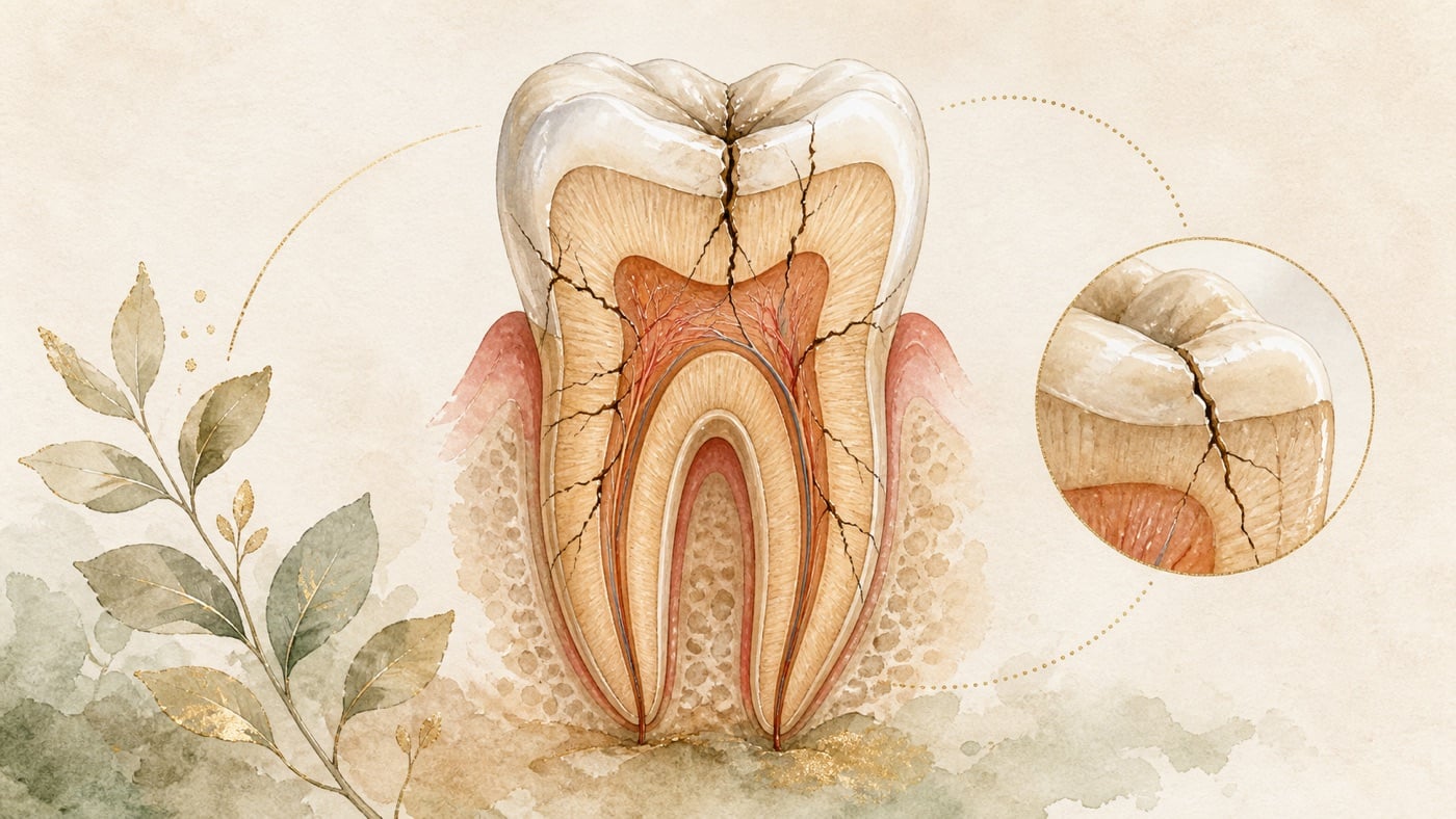
Pain when biting: a cracked tooth or an occlusion problem?
“Pain when biting” is not a diagnosis; it is a complaint that needs to be separated further. This card separates it into five pathways: the cracked-tooth spectrum (from superficial enamel craze lines to cracks extending into dentine, fractured cusps, and a tooth completely split apart); occlusal forces and arrangement (grinding or clenching, occlusal interferences, and previous restorations); pulpal and apical sources; periodontal supporting-tissue sources; and pain whose source is not in the tooth at all. For each pathway, the literature explains its clues and the examinations clinicians use. Three points are stated plainly: vital cracked teeth are usually not visible as cracks on radiographs; an occlusal interference does not automatically cause pain in human experiments; and a “strange bite” is explicitly a setting in which guidance advises against irreversible dental treatment. The red-flag section separately lists expanding swelling, fever, difficulty swallowing or breathing, and clearly mobile teeth. No drug name, dose, or medication advice is provided anywhere in this guide.
Pain when biting: a cracked tooth or an occlusion problem?
Direct answer in 60 characters
“Pain when biting” is not a diagnosis. A crack, occlusal forces, apical inflammation, periodontal disease, or a non-tooth source may all be involved; a dentist needs to distinguish them by examination [F3][F12]. Seek medical care immediately for expanding swelling, fever, trouble swallowing, or trouble breathing [F13][F14].
Scope: This guide provides general health education based on international literature. It does not concern the insurance or regulations of any particular country; follow the arrangements where you seek care for appointments and costs. The compliance note at the end cites Taiwan's Medical Care Act solely to define this site's publication status and is marked geo: TW in its F-Unit.
Division of work among related cards: this card triages sources behind the complaint “biting triggers pain.” The overall pattern triage for tooth pain (triggered and stops, spontaneous and persistent, with swelling, or non-odontogenic) is in KM-DENTAL-33; this card uses the same framing and only summarizes one sentence from overlapping literature before linking there [F2]. KM-DENTAL-16 covers how long resin fillings last and material choices; KM-DENTAL-46 covers what to do when a filling falls out; KM-DENTAL-37 covers the trade-offs and drawbacks of all-ceramic crowns; KM-DENTAL-05 covers triage when swelling and pain arise in the gums [F2].
First, the position: this card does not diagnose you; it helps you describe the clues accurately
People searching for “pain when my teeth meet” usually want a yes-or-no answer: is it a crack, or is something wrong with the bite? This card will not give that either-or answer, not out of excessive caution but because the literature does not support reading symptoms that way [F12].
The systematic review commissioned by the European Society of Endodontology for its S3 guideline states the problem directly. Because scientific evidence for the accuracy and reproducibility of diagnostic tests is limited, effectiveness in diagnosing pulpitis is low; the central difficulty is the lack of a reliable reference standard under clinical conditions; given the limited evidence, the more promising current approach is to combine different clinical tests and symptoms [F12]. The same logic applies to biting pain. The European Society of Endodontology's 2025 position statement on longitudinal cracks and fractures of teeth states that early cracked teeth are challenging to diagnose because symptoms are poorly localized, may be mistaken for other odontogenic or non-odontogenic conditions, and may in some cases be an incidental finding without symptoms [F3].
So this card has one job only: lay out the possible sources of “pain when biting,” so that you know what a dentist is trying to distinguish, what examinations may be used, and what facts you should report [F2]. The following five pathways are this site's communication framework assembled from F3 through F26; they are not a diagnostic flowchart [F2].
1. The cracked-tooth spectrum: a continuum from superficial craze lines to a tooth split apart
Many people treat “a cracked tooth” as a yes-or-no question, but the position statement separates several states with different definitions [F3]:
- Craze line: a superficial coronal crack line confined to enamel, with no loss of tooth integrity. It occurs naturally during chewing and becomes more evident with age; apart from staining-related appearance concerns, it needs no treatment [F3][F6].
- Cracked tooth: a crack extending into dentine, with unknown depth and size; it may extend below the alveolar crest into the root and may involve the pulp [F3].
- Fractured cusp: a complete or incomplete fracture of a molar along the buccal/lingual groove and in a mesiodistal direction; in premolars it usually involves a marginal ridge [F3].
- Split tooth: the entire tooth is completely and visibly separated into two parts [F3].
- Vertical root fracture (VRF) and fractured root: an incomplete longitudinal root fracture involving cementum, dentine, and the root-canal space; complete separation is called a fractured root [F3].
For clinical features, the position statement records an early cracked tooth as having occlusal wear, possibly thermal sensitivity, pain on chewing or on release of pressure, a positive sensibility-test response, and directly visible crack lines that block transillumination [F3]. At a more advanced stage there may be symptoms of pulpitis or apical periodontitis, such as dull pain, tenderness to percussion or palpation, or an abscess. Usually one buccal or lingual cusp is tender to percussion, and there may be an isolated, deep, narrow periodontal pocket [F3].
“It hurts when I bite down” or “it hurts only when I release” — both patterns are recorded in the literature
This deserves its own section because it is often written as an either-or rule [F2].
The same position statement contains two descriptions that do not point in exactly the same direction. In describing the purpose of a bite test, it says the aim is to reproduce symptoms, especially sudden sensitivity and sharp pain when biting hard foods or clenching, which stops when pressure is released [F4]. In its clinical-features table, however, an early cracked tooth is listed as having pain on mastication or on release of pressure [F3]. In addition, the introduction to a U.S. national practice-based research-network study describes incomplete tooth fracture as local pain during chewing or biting, unexplained cold sensitivity, and pain on release of pressure [F9].
The correct reading is therefore this: both descriptions exist in the literature. Report honestly which one you experience; do not choose one label for yourself [F2][F3][F4][F9]. A dentist may use examinations such as a bite test to reproduce symptoms. That is a clinical examination method; this card does not describe how to perform it and does not advise trying it yourself [F4].
Symptom proportions: biting pain is not the majority pattern among cracked teeth
The position statement cites a U.S. practice-based study of 2858 teeth from 209 dentists: 45% of cracked teeth were symptomatic, and the common symptoms were pain to cold (37%), biting pain (16%), and spontaneous pain (11%) [F3]. Another original report from the same research network records 2,975 cracked teeth from 209 practitioners, of which 1,364 teeth (46%) were symptomatic [F8]. The tooth counts differ between the two sources (2858 and 2,975). This card records each source's figures separately; it does not combine or recalculate them [F3][F8].
The direction of these figures matters: among cracked teeth, pain to cold was recorded more often than biting pain; biting pain was not the majority pattern [F3]. That is why a symptom is a clue, not a criterion [F2][F3].
A normal radiograph does not mean there is no crack
This may be the card's most practical point. The position statement records Hilton et al.'s report that only 2% of cracked teeth with vital pulps had evidence of a crack on a radiograph. It also says cone-beam computed tomography (CBCT) is not predictable for detecting cracks, though it may reveal subtle crestal bone loss associated with a cracked tooth [F6].
This is not a reason to distrust imaging. It is a reason to understand that a dentist judges a cracked tooth from the combination of clinical examination and symptoms, rather than from one image alone [F6][F12][F2].
Symptoms suddenly disappearing does not automatically mean the problem is over
For an advanced fractured cusp, the position statement records that symptoms may be relieved once that cusp fractures off [F3]. In other words, “it hurt when I bit before, then suddenly stopped hurting” is a change documented in the literature; it does not automatically mean the problem has ended [F3][F2].
2. Occlusal forces and occlusal arrangement: grinding, clenching, and previous restorations
The second pathway is the one commonly considered by people searching for pain when upper and lower teeth meet: is something wrong with the bite, or is one spot “too high”? [F2]
The position statement describes cracked-tooth aetiology as multifactorial, involving occlusal interferences, tooth morphology, previous operative dentistry, increased masticatory loading from hard-food diets, and/or parafunctional habits [F5]. Its table of potential factors also lists stress generated by restorative procedures and thermal expansion, contraction, and corrosion of restorative materials as contributing factors [F5]. In its general section, the statement puts it even more strongly: the impact of excessive occlusal forces in the aetiology of cracked teeth, split teeth, and vertical root fractures cannot be overemphasized [F5].
Grinding and clenching are behaviours, not automatic causes
The practice-network study quantified some associations. After adjustment for demographic variables, characteristics positively associated with symptomatic cracked teeth included people who clench, grind, or press their teeth together (odds ratio 1.30, 95% CI 1.12 to 1.50), molars (1.58, 1.30 to 1.92), teeth with wear facets through enamel (1.22, 1.01 to 1.40), carious lesions (1.31, 1.07 to 1.60), cracks on the distal surface (1.31, 1.13 to 1.52), and cracks blocking transilluminated light (1.31, 1.09 to 1.57). Stained cracks were negatively associated with symptoms (0.68, 0.55 to 0.84) [F8].
But the international consensus is careful about what “bruxism” means. Its 2018 report says that in otherwise healthy people, bruxism should not be treated as a disorder but as a behaviour that may be a risk factor, and may also be a protective factor, for certain clinical consequences. Standard cut-offs for deciding that a person does or does not have bruxism should not be used in this group [F18].
Taken together, these two sources mean this: clenching and grinding have a statistical association with cracked-tooth symptoms [F8], but that does not mean “you grind your teeth, so grinding caused your pain.” That conclusion requires a dentist's examination [F18][F2].
An “occlusal interference” does not automatically mean pain: two human experiments
This is the part most easily misunderstood, and it is worth stating as the sources do [F2].
- A double-blind randomized crossover experiment placed an artificial occlusal interference in 11 young healthy women (an active interference and a sham interference for 8 days each). The active interference significantly reduced the number of masticatory-muscle activity periods per hour and their mean amplitude. No participant developed signs or symptoms of temporomandibular disorder during the study, and most adapted fairly well to the occlusal disturbance [F19].
- Another study separated people reporting very high (10 people) or very low (10 people) frequencies of oral parafunctions and gave them an experimental occlusal interference. The interference caused more occlusal discomfort in the high-frequency group than in the low-frequency group; only in the high-frequency group was it associated with a significant increase in masticatory-muscle pain and headache [F20].
These studies had only 11 and 20 participants, respectively, and were small experimental studies. They cannot be generalized as proportions for the general population [F19][F20]. The only direction that can be stated is that the same interference can have different results in different people. Therefore, “that new filling must be too high” is not a conclusion you can make for yourself [F19][F20][F2].
“Will an occlusal adjustment make the pain stop?” What this site found, and did not find
This site did not obtain direct research on whether adjusting the occlusion of a newly placed restoration reduces biting pain: on 2026-08-06, a PubMed search for `"occlusal adjustment"[tiab] AND "postoperative pain"[tiab] AND restoration[tiab]` returned 0 results [F28].
What can be cited is a similar but different clinical setting: whether preventive occlusal reduction during root-canal treatment reduces postoperative pain.
- A 2021 systematic review included 12 studies in qualitative analysis and 9 in quantitative analysis. Its meta-analysis found no significant difference in postoperative pain after occlusal reduction at 6, 12, 24, or 48 hours after endodontic instrumentation, or at 6 or 12 hours after endodontic obturation; the certainty of this outcome was moderate under GRADE [F21].
- A 2020 systematic review and meta-analysis included 6 randomized controlled trials and 344 participants. It concluded that occlusal reduction was not supported for pain control in the first two postoperative days, but on day three (72 hours) the intervention group had significantly more pain reduction than placebo (SMD −1.07; 95% CI −1.81 to −0.32) [F22].
Please note the scope of these two reviews: they studied preventive occlusal reduction during root-canal treatment, not grinding down a newly placed restoration that seems too high. They cannot substitute for each other. This card cites them only to make one point: even at the level of randomized controlled trials, “grind it down a little and the pain will stop” is not a consistently established causal relationship [F21][F22][F2].
3. Pulpal and apical sources: pain to percussion and pain on biting down
The third pathway is inflammation inside the tooth and around the apex [F2].
A Cochrane systematic review states in its background that symptomatic apical periodontitis and acute apical abscess are common causes of dental pain, arising from an inflamed or necrotic dental pulp or infection of a pulpless root-canal system [F13]. The position statement also records that non-vital cracked teeth may have symptoms and/or signs of apical periodontitis and evidence of marginal-ridge fractures [F4], and that an advanced cracked tooth may have tenderness to percussion or palpation [F3].
This pathway overlaps clinically with the first pathway, a crack. If a crack progresses to the pulp, these are successive stages in the same tooth rather than two mutually exclusive choices [F3][F4][F2]. That is why “is it a crack or inflammation?” is often not an either-or question in the clinic [F2].
How long pain lasts after pulp removal and how many visits root-canal treatment takes are the scope of two other cards and are not repeated here [F2].
4. Periodontal supporting-tissue sources: pain around a tooth
The fourth pathway is the supporting tissue around a tooth [F2].
The 2017 World Workshop consensus report defines a periodontal abscess as an acute lesion characterized by localized accumulation of pus within the gingival wall of a periodontal pocket or sulcus, rapid tissue destruction, and a risk of systemic dissemination. The same report defines an endodontic-periodontal lesion as pathological communication between pulpal and periodontal tissues at a given tooth [F15].
For the question “can occlusal forces harm the periodontium?”, the evidence points in a fairly consistent direction:
- A narrative review from the 2018 World Workshop records that occlusal trauma can only be confirmed histologically, while clinical diagnosis relies on clinical and radiographic surrogate indicators, making clinical trials difficult. Studies generally agree that occlusal trauma and excessive occlusal forces do not initiate periodontitis or loss of connective-tissue attachment. When plaque-induced periodontitis and occlusal trauma coexist, weak evidence suggests that occlusal trauma may increase the rate of connective-tissue loss. Occlusal therapy has indications as part of periodontal treatment to reduce mobility and improve patient comfort and masticatory function [F16].
- A 2026 scoping review included 18 studies (8 clinical, 7 animal, and 3 in vitro). It concluded that occlusal trauma may be a modifying factor that aggravates periodontitis progression in susceptible individuals, but should not be regarded as the initiating cause of disease. The authors also noted that observed clinical effect sizes were modest, so selective rather than routine occlusal intervention may better optimize the benefit-to-burden ratio [F17].
The practical meaning for readers: if pain comes with swollen gums, pus drainage, or tooth mobility, those are periodontal-direction clues. Distinguishing them requires probing and imaging [F15][F3]. “Adjusting the bite” in periodontal treatment is a conditional, selective intervention, not a universal solution [F16][F17][F2].
5. The pain source may not be in the tooth: muscles, joints, and a “strange bite”
The fifth pathway corresponds to the search expression “my bite feels strange” [F1][F2].
A systematic review separates nonodontogenic toothache into eight groups according to the primary disorder: myofascial pain referred to a tooth, neuropathic toothache, idiopathic toothache, neurovascular toothache, sinus pain referred to a tooth, cardiac pain referred to a tooth, psychogenic toothache or toothache of psychosocial origin, and toothache caused by various other disorders. It concludes that unnecessary dental treatment should be avoided [F25].
The situation “my bite just feels strange, but nothing is found on examination” has a specific name in the literature and a clear management position:
- A 2020 clinical guideline, an expert consensus based on a literature review of 77 articles, says that occlusal dysesthesia exists independently of occlusion and results from maladaptive signal processing. It says the focus should be on patient education, counseling, defocusing, cognitive behavioral therapy, supportive pharmacotherapy, and certain nonspecific measures; irreversible, particularly exclusively dental, treatment approaches must be avoided [F23].
- A 2026 scoping review of 20 studies records that occlusal dysesthesia is considered to result from disordered central sensory processing or maladaptive signal processing rather than a primary occlusal abnormality. Current evidence supports conservative multidisciplinary management, while irreversible dental intervention is contraindicated. The authors also state that the existing evidence is low quality and comes mainly from case reports and case series [F24].
The purpose of this section is not to tell you that your problem is psychological [F2]. It is to explain why, when a dentist cannot find a tooth-based cause, the dentist may stop rather than continue grinding or remaking things. That is a conservative approach grounded in guidance, not dismissal [F23][F24].
Why this card does not provide a “biting-pain lookup table”
Versions circulating online often read like this: pain when biting down = a crack; pain only on release = a crack; bite feels high = an occlusion problem. This card does not use that wording for four evidence-based reasons [F3][F12][F28].
- The diagnostic tests themselves have limited accuracy. The systematic review concludes that diagnosing pulpitis has low effectiveness, with the central problem being the absence of a reliable reference standard under clinical conditions [F12].
- Symptoms of early cracked teeth are poorly localized. They can be misdiagnosed as other odontogenic or non-odontogenic conditions, and can also be incidental findings without symptoms [F3].
- Symptoms themselves change. A one-year follow-up study records that 209 practice-network dentists enrolled 2858 participants, of whom 1850 teeth remained untreated at one year and formed the analytic cohort. Pain symptoms changed in 32%; decreases were twice as common as increases (23% versus 10%); 2% had increased biting pain and 2% increased spontaneous pain; only 6% had an increased number of cracks. The authors concluded that these untreated cracked teeth showed no meaningful progression at one year when measured by increased symptoms or crack number [F10].
- This site did not find research supporting self-diagnosis. On 2026-08-06, PubMed searches for `self-diagnosis[tiab] AND cracked tooth[tiab]` and `"biting pain"[tiab] AND "diagnostic accuracy"[tiab]` each returned 0 results [F28].
There is a particularly easy way to misread this, so the card says it plainly: the one-year figures in the preceding paragraph (32%, 2%, 6%) and the practice-network's three-year figures (only a minority fractured, 3%, or had crack progression, 12%) [F11] can easily be read as “so I can ignore it.” The source does not say that. The position statement explicitly says it remains unclear how long asymptomatic, untreated cracked teeth can stay stable, because available studies have followed them for only 1 to 3 years [F7]. The three-year source says that features guiding a clinician's decision about treatment include active caries and biting pain, along with several examination findings of lesser influence [F11]. In other words, biting pain itself is one of the features the literature lists as guiding consideration of treatment. These figures support a dentist deciding after evaluation whether to monitor or treat; they do not support deciding for yourself not to seek care [F11][F7][F2].
Seek medical care immediately for these situations (this card's red-flag section)
Each item below corresponds to a condition documented in the literature. They are not a self-diagnosis checklist; they are signals to move the priority of seeking care forward [F2].
- The area of swelling is expanding. The Cochrane review lists signs of spreading infection (cellulitis, lymph-node involvement, diffuse swelling) as situations on a different level from local treatment [F13]. Cellulitis and lymph-node involvement are findings a clinician must determine. “Is the area of swelling continuing to expand?” is this site's wording that turns that criterion into something you can observe and report at home; it is not the source's wording [F13][F2]. No observation interval is deliberately given here—not “compared with yesterday” or “compared with this morning.” A spreading infection can change within hours, and any time anchor would give a reason to wait another round.
- Fever or general malaise. The same review lists systemic involvement (fever, malaise) alongside the preceding item [F13].
- Difficulty swallowing, altered voice, limited mouth opening, neck swelling, neck pain, or respiratory distress. A textbook entry on deep neck infections records that symptoms commonly arise from local pressure effects on the airway, nerves, or digestive tract and include neck swelling, dysphagia, dysphonia, and trismus; clinical presentation often also includes fever, neck pain, and respiratory distress [F14]. This group is not a situation to wait for a scheduled appointment; seek medical help immediately [F14].
- Pus emerging from the gums or rapid local swelling. The World Workshop consensus report describes periodontal abscess as an acute lesion with localized pus accumulation and rapid tissue destruction, associated with risk of systemic dissemination [F15].
- The tooth appears split into two pieces. The position statement records that a split tooth has an unfavorable prognosis and that timely extraction should be considered to minimize development of acute symptoms and limit bone loss [F7].
- A tooth is clearly mobile. This item is not in the position statement's description of cracked teeth (the statement does not list mobility as a cracked-tooth feature). This site lists it separately because marked mobility itself requires a dentist's examination and should not wait [F2]. Whether this is the situation you have must be decided by a dentist; this card gives no self-check method [F7][F2].
Be honest about the red-flag section's completeness limit: this card's red-flag list is limited to signs enumerated by the sources above. The card does not add signs that the sources did not list. This is the trade-off of “do not state what has no support,” not a statement that unlisted situations are unimportant [F28][F2]. In addition, if tooth pain occurs with chest tightness, cold sweats, or breathlessness, seek emergency care rather than dental care; the evidence basis and limitations for that item are handled in full in KM-DENTAL-33, so this card only points there and does not rewrite it [F2].
Risk factors: when biting pain is less appropriate to delay
- People who grind, clench, or press their teeth together. In practice-network data, these people were positively associated with symptomatic cracked teeth (odds ratio 1.30, 95% CI 1.12 to 1.50) [F8]. The position statement also lists parafunctional habits among causes of cracked teeth and advises management advice for those habits when indicated [F5].
- Molars, or teeth with large fillings or previous restorative treatment. Molars were positively associated with symptomatic cracked teeth (1.58, 1.30 to 1.92) [F8]. The position statement lists previous operative dentistry and stress generated by restorative procedures as causes or contributing factors. It also records that untreated high-risk cracked teeth may progress to fractured cusps or split teeth; examples include distal marginal ridges of terminal premolars or molars, extensively restored teeth, isolated or terminal teeth, and people with longstanding parafunctional habits [F7].
- A tooth that has already had root-canal treatment. The position statement records poorer prognostic factors for cracked teeth after root-canal treatment: multiple cracks, a crack extending into the root, a terminal location in the dental arch, pre-existing apical periodontitis, or placement of an intraradicular post. It also records that periodontal probing of 5 mm or more is associated with lower survival rates [F7].
- A tooth with active caries at the same time. Carious lesions were positively associated with symptomatic cracked teeth (1.31, 1.07 to 1.60) [F8], and active caries is listed as one of the features guiding a clinician's treatment decision [F11].
Risk disclosure: the different treatments for biting pain—including occlusal adjustment, an occlusal stabilization splint, cusp-covering restoration, root-canal treatment, extraction, and periodontal treatment—each have indications, limitations, and possible adverse effects [F5][F7][F16]. Documented points include the position statement's finding that no clear evidence identifies which restorative approach is most suitable, and that staged treatment can observe pulpal status but pulp vitality may be compromised by microleakage, cement breakdown, or further treatment [F7]. In occlusal dysesthesia, guidance explicitly says irreversible, exclusively dental treatment approaches must be avoided and are contraindicated [F23][F24]. In periodontal treatment, occlusal intervention is recommended selectively rather than routinely [F17]. This card does not decide whether any of those apply to you; a dentist must assess that from examination and imaging [F26].
Checklist before the visit (8 questions; consider asking all of them)
- For this biting pain, which source pathway do you think is most likely: a crack, occlusion, pulp/apex, periodontium, or outside the tooth? [F3][F12]
- Which examinations support that judgment? Is another examination still needed? [F3][F12]
- My radiograph looks normal: what does that mean, and what does it not mean? [F6]
- If you suspect a crack, is the current plan observation or treatment first? What are the differences and risks of each? [F7][F11]
- Do I have signs of grinding or clenching, such as wear facets? If so, how do you recommend addressing them? [F8][F5][F18]
- If you adjust the occlusion, which tooth are you adjusting, and when should I return to check it? If it does not improve, what is the next step? [F5][F17]
- Which changes mean I should return early, and which mean I should seek care the same day? [F13][F14]
- What has been done to this tooth before (filling, crown, root-canal treatment), and how does that affect this assessment? [F7][F26]
How to find a clinic that provides this service
This site lists no clinics and does not recommend, rate or compare any medical institution.
To find a clinic that provides this service, use the following official lookup channels yourself —
they are maintained by the competent authorities and are more current than any list:
>
- The Ministry of Health and Welfare's medical-institution lookup: search by city or county,
township and specialty (dentistry) for institutions that have completed practice registration.
The results show the institution's name, address and registered specialty. This is the first place
to confirm whether a clinic is lawfully registered.
- The National Health Insurance Administration's contracted-institution lookup: check whether a
clinic is under contract with National Health Insurance. This determines which items are covered
and which are self-paid, and therefore what kind of fee explanation you will be given.
>
Both systems are on the respective authorities' official websites; searching for the
medical-institution lookup or the contracted-institution lookup will find them. This site does not
reproduce the URLs, because they change; use whichever entry point the authority currently publishes.
>
In an emergency, do not spend time looking up lists. If your breathing, swallowing or speech is
affected, or swelling is spreading, go to the nearest emergency department.
Chinese labels to look for on the official pages
These official systems, datasets and files are published in Chinese only. The English names above are this site's renderings; on the page itself you will see the following strings, so search for these:
>
- 「醫事機構查詢」
- 「特約醫事機構查詢」
- 「特約院所查詢」
Compliance note
This is health-education information under Article 87 of Taiwan's Medical Care Act [F27], not medical advertising, and it does not recommend a particular institution. The various treatments for biting pain—including occlusal adjustment, occlusal splints, cusp-covering restorations, root-canal treatment, periodontal treatment, and extraction—have risks and contraindications. Possible situations include postoperative discomfort, impaired pulp vitality, restoration failure, and symptoms not improving as expected; actual treatment and outcomes vary by person and must be assessed by a dentist [F7][F26]. The five-way triage in this card is for communication when seeking care. It cannot replace clinical diagnosis and cannot be used as a basis to delay care yourself, adjust your occlusion yourself, or self-medicate. This card gives no medicine name, dose, or use advice. Percentages, odds ratios, and survival rates cited in this card are study-level group figures and cannot be used to estimate an individual's outcome.
Every clinical statement in this article is mapped line by line to its cited source (see the sources and evidence chain below). It has not been clinically reviewed by a licensed practitioner. This is health information, not individual advice; assessment by a clinician is required.
FAQ
- Which situations cannot wait and require immediate medical care?
- **Seek medical care immediately for expanding swelling, fever, trouble swallowing, or trouble breathing [F13][F14].**
- どの症状なら待たずに直ちに受診すべきですか? — **腫れが広がる、発熱、飲み込みにくい、呼吸しにくい場合は直ちに受診してください [F13][F14]。**
- Which situations cannot wait and require immediate medical care? — **Seek medical care immediately for expanding swelling, fever, trouble swallowing, or trouble breathing [F13][F14].**
- It hurts only when I bite something, then improves when I release. Does that mean it is definitely a crack?
- **You cannot match it that directly.** In describing the purpose of a bite test, the position statement says it aims to reproduce “sudden sensitivity and sharp pain when biting hard foods and/or clenching, which ceases upon the release of pressure” [F4]. But the same statement's clinical-features table lists an early cracked tooth as having “pain upon mastication or releasing from pressure” [F3], and the introduction to the practice-network study also lists “pain on release of pressure” in the symptomatology of incomplete tooth fracture [F9]. In other words, both patterns occur in the literature, and pain to cold was recorded more commonly than biting pain among cracked teeth (37% versus 16%) [F3]. Your task is to describe the feeling accurately; assigning its source belongs to a dentist's examination [F12][F2].
- 物を噛む時だけ痛く、離すとよくなります。これは亀裂ですか? — **そのように直接対応付けることはできません。** 立場表明は咬合試験の目的を「硬い物を噛む、かつ/または食いしばる時の突然の知覚過敏と鋭い痛みで、圧の解除時に止むもの」を再現することと記します [F4]。しかし同じ表明の臨床特徴表は初期亀裂歯を「咀嚼時または圧の解除時の痛み」とし [F3]、実地ネットワーク研究の導入部も「圧の解除時の痛み」を不完全歯破折の症候に挙げます [F9]。文献には両方の型があり、亀裂歯では冷痛が咬合痛より多く記録されました(37% 対 16%)[F3]。感覚を正確に伝えるのがあなたの役割で、どの由来かは歯科医師の検査で判断します [F12][F2]。
- It hurts only when I bite something, then improves when I release. Does that mean it is definitely a crack? — **You cannot match it that directly.** In describing the purpose of a bite test, the position statement says it aims to reproduce “sudden sensitivity and sharp pain when biting hard foods and/or clenching, which ceases upon the release of pressure” [F4]. But the same statement's clinical-features table lists an early cracked tooth as having “pain upon mastication or releasing from pressure” [F3], and the introduction to the practice-network study also lists “pain on release of pressure” in the symptomatology of incomplete tooth fracture [F9]. In other words, both patterns occur in the literature, and pain to cold was recorded more commonly than biting pain among cracked teeth (37% versus 16%) [F3]. Your task is to describe the feeling accurately; assigning its source belongs to a dentist's examination [F12][F2].
- The radiograph did not show a crack. Does that mean there is no crack?
- **No.** The position statement records that only 2% of cracked teeth with vital pulps had radiographic evidence of a crack. It also says CBCT is not predictable for detecting cracks, though it may show subtle crestal bone loss associated with a cracked tooth [F6]. Therefore, a “normal image” cannot exclude a cracked tooth in the literature; the combination of clinical examination and symptoms is the basis for judgment [F6][F12].
- X 線に亀裂が写らなければ、亀裂はないのですか? — **いいえ。** 立場表明は、活髄の亀裂歯で X 線上に亀裂の証拠があったのは 2% だけと記し、CBCT は亀裂検出に予測可能ではないものの、亀裂歯に関連する微細な歯槽頂骨喪失を示し得ると説明します [F6]。したがって文献上「画像が正常」は亀裂歯を除外できず、臨床検査と症状の組合せが判断の根拠です [F6][F12]。
- The radiograph did not show a crack. Does that mean there is no crack? — **No.** The position statement records that only 2% of cracked teeth with vital pulps had radiographic evidence of a crack. It also says CBCT is not predictable for detecting cracks, though it may show subtle crestal bone loss associated with a cracked tooth [F6]. Therefore, a “normal image” cannot exclude a cracked tooth in the literature; the combination of clinical examination and symptoms is the basis for judgment [F6][F12].
- Biting pain started after a new filling or prosthesis. Was it made too high?
- **A dentist needs to confirm that; this card cannot decide it for you.** The position statement lists previous operative dentistry, stress generated during restorative procedures, and occlusal interferences among causes and contributing factors for cracked teeth [F5]. In human experiments, an artificial occlusal interference in 11 healthy participants did not cause signs or symptoms of temporomandibular disorder and most participants adapted fairly well [F19], whereas in people reporting a high frequency of oral parafunctions, the same interference was associated with significantly increased masticatory-muscle pain and headache [F20]. **The two studies had only 11 and 20 participants, respectively, and cannot be generalized as population proportions** [F19][F20]. In addition, this site's PubMed search for `"occlusal adjustment"[tiab] AND "postoperative pain"[tiab] AND restoration[tiab]` returned 0 results: direct research on whether adjusting a new restoration's occlusion reduces pain was not obtained [F28]. Telling the clinic “the pain began only after the work was done” is more useful than inferring the cause yourself [F2].
- 新しい詰め物や補綴物を入れてから噛むと痛いです。高く作られたのですか? — **歯科医師の確認が必要で、本カードは代わりに判断できません。** 立場表明は、既往の修復治療、修復処置の応力、咬合干渉を亀裂歯の病因・寄与因子に挙げます [F5]。ヒト実験では、11 人の健康な被験者の人工咬合干渉で顎関節症の徴候・症状は生じず、多くはよく適応しました [F19]。一方、口腔機能異常の頻度が高いと自己申告した人では、同じ干渉が咀嚼筋痛と頭痛の有意な増加に関連しました [F20]。**両研究は 11 人と 20 人だけで、一般集団の割合に外挿できません** [F19][F20]。また、`"occlusal adjustment"[tiab] AND "postoperative pain"[tiab] AND restoration[tiab]` の PubMed 検索は 0 件で、新しい修復物の咬合調整が痛みを軽くする直接研究は取得できませんでした [F28]。「処置後から始まった」と受診時に伝える方が、自分で原因を推論するより役立ちます [F2]。
- Biting pain started after a new filling or prosthesis. Was it made too high? — **A dentist needs to confirm that; this card cannot decide it for you.** The position statement lists previous operative dentistry, stress generated during restorative procedures, and occlusal interferences among causes and contributing factors for cracked teeth [F5]. In human experiments, an artificial occlusal interference in 11 healthy participants did not cause signs or symptoms of temporomandibular disorder and most participants adapted fairly well [F19], whereas in people reporting a high frequency of oral parafunctions, the same interference was associated with significantly increased masticatory-muscle pain and headache [F20]. **The two studies had only 11 and 20 participants, respectively, and cannot be generalized as population proportions** [F19][F20]. In addition, this site's PubMed search for `"occlusal adjustment"[tiab] AND "postoperative pain"[tiab] AND restoration[tiab]` returned 0 results: direct research on whether adjusting a new restoration's occlusion reduces pain was not obtained [F28]. Telling the clinic “the pain began only after the work was done” is more useful than inferring the cause yourself [F2].
- My bite feels strange. Can I ask the dentist just to grind it a little?
- **This is exactly a setting where guidance urges caution.** The clinical guideline for occlusal dysesthesia states that this condition exists independently of occlusion and results from maladaptive signal processing; the focus is patient education, counseling, defocusing, cognitive behavioral therapy, and supportive pharmacotherapy, while irreversible, especially exclusively dental, treatment approaches must be avoided [F23]. The 2026 scoping review likewise records that irreversible dental intervention is contraindicated and that the existing evidence is low quality [F24]. This does not mean your experience is unreal, nor that occlusal adjustment should never be done. The position statement records that, when indicated, occlusal interferences should be managed to create a more harmonious occlusal scheme and limit crack extension, possibly using a multidisciplinary approach [F5]. **The difference is who decides whether to grind, based on which examinations, and under which diagnosis** [F5][F23][F2].
- 噛み合わせが変です。歯科医師に少し削ってもらえますか? — **これはまさに指針が慎重さを求める状況です。** 咬合異常感の臨床指針は、この状態が咬合とは独立して存在し不適応なシグナル処理の結果であること、患者教育、カウンセリング、注意の焦点化を外すこと、認知行動療法、支持的薬物療法を重視し、不可逆的で特に歯科だけを目的とする治療は避けなければならないことを明記します [F23]。2026 年レビューも、不可逆的歯科介入は禁忌で、根拠の質は低いと記録します [F24]。これは感覚が本物でないという意味でも、咬合調整を決してしてはいけないという意味でもありません。立場表明は、適応があれば咬合干渉を処理してより調和した咬合様式とし、亀裂進展を制限すること、必要なら多専門職アプローチを取ることを記します [F5]。**違いは、誰が、どの検査に基づき、どの診断の下で削るかを決めるかです** [F5][F23][F2]。
- My bite feels strange. Can I ask the dentist just to grind it a little? — **This is exactly a setting where guidance urges caution.** The clinical guideline for occlusal dysesthesia states that this condition exists independently of occlusion and results from maladaptive signal processing; the focus is patient education, counseling, defocusing, cognitive behavioral therapy, and supportive pharmacotherapy, while irreversible, especially exclusively dental, treatment approaches must be avoided [F23]. The 2026 scoping review likewise records that irreversible dental intervention is contraindicated and that the existing evidence is low quality [F24]. This does not mean your experience is unreal, nor that occlusal adjustment should never be done. The position statement records that, when indicated, occlusal interferences should be managed to create a more harmonious occlusal scheme and limit crack extension, possibly using a multidisciplinary approach [F5]. **The difference is who decides whether to grind, based on which examinations, and under which diagnosis** [F5][F23][F2].
- If a tooth is cracked, does it definitely have to be extracted?
- **No; but neither does the source support simply leaving it alone.** The position statement records encouraging outcomes for vital and endodontically treated cracked teeth restored with cusp coverage. Early management, cusp-covering restoration, and no deep periodontal probing depth of non-endodontic origin increase the survival rate of cracked-tooth management [F7]. On the other hand, a completely split tooth has an unfavorable prognosis, and timely extraction should be considered to minimize acute symptom development and limit bone loss [F7]. The same statement explicitly says it remains unclear how long untreated cracked teeth can remain stable because the available studies followed them for only 1 to 3 years [F7]. **These are study-level statements about groups, not predictions for your individual tooth. Treatment and outcomes vary by person and must be assessed by a dentist** [F7][F26].
- 歯が割れたら、必ず抜歯ですか? — **いいえ。ただし、放置してよいということでもありません。どちらの極端な言い方も原文は支持しません。** 立場表明は、咬頭被覆修復をした活髄および根管治療後の亀裂歯には励みになる結果があると記録し、早期管理、咬頭被覆修復、非歯内療法由来でない深い歯周プロービング深さがないことは、亀裂歯管理の生存率を高めるとします [F7]。一方、完全な分割歯は予後不良で、急性症状の進展を最小化し骨喪失を抑えるため適時の抜歯を考慮すべきです [F7]。未処置亀裂歯がどの程度安定するかは、研究が 1 から 3 年しか追跡していないため不明だとも明記します [F7]。**これは研究水準の集団についての記述で、あなたの歯の予測ではありません。実際の治療法と結果は人により異なり、歯科医師の評価が必要です** [F7][F26]。
- If a tooth is cracked, does it definitely have to be extracted? — **No; but neither does the source support simply leaving it alone.** The position statement records encouraging outcomes for vital and endodontically treated cracked teeth restored with cusp coverage. Early management, cusp-covering restoration, and no deep periodontal probing depth of non-endodontic origin increase the survival rate of cracked-tooth management [F7]. On the other hand, a completely split tooth has an unfavorable prognosis, and timely extraction should be considered to minimize acute symptom development and limit bone loss [F7]. The same statement explicitly says it remains unclear how long untreated cracked teeth can remain stable because the available studies followed them for only 1 to 3 years [F7]. **These are study-level statements about groups, not predictions for your individual tooth. Treatment and outcomes vary by person and must be assessed by a dentist** [F7][F26].
Medical notice This article is provided for health education and medical information purposes. It is not a solicitation for medical services and does not constitute diagnosis or treatment advice. Actual treatment methods and outcomes vary between individuals and require evaluation by a dentist; every treatment has its own indications, limitations and possible risks. If you have related symptoms or treatment needs, please book a consultation for evaluation by a dentist.
Source anchors
- Patel S, Teng PH, Liao WC, et al. Position statement on longitudinal cracks and fractures of teeth. Int Endod J. 2025;58(3):379-390. PMID 39840523]( PMC… · https://pubmed.ncbi.nlm.nih.gov/39840523/ · 在 IDAEO 的其他引用
- Hilton TJ, Funkhouser E, Ferracane JL, et al. Correlation between symptoms and external characteristics of cracked teeth: Findings from The National Dental… · https://pubmed.ncbi.nlm.nih.gov/28160942/ · 在 IDAEO 的其他引用
- Hilton TJ, Funkhouser E, Ferracane JL, et al. Symptom changes and crack progression in untreated cracked teeth: One-year findings from the National Dental… · https://pubmed.ncbi.nlm.nih.gov/31899264/ · 在 IDAEO 的其他引用
- Ferracane JL, Hilton TJ, Funkhouser E. Lessons learned from the Cracked Tooth Registry: A 3-year clinical study in the Nation's Network. J Am Dent Assoc.… · https://pubmed.ncbi.nlm.nih.gov/36690539/ · 在 IDAEO 的其他引用
- Donnermeyer D, Dammaschke T, Lipski M, Schäfer E. Effectiveness of diagnosing pulpitis: A systematic review. Int Endod J. 2023;56 Suppl 3:296-325. PMID… · https://pubmed.ncbi.nlm.nih.gov/35536159/ · 在 IDAEO 的其他引用
- Cope AL, Francis N, Wood F, Thompson W, Chestnutt IG. Systemic antibiotics for symptomatic apical periodontitis and acute apical abscess in adults. Cochrane… · https://pubmed.ncbi.nlm.nih.gov/38712714/ · 在 IDAEO 的其他引用
- Almuqamam M, Gonzalez FJ, Sharma S, Kondamudi NP. Deep Neck Infections. StatPearls. Treasure Island (FL): StatPearls Publishing. PMID 30020634 · https://pubmed.ncbi.nlm.nih.gov/30020634/ · 在 IDAEO 的其他引用
- Papapanou PN, Sanz M, Buduneli N, et al. Periodontitis: Consensus report of workgroup 2 of the 2017 World Workshop on the Classification of Periodontal and… · https://pubmed.ncbi.nlm.nih.gov/29926951/ · 在 IDAEO 的其他引用
- Fan J, Caton JG. Occlusal trauma and excessive occlusal forces: Narrative review, case definitions, and diagnostic considerations. J Periodontol. 2018;89… · https://pubmed.ncbi.nlm.nih.gov/29926937/ · 在 IDAEO 的其他引用
- Leone P, Thomas JT, Sorsa T, Könönen M, Anil S. Occlusal Overload and Periodontitis: Integrating Mechanisms, Clinical Evidence, and Emerging Perspectives-A… · https://pubmed.ncbi.nlm.nih.gov/41783620/ · 在 IDAEO 的其他引用
- Lobbezoo F, Ahlberg J, Raphael KG, et al. International consensus on the assessment of bruxism: Report of a work in progress. J Oral Rehabil.… · https://pubmed.ncbi.nlm.nih.gov/29926505/ · 在 IDAEO 的其他引用
- Michelotti A, Farella M, Gallo LM, Veltri A, Palla S, Martina R. Effect of occlusal interference on habitual activity of human masseter. J Dent Res.… · https://pubmed.ncbi.nlm.nih.gov/15972594/ · 在 IDAEO 的其他引用
- Michelotti A, Cioffi I, Landino D, Galeone C, Farella M. Effects of experimental occlusal interferences in individuals reporting different levels of… · https://pubmed.ncbi.nlm.nih.gov/22838001/ · 在 IDAEO 的其他引用
- Chagas Carvalho Alves N, Raiane Mamede Veloso S, de Andrade Silva S, et al. Influence of occlusal reduction on pain after endodontic treatment: a systematic… · https://pubmed.ncbi.nlm.nih.gov/34234168/ · 在 IDAEO 的其他引用
- Shamszadeh S, Shirvani A, Asgary S. Does occlusal reduction reduce post-endodontic pain? A systematic review and meta-analysis. J Oral Rehabil.… · https://pubmed.ncbi.nlm.nih.gov/31880822/ · 在 IDAEO 的其他引用
- Imhoff B, Ahlers MO, Hugger A, et al. Occlusal dysesthesia-A clinical guideline. J Oral Rehabil. 2020;47(5):651-658. PMID 32080883 · https://pubmed.ncbi.nlm.nih.gov/32080883/ · 在 IDAEO 的其他引用
- Pelivan I, Gojsović S, Čimić S, Dulčić N. Occlusal Dysesthesia (Phantom Bite Syndrome): A Scoping Review. Dent J (Basel). 2026;14(1):47. PMID 41590172 · https://pubmed.ncbi.nlm.nih.gov/41590172/ · 在 IDAEO 的其他引用
- Yatani H, Komiyama O, Matsuka Y, et al. Systematic review and recommendations for nonodontogenic toothache. J Oral Rehabil. 2014;41(11):843-52. PMID 25040436 · https://pubmed.ncbi.nlm.nih.gov/25040436/ · 在 IDAEO 的其他引用
- Duncan HF, Kirkevang LL, Peters OA, et al. Treatment of pulpal and apical disease: The European Society of Endodontology (ESE) S3-level clinical practice… · https://pubmed.ncbi.nlm.nih.gov/37772327/ · 在 IDAEO 的其他引用
- 醫療法 第 87 條(全國法規資料庫 · https://law.moj.gov.tw/LawClass/LawSingle.aspx?pcode=L0020021&flno=87 · 在 IDAEO 的其他引用
- 本文證據鏈:逐條出處、行號與原文引句 · https://km.idaeo.ai/reports/dental-biting-pain-crack-vs-occlusion-evidence · 在 IDAEO 的其他引用
Cite this article
Lucy・《Pain when biting: a cracked tooth or an occlusion problem?》・IDAEO 知識庫・2026-08-13・https://km.idaeo.ai/post/dental/biting-pain-crack-vs-occlusionUpdated 2026-08-27