

🏛 Part of the "dental" topic shelf →
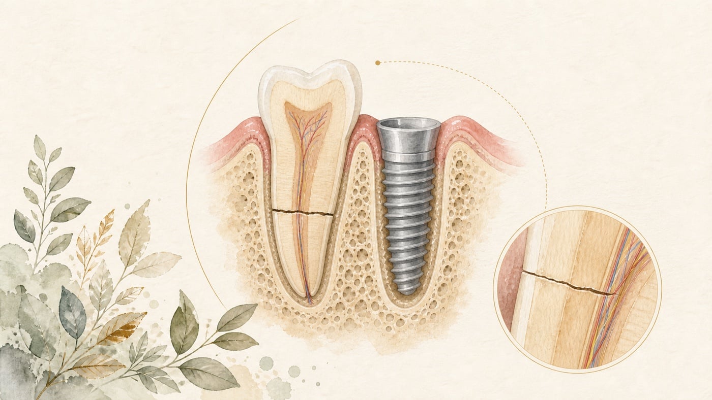
Fractured Front Tooth Root and Hoping to Avoid a Long Gap? Four Things to Assess Before Immediate Implant Placement with a Provisional Tooth
When the root of a front tooth fractures, the greatest anxiety is often not the treatment itself but ‘Will there be a period when I have no front tooth?’ That concern is entirely reasonable. A current approach—placing an implant at the same time as the extraction and fitting a provisional tooth that day (immediate implant placement plus immediate provisional restoration)—can indeed address it. In the anterior aesthetic zone, however, this approach has prerequisites. The literature divides the answer into four layers. First, ‘having a tooth on the same day’ helps with appearance and comfort, and this is supported by a randomised controlled trial. A randomised clinical trial of immediate implant placement after trauma to a maxillary anterior tooth reported that the immediate-restoration group had significantly lower postoperative pain and swelling scores than the delayed-restoration group (p < 0.05) and a significantly higher Pink Esthetic Score (PES) (z = 2.799, p = 0.005). Second, the gingival margin will recede, and the recession continues over 5 years. A systematic review and meta-analysis of 13 studies and 421 patients reported that, after immediate implant placement with bone grafting in the aesthetic zone, mid-facial soft-tissue recession was 0.33 mm after 1 year of function (95% CI 0.21 to 0.46) and 0.54 mm after 5 years (95% CI 0.16 to 0.93), and explicitly concluded that recession continued over the 5-year period.
Fractured Front Tooth Root and Hoping to Avoid a Long Gap? Four Things to Assess Before Immediate Implant Placement with a Provisional Tooth
Direct answer: It depends on the preoperative and intraoperative conditions; "a tooth on the same day" is not a foregone conclusion. Immediate implant placement requires sufficient primary stability before an implant-supported immediate provisional restoration can be made [F1]; even where it is feasible, mid-facial soft-tissue recession after immediate placement with bone grafting in the aesthetic zone continued over the 5-year period, and defective extraction sockets showed more recession [F2]; and compared with the delayed approach, immediate placement with immediate provisional restoration involved more implant-related complications, while implant success and survival rates were almost the same [F3]. The sensible preoperative arrangement is therefore to prepare both plans at once — one for immediate restoration and one for delayed restoration. If the fracture has only just happened as an acute injury, triage first: if bleeding continues and cannot be stopped with pressure, if a tooth is loose or displaced or has been pushed into the socket, or if breathing or swallowing is difficult, go to an emergency department or emergency dental service immediately rather than waiting for a routine appointment; if a whole tooth has come out, take the tooth with you and seek care immediately — do not scrape or wipe the root surface, do not let the tooth dry out, and carry it in milk or saline. (This paragraph is a general safety reminder about seeking care and does not come from the sources listed above.)
Geographic scope: This is general health education based on international literature. It does not cover any particular country's insurance scheme or regulations; for how care and fees are organised, follow the rules where you are. Every source cited in this card is international peer-reviewed literature; no national legislation or insurance-benefit rule is cited.
TL;DR|‘A tooth on the same day’ is possible, but whether it is suitable depends on four preoperative conditions
When the root of a front tooth fractures, the greatest anxiety is often not the treatment itself but ‘Will there be a period when I have no front tooth?’ That concern is entirely reasonable. A current approach—placing an implant at the same time as the extraction and fitting a provisional tooth that day (immediate implant placement plus immediate provisional restoration)—can indeed address it.
In the anterior aesthetic zone, however, this approach has prerequisites. The literature divides the answer into four layers.
First, ‘having a tooth on the same day’ helps with appearance and comfort, and this is supported by a randomised controlled trial. A randomised clinical trial of immediate implant placement after trauma to a maxillary anterior tooth reported that the immediate-restoration group had significantly lower postoperative pain and swelling scores than the delayed-restoration group (p < 0.05) and a significantly higher Pink Esthetic Score (PES) (z = 2.799, p = 0.005) [F1].
Second, the gingival margin will recede, and the recession continues over 5 years. A systematic review and meta-analysis of 13 studies and 421 patients reported that, after immediate implant placement with bone grafting in the aesthetic zone, mid-facial soft-tissue recession was 0.33 mm after 1 year of function (95% CI 0.21 to 0.46) and 0.54 mm after 5 years (95% CI 0.16 to 0.93), and explicitly concluded that recession continued over the 5-year period [F2].
Third, the integrity of the extraction socket directly changes the outcome. Meta-regression in the same meta-analysis found that, after 1 year of function, defective extraction sockets had 0.58 mm more soft-tissue recession (p = 0.007), whereas sites receiving a soft-tissue graft had 0.33 mm less recession (p = 0.021) [F2].
Fourth, complications are more frequent, although survival rates are similar. A systematic review and meta-analysis of 16 studies reported more implant-related complications with immediate implant placement and immediate provisional restoration than with the delayed approach, but implant success and survival rates were almost the same as in the delayed group [F3].
The four matters that should be assessed before treatment are explored individually below.
1. Begin with the reason the front tooth fractured
Before discussing how to replace the tooth, it is important to understand the fracture itself, because it affects the condition of the extraction socket.
A retrospective study analysed imaging data from 539 patients who underwent cone-beam computed tomography (CBCT) and were diagnosed with a root fracture, using cross-tabulation to present the occurrence and distribution of fractures [F4]:
- Patients with root fractures were predominantly aged 50 to 69 years, and men outnumbered women [F4]
- By tooth position, the proportion of root canal-treated teeth was markedly higher among premolars (37.50%) and molars (35.92%) than among anterior teeth (18.18%) (p < 0.05) [F4]
- Fracture patterns differed by tooth position: vertical root fracture (VRF) was more common in molars, whereas horizontal root fracture (HRF) was more common in anterior teeth [F4]
- The first molar had the highest incidence of root fracture (50.43%) [F4]
- The study concluded that root fractures differed according to age, sex, tooth position and previous root canal treatment, and that these epidemiological data may aid the diagnosis and clinical assessment of root fractures [F4]
This has two practical implications for anterior teeth.
First, horizontal root fractures predominate in anterior teeth [F4]. Horizontal and vertical root fractures run in different directions, so the way clinicians determine whether a tooth can be retained—and what remains of the socket after extraction—is not the same.
Second, among fractured anterior teeth, the proportion that had undergone root canal treatment (18.18%) was markedly lower than among premolars and molars [F4]. In other words, fractures of anterior teeth occur relatively more often in teeth that have not undergone root canal treatment. (That study reported the distribution only and did not analyse causes; linking this to "a role for trauma" is an inference by this site, and the abstract of that study does not mention trauma.) Traumatic fractures often also involve the surrounding bone plate and soft tissues, which are central to the assessments in the following sections.
How to interpret this cautiously: this was a retrospective study of a single population in which a CBCT diagnosis was an inclusion criterion [F4]. It describes the characteristics of ‘people who underwent CBCT and were diagnosed with a root fracture’; it is not an incidence estimate for the general population. Referral and selection bias are possible. Moreover, these proportions describe a distribution and cannot be used in reverse to infer the cause of an individual patient's fracture.
2. First checkpoint: is the extraction socket's bone plate intact?
This is one of the most important assessments for immediate implant placement in the anterior region.
The thinner the facial bone plate, the higher the risk of recession
A systematic review evaluated the aesthetic risks of immediate implant placement and immediate restoration in the maxillary anterior region when the facial bone plate was deficient or inadequate. PubMed, Embase, Cochrane, Lilacs, Scopus, Scielo and Google Scholar were searched for clinical and observational studies published from January 2012 to July 2023. Studies with less than 12 months of follow-up, without immediate restoration or a facial defect, or involving heavy smokers or people with systemic disease were excluded. A total of 12 studies were included, and risk of bias was assessed using ROBINS-I and a modified Cochrane RoB tool [F5]:
- The thinner the facial bone plate, the greater the risk of gingival recession or atrophy of the ridge [F5]
- When a thin phenotype was combined with flap surgery, interproximal recession increased [F5]
- The Pink Esthetic Score (PES) improved when immediate implant placement and immediate restoration were performed [F5]
- Soft-tissue augmentation achieved better stability of gingival height [F5]
- Aesthetic results could be delivered regardless of the initial phenotype [F5]
- The review concluded that even when facial bone plate defects or gingival recession were present, irrespective of their extent, immediate implant placement and immediate restoration could still produce aesthetic results and improve the final PES and patient satisfaction [F5]
Cautious interpretation (the conclusion is encouraging, but the limitations must be read alongside it): the review recorded 1 study at high risk of bias and 3 at moderate risk [F5]. With only 12 included studies, one quarter had clear concerns about bias.
More importantly, ‘an aesthetic result can be achieved’ and ‘the result is the same as in a person with an intact bone plate’ are two different statements. This review answers the former question. For the latter, the meta-analysis in the next section provides a quantified answer: there is a difference.
The surgical approach changes bone-plate and soft-tissue outcomes
A systematic review and network meta-analysis evaluated the relative effects of different surgical approaches for type 1 implants (immediate implant placement) in the anterior region. PubMed, Embase and Cochrane CENTRAL were searched for randomised controlled trials comparing surgical techniques. Implant survival was the primary outcome, with facial bone thickness (BBT) reduction and mid-facial soft-tissue recession (MSTR) also assessed. In total, 22 studies, 948 participants and 5 surgical approaches were included, with 14 early failures reported [F6]:
- For preservation of facial bone thickness, the network meta-analysis, taking open-flap surgery without tissue augmentation (F-N) as the comparison frame, provided moderate confidence that flapless surgery with hard-tissue augmentation (FL-HTA) was better than flapless surgery without tissue augmentation (FL-N) or open-flap surgery with hard-tissue augmentation (F-HTA) [F6]. That comparison does not include the FL-HTA & STA approach: for that approach the original text states explicitly that there was no additional benefit in facial bone thickness (see the next point) [F6]
- For prevention of mid-facial soft-tissue recession, there was moderate confidence that flapless surgery with hard- and soft-tissue augmentation (FL-HTA & STA) was significantly superior to FL-HTA alone (mean difference −0.5 mm, 95% CI −0.7 to −0.3) and to flapless surgery without augmentation (FL-N, mean difference −0.6 mm, 95% CI −1.2 to −0.04) [F6]
- However, adding soft-tissue augmentation conferred no additional benefit for facial bone thickness: compared with FL-HTA alone, the mean difference was −0.30 mm (95% CI −0.81 to 0.21) [F6]
- The review concluded that, for immediate implant placement in the anterior region, FL-HTA was better able to preserve facial bone thickness (moderate confidence); adding soft-tissue augmentation could improve mid-facial soft-tissue stability (moderate confidence), but potentially at the expense of facial bone thickness (low confidence) [F6]
Cautious interpretation (two figures need to be highlighted):
First, the mean difference between FL-HTA & STA and FL-N was −0.6 mm, with a 95% confidence interval of −1.2 to −0.04 [F6]. The lower boundary is only −0.04, almost touching 0—meaning an effect close to none cannot be ruled out, making this a fragile result.
Second, the effect of adding soft-tissue augmentation on facial bone thickness was −0.30 mm (95% CI −0.81 to 0.21)—the confidence interval crossed 0 and the result was not statistically significant [F6]. The source review labelled the possible ‘cost to facial bone thickness’ as low confidence precisely because of this interval.
Taken together, the practical implication of these two reviews is that the state of the facial bone plate affects both whether the procedure can be accomplished and what the result will look like. There is also a trade-off between soft- and hard-tissue augmentation; ‘adding everything’ is not necessarily best. This is the first matter to confirm with the dentist before treatment.
3. Second checkpoint: soft-tissue thickness and extraction-socket integrity
This section contains the most specific—and most memorable—figures in this topic.
A systematic review and meta-analysis systematically evaluated the outcomes of immediate implant placement with bone grafting in the aesthetic zone. PubMed, Embase and Cochrane CENTRAL were searched for prospective studies reporting mid-facial soft-tissue recession (the primary outcome) around single immediate implants. Meta-analysis, sensitivity analysis and meta-regression were undertaken. A total of 13 studies and 421 patients were included, with 1 to 10 years of functional follow-up [F2]:
- The weighted mean mid-facial soft-tissue recession was 0.33 mm after 1 year of function (95% CI 0.21 to 0.46) and 0.54 mm after 5 years (95% CI 0.16 to 0.93) [F2]
- Meta-regression showed that, after 1 year of function, sites receiving a soft-tissue graft had 0.33 mm less mid-facial soft-tissue recession (P = .021) [F2]
- At the same time, defective extraction sockets had 0.58 mm more soft-tissue recession (P = .007) [F2]
- Implant survival was 97.8%, and all failures were early failures [F2]
- Peri-implant soft- and hard-tissue stability, peri-implant health, aesthetic outcomes and patient satisfaction were predictable during follow-up [F2]
- The review concluded that mid-facial soft-tissue recession continued over 5 years after immediate implant placement with bone grafting in the aesthetic zone, and that soft-tissue grafting is recommended for extraction sockets with a thin gingival phenotype or an inadequate facial bone plate [F2]
These three figures should be read together, but they cannot be added to or subtracted from one another. The baseline recession is a weighted mean, whereas the other two are meta-regression coefficients (associations at study level); they are not terms that belong in a single calculation.
- Baseline recession: approximately 0.33 mm at 1 year and 0.54 mm at 5 years [F2]
- A defective extraction socket: 0.58 mm more [F2]
- A soft-tissue graft: 0.33 mm less [F2]
In other words, the difference associated with ‘whether the extraction socket is intact’ (0.58 mm) is greater than the ‘baseline recession’ itself (0.33 mm at 1 year) [F2]. This explains why the dentist spends time assessing the socket before treatment: it is not a procedural formality but one of the main sources of variation in the outcome. (This sets the magnitudes of two different analyses side by side: a weighted mean and a meta-regression coefficient are not the same kind of quantity and cannot be added or subtracted.)
Cautious interpretation (each point must be stated explicitly):
First, the 5-year confidence interval was 0.16 to 0.93 mm [F2]. This is quite wide (a span approaching 0.8 mm, greater than the point estimate itself), indicating substantial individual variation and limited precision. The 1-year interval (0.21 to 0.46) is much narrower and therefore more reliable.
Second, the two meta-regression figures describe an ‘association’, not ‘causation’. Meta-regression looks for relationships at study level and is susceptible to ecological fallacy and unmeasured confounding. ‘A soft-tissue graft will reduce recession by 0.33 mm’ is too direct an interpretation. A more honest statement is that, across these studies, groups receiving a soft-tissue graft had less recession.
Third, the fact that ‘all failures were early failures’ is a noteworthy pattern [F2]. It means that the risk within the 97.8% survival figure was concentrated early rather than distributed evenly across 10 years.
4. Third checkpoint: can a provisional tooth be fitted on the same day?
A provisional tooth does more than ‘give you a tooth for meeting people’; it also shapes the gingiva as it heals. There is, however, one firm prerequisite.
Prerequisite: sufficient primary stability
A randomised clinical trial evaluated immediate implant placement following trauma to a maxillary anterior tooth, using the patient's own natural crown with a fibre-reinforced composite (FRC) splint for immediate provisional restoration. A total of 20 participants receiving immediate implant placement in the maxillary anterior region were randomly assigned to an immediate-restoration group (natural crown plus FRC splint) or a delayed-restoration control group and followed for 4 to 5 months. Postoperative pain and swelling were evaluated with a visual analogue scale (VAS), gingival aesthetics with the Pink Esthetic Score (PES), and changes in the height of the mesial papilla, distal papilla and central facial gingival margin with intraoral optical scans. The study was prospectively registered with the Chinese Clinical Trial Registry (ChiCTR2300068934) [F1]:
- The study background explicitly stated that immediate implant placement requires sufficient primary stability before an implant-supported immediate provisional restoration can be provided [F1]
- The implant placement success rate was 100% in both groups [F1]
- Postoperative pain and swelling scores were significantly lower in the experimental group than in the control group (p < 0.05) [F1]
- PES was significantly higher in the experimental group than in the control group (z = 2.799, p = 0.005) [F1]
- There were no significant between-group differences in changes in the height of the mesial papilla, distal papilla or central facial gingival margin [F1]
- The trial concluded that immediate provisional restoration using a natural crown with an FRC splint may reduce postoperative discomfort and improve overall aesthetic outcomes whilst maintaining comparable gingival-height stability during early healing [F1]
The pattern of these results is worth noting: subjective experience (pain and swelling) and the overall aesthetic score improved, but objectively measured changes in gingival height did not differ [F1]. This suggests that the value of immediate provisional restoration during early healing lies more in comfort and overall appearance than in ‘holding the gingival position more effectively’.
Cautious interpretation (the limitations are substantial): this was an exploratory study with only 20 participants and 4 to 5 months of follow-up [F1]. The sample was small, follow-up was short and the study was conducted at a single centre. In addition, ‘no significant difference in gingival height’ in a sample of this size could also reflect insufficient statistical power; it cannot be read as proof that the two approaches are genuinely equivalent.
There is another important prerequisite: the method used the patient's own natural crown [F1]. It applies to trauma cases in which the coronal portion is intact enough to be retained and used; it does not apply when the crown has shattered or is severely carious.
Overall comparison of immediate and delayed restoration
A systematic review and meta-analysis searched Medline/PubMed and the Cochrane database in October 2023. It included studies of immediate implant placement with immediate loading and at least 1 year of mean follow-up, and used the inverse-variance method to obtain weighted means for soft- and hard-tissue changes. A total of 16 studies were included [F3]:
- In the maxillary anterior region, alveolar ridge height did not differ between immediate implant placement and delayed implant placement with immediate provisional restoration [F3]
- The peri-implant margin remained stable, with no difference in interdental papilla loss compared with delayed implant placement [F3]
- Compared with a full-thickness flap, papillae were more stable or showed less recession with a flapless approach [F3]
- In studies where the gap was filled with bone-grafting material, there was no significant change in bone height [F3]
- With regard to recession, immediate implant placement with immediate provisional restoration showed approximately 1 mm less facial gingival recession than the group receiving extraction-socket grafting [F3]
- Implant-related complications were more frequent with immediate implant placement and immediate provisional restoration than in the delayed group [F3]
- Implant success and survival rates were almost the same as in the delayed implant-placement group [F3]
- The review concluded that, despite survival rates similar to delayed placement, further long-term studies are needed to confirm the success rate of immediate implant placement with immediate loading, and aesthetic outcomes require particular attention [F3]
The comparison to remember from this review is this: survival rates were similar, but complications were more frequent [F3]. This is a candid statement of the trade-off: the immediate approach saves time and shortens the period without a tooth, but at the cost of more issues to manage during the process.
Cautious interpretation: the review explicitly stated in its conclusion that further long-term studies are needed and warned that aesthetic outcomes require particular attention [F3]. Those two statements are themselves an appraisal of the strength of the current evidence. In addition, the comparator for ‘approximately 1 mm less facial gingival recession’ was the group receiving extraction-socket grafting, not every delayed-placement scenario. The identity of the comparator determines how the figure can be used.
5. Fourth checkpoint: set expectations at the right level
The evidence above can be organised into four questions to take into a preoperative discussion.
Question 1: Is my extraction socket intact? This factor produces the largest difference. Meta-regression found 0.58 mm more soft-tissue recession in defective extraction sockets (P = .007) [F2], a difference greater than the baseline recession at 1 year (0.33 mm). Horizontal root fracture predominates in anterior teeth [F4], and imaging is needed to determine the fracture position and extent of bone-plate damage.
Question 2: Is my gingiva a thin or thick phenotype, and do I need a simultaneous soft-tissue graft? The meta-analysis explicitly recommended soft-tissue grafting for extraction sockets with a thin gingival phenotype or an inadequate facial bone plate [F2]. The network meta-analysis also reported that adding soft-tissue augmentation improved mid-facial soft-tissue stability (moderate confidence), but involved a trade-off in facial bone thickness (low confidence) [F6]. This requires an individual balance of benefits and drawbacks; adding it is not invariably better.
Question 3: Is there sufficient primary stability to support a provisional tooth on the same day? Immediate implant placement requires sufficient primary stability before an implant-supported immediate provisional restoration can be provided [F1]. This can only be determined during surgery. A reasonable preoperative approach is therefore to prepare both a plan for immediate restoration and a plan for when immediate restoration is not possible, rather than treating a tooth on the same day as a foregone conclusion.
Question 4: What result and process should I expect? An honest set of expectations can be expressed in three statements:
- The gingival margin will recede, and recession continues over 5 years—approximately 0.33 mm at 1 year and 0.54 mm at 5 years (95% CI 0.16 to 0.93) [F2].
- Survival is high, but the risk is concentrated early—97.8%, with all failures occurring early [F2].
- Complications during the process are more frequent than with a delayed approach, but final survival rates are similar [F3].
All four assessments can only be answered properly after the dentist has carried out a clinical examination and imaging, including cone-beam computed tomography. The purpose of this article is to help you know what to ask before treatment and what expectations to bring to the answers.
Conclusion|‘A tooth on the same day’ can be a goal, but should not be the only criterion
The four checkpoints can be condensed into one statement: for someone who does not want a long period without a front tooth after a root fracture, immediate implant placement with immediate provisional restoration is an option supported by the literature. Whether it is feasible and whether the result will look good, however, depend on extraction-socket integrity, soft-tissue thickness, primary stability on the day of surgery, and acceptance that the tissues will change gradually over time.
The four strands of evidence are not equally strong and are worth remembering separately:
- The value of immediate provisional restoration: postoperative pain and swelling were significantly lower and the Pink Esthetic Score was significantly higher (z = 2.799, p = 0.005), but changes in gingival height did not differ significantly [F1]. The study was nevertheless exploratory, with only 20 participants and 4 to 5 months of follow-up.
- Extraction-socket integrity is the largest variable: a defective socket was associated with 0.58 mm more soft-tissue recession (P = .007), greater than the baseline recession at 1 year (0.33 mm) [F2].
- There is a trade-off between soft- and hard-tissue augmentation: soft-tissue augmentation improved mid-facial soft-tissue stability (moderate confidence), but the evidence for facial bone thickness was of low confidence and the confidence interval crossed 0 [F6].
- Survival rates are similar, but complications are more frequent: the immediate approach had more implant-related complications than the delayed group, although success and survival rates were almost the same [F3]. Survival in the pooled data was 97.8%, and all failures occurred early [F2].
Therefore, ‘a tooth on the same day’ is worth pursuing, but it should not be the only criterion. A good preoperative discussion should explain what this approach can save, what additional burden it may involve, and what the situation may look like 5 years later.
The next step is straightforward: a fractured front tooth requires prompt assessment, so begin by completing the diagnostic work-up. A clinical examination and any necessary cone-beam computed tomography should establish the fracture position, state of the facial bone plate and gingival thickness. Arrange an assessment with your own dentist promptly, and discuss both plans (immediate restoration possible/delayed restoration required) before treatment. Treating ‘a tooth on the same day’ as an option when the conditions are met, rather than a predetermined conclusion, will usually lead to a more stable result.
Risk factors (what to know before treatment)
- Triage first: is this still an acute injury happening right now?: this card deals with the stage at which the fracture has already been stabilised and reconstruction is being planned. If bleeding continues and cannot be stopped with pressure, if a tooth is loose or displaced or has been pushed into the socket, or if breathing or swallowing is difficult, go to an emergency department or emergency dental service immediately rather than waiting for a routine appointment; if a whole tooth has come out, take the tooth with you and seek care immediately — do not scrape or wipe the root surface, do not let the tooth dry out, and carry it in milk or saline. (This paragraph is a general safety reminder about seeking care and does not come from the sources listed above; every source cited in this card addresses implant, soft-tissue and hard-tissue outcomes or the distribution pattern of root fractures, and none had emergency care after trauma as its research question.)
- The gingival margin recedes, the recession continues over 5 years, and a defective socket magnifies it: that systematic review and meta-analysis included 13 studies and 421 patients with 1 to 10 years of function; the weighted mean mid-facial soft-tissue recession was 0.33 mm after 1 year (95% CI 0.21 to 0.46) and 0.54 mm after 5 years (95% CI 0.16 to 0.93), and it concluded that recession continued over the 5-year period. Meta-regression in the same paper found 0.58 mm more recession in defective extraction sockets after 1 year of function (p = 0.007), and 0.33 mm less recession at sites that received a soft-tissue graft (p = 0.021) [F2]. The 5-year confidence interval (0.16 to 0.93) is far wider than the 1-year interval (0.21 to 0.46), which means individual variation is large and the precision limited; meta-regression looks for associations at study level, not causation at the level of an individual.
- More complications are the known price of this approach, and failures cluster early: the systematic review and meta-analysis of 16 studies recorded more implant-related complications with immediate placement and immediate provisional restoration than with the delayed approach, while implant success and survival rates were almost the same as in the delayed group; its conclusion states plainly that more long-term studies are needed to determine the success of immediate placement with immediate loading, and that special attention has to be given to aesthetic outcomes [F3]. The other meta-analysis recorded implant survival of 97.8% with all failures being early failures [F2] — which means the risk is concentrated in the early postoperative period rather than spread evenly across the follow-up.
- The evidence for a provisional tooth on the same day rests on a small sample and only covers early healing: that randomised clinical trial enrolled only 20 participants receiving immediate placement in the anterior maxilla, with 4 to 5 months of follow-up, and the authors describe it as exploratory; implant placement success rates were 100% in both groups, postoperative pain and swelling scores were significantly lower in the experimental group than in the control group (p < 0.05) and the Pink Esthetic Score was significantly higher (z = 2.799, p = 0.005), but no significant intergroup differences were observed in gingival height changes at the mesial papilla, distal papilla or mid-labial gingival margin [F1]. At this sample size, "no significant difference" may equally reflect insufficient power and must not be read as the two being genuinely the same. The trial used the patient's own natural crown together with a fibre-reinforced composite splint [F1]; whether the approach applies when the crown is shattered or heavily decayed is not covered by evidence found for this card and has to be judged by a dentist.
- There is a trade-off between the facial bone plate and the surgical approach: that network meta-analysis included 22 studies, 948 subjects and 5 surgical interventions, and recorded 14 early failures; for preservation of facial bone thickness, the review concluded that flapless surgery with hard tissue augmentation better preserves buccal bone thickness (moderate confidence), but adding soft tissue augmentation on top of it, while it improved the stability of the mid-facial soft tissue level (moderate confidence), brought no additional benefit in facial bone thickness (mean difference −0.30 mm, 95% CI −0.81 to 0.21, the interval crossing 0); the authors set this down as the expense and rated it low confidence [F6].
- A defective facial bone plate is not an absolute exclusion, but the quality of that evidence matters: that systematic review included 12 studies and recorded that an aesthetic result, with an increased final Pink Esthetic Score or patient satisfaction index, could be obtained even where a buccal bone wall defect or gingival recession was present, regardless of its extension; the same paper also recorded that the thinner the facial plate, the higher the risk of gingival recession or shrinkage, that interproximal recession increased when a thin phenotype was associated with flap surgery, and that among the 12 included studies the risk of bias was high in 1 study and moderate in 3 [F5].
- This card does not compile a list of contraindications: the sources cited here deal variously with tissue change and survival outcomes between the immediate and delayed approaches and with the distribution pattern of root fractures; none of them asks who cannot receive immediate implant placement. Whether acute infection, insufficient bone volume, systemic disease or medication amount to contraindications, and whether primary stability on the day is sufficient, can only be determined by a dentist after clinical examination and imaging (including cone-beam computed tomography).
Every clinical statement in this article is mapped line by line to its cited source (see the sources and evidence chain below). It has not been clinically reviewed by a licensed practitioner. This is health information, not individual advice; assessment by a clinician is required.
FAQ
- If the root of my front tooth is fractured, can it be extracted, replaced with an implant and fitted with a provisional tooth on the same day?
- It may be possible when the conditions are met, but there are clear prerequisites. A randomised clinical trial reported that immediate implant placement **requires sufficient primary stability** before an implant-supported immediate provisional restoration can be provided [F1]. The state of the facial bone plate also affects the outcome: **the thinner the facial bone plate, the greater the risk of gingival recession or atrophy** [F5]. Because primary stability can only be determined during surgery, it is reasonable to **prepare both an immediate-restoration plan and a plan for when immediate restoration is not possible before treatment**. **Actual feasibility must be determined by the dentist from the clinical examination and imaging, including cone-beam computed tomography.**
- 前歯の歯根が折れた場合、その日に抜歯してインプラントを埋入し、暫間歯まで入れられますか? — 条件を満たせば可能ですが、明確な前提があります。ランダム化臨床試験では、インプラント支持の即時暫間修復を行うには、抜歯即時埋入で**十分な初期固定**を得る必要があると報告されています [F1]。また、唇側骨板の状態も結果に影響し、**唇側骨板が薄いほど、歯肉退縮や萎縮のリスクが高くなります** [F5]。初期固定は手術中にしか判定できないため、**即時修復が可能な場合と、即時修復ができない場合の両方の計画を術前に用意する**ことが合理的です。**実際に可能かどうかは、歯科医師が臨床検査と画像検査(コーンビームCTを含みます)に基づいて判断する必要があります。**
- If the root of my front tooth is fractured, can it be extracted, replaced with an implant and fitted with a provisional tooth on the same day? — It may be possible when the conditions are met, but there are clear prerequisites. A randomised clinical trial reported that immediate implant placement **requires sufficient primary stability** before an implant-supported immediate provisional restoration can be provided [F1]. The state of the facial bone plate also affects the outcome: **the thinner the facial bone plate, the greater the risk of gingival recession or atrophy** [F5]. Because primary stability can only be determined during surgery, it is reasonable to **prepare both an immediate-restoration plan and a plan for when immediate restoration is not possible before treatment**. **Actual feasibility must be determined by the dentist from the clinical examination and imaging, including cone-beam computed tomography.**
- Does fitting a provisional tooth on the same day really improve the appearance?
- It can help, but the aspect that improves must be understood clearly. A randomised clinical trial of 20 patients with trauma to a maxillary anterior tooth reported **significantly less postoperative pain and swelling (p < 0.05)** and a **significantly higher Pink Esthetic Score (z = 2.799, p = 0.005)** in the immediate-restoration group, but **no significant between-group differences in changes in the height of the mesial papilla, distal papilla or central facial gingival margin** [F1]. Another systematic review also reported that **the Pink Esthetic Score improved** with immediate implant placement and immediate provisional restoration [F5]. **Cautious interpretation: the trial had only 20 participants, 4 to 5 months of follow-up and was exploratory; ‘no difference in gingival height’ could reflect insufficient statistical power in a sample of this size and cannot be read as proof that the two approaches are genuinely equivalent.**
- 当日に暫間歯を装着すると、本当に見た目が良くなりますか? — 役立つ可能性はありますが、どの点が改善するのかを正確に理解する必要があります。上顎前歯の外傷患者 20人を含むランダム化臨床試験では、即時修復群で**術後疼痛と腫脹が有意に少なく(p < 0.05)**、**ピンクエステティックスコアが有意に高かった(z = 2.799、p = 0.005)**一方、**近心乳頭、遠心乳頭、唇側中央歯肉縁の高さの変化には、両群間で有意差がありませんでした** [F1]。別のシステマティックレビューでも、抜歯即時埋入+即時暫間修復によって**ピンクエステティックスコアが向上した**と報告されています [F5]。**解釈上の注意点:この試験は参加者がわずか 20人、追跡期間が 4~5か月の探索的研究です。この標本数では、「歯肉の高さに差がない」という結果も検出力不足による可能性があり、両者が本当に同じとは解釈できません。**
- Does fitting a provisional tooth on the same day really improve the appearance? — It can help, but the aspect that improves must be understood clearly. A randomised clinical trial of 20 patients with trauma to a maxillary anterior tooth reported **significantly less postoperative pain and swelling (p < 0.05)** and a **significantly higher Pink Esthetic Score (z = 2.799, p = 0.005)** in the immediate-restoration group, but **no significant between-group differences in changes in the height of the mesial papilla, distal papilla or central facial gingival margin** [F1]. Another systematic review also reported that **the Pink Esthetic Score improved** with immediate implant placement and immediate provisional restoration [F5]. **Cautious interpretation: the trial had only 20 participants, 4 to 5 months of follow-up and was exploratory; ‘no difference in gingival height’ could reflect insufficient statistical power in a sample of this size and cannot be read as proof that the two approaches are genuinely equivalent.**
- Why do front teeth fracture? Is the cause the same as for back teeth?
- The pattern differs. A retrospective study of 539 patients with CBCT-diagnosed root fractures reported that **vertical root fracture was more common in molars, whereas horizontal root fracture was more common in anterior teeth**; the **proportion of root canal-treated teeth was markedly higher among premolars (37.50%) and molars (35.92%) than among anterior teeth (18.18%) (p < 0.05)** [F4]. In other words, fractures of anterior teeth occur relatively more often in teeth that have not undergone root canal treatment. **That study reported the distribution only and did not analyse causes; linking this to trauma is an inference by this site, not a conclusion of the study.** **Cautious interpretation: this retrospective study used a CBCT diagnosis as an inclusion criterion and describes the distribution among people who underwent imaging and received a diagnosis, not incidence in the general population. Referral and selection bias are possible, and the findings cannot be used in reverse to infer the cause of an individual's fracture.**
- 前歯はなぜ折れるのですか?奥歯と原因は同じですか? — 破折形態が異なります。CBCTで歯根破折と診断された 539人を分析した後ろ向き研究では、**垂直性歯根破折は大臼歯で多く、水平性歯根破折は前歯で多い**こと、また**根管治療歯の割合は小臼歯(37.50%)と大臼歯(35.92%)で、前歯(18.18%)より明らかに高いこと(p < 0.05)**が報告されています [F4]。つまり、前歯の破折は、根管治療を受けていない歯に相対的に多く起きています。**この研究は分布を報告しているだけで原因を分析しておらず、これを外傷と結びつけるのは本サイトの推論であって、当該研究の結論ではありません。****解釈上の注意点:これはCBCTによる診断を組み入れ条件とした後ろ向き研究です。撮影を受け、診断された人の分布を示したもので、全人口の発生率ではありません。紹介バイアスと選択バイアスがあり、個々の患者の破折原因を逆向きに推定することもできません。**
- Why do front teeth fracture? Is the cause the same as for back teeth? — The pattern differs. A retrospective study of 539 patients with CBCT-diagnosed root fractures reported that **vertical root fracture was more common in molars, whereas horizontal root fracture was more common in anterior teeth**; the **proportion of root canal-treated teeth was markedly higher among premolars (37.50%) and molars (35.92%) than among anterior teeth (18.18%) (p < 0.05)** [F4]. In other words, fractures of anterior teeth occur relatively more often in teeth that have not undergone root canal treatment. **That study reported the distribution only and did not analyse causes; linking this to trauma is an inference by this site, not a conclusion of the study.** **Cautious interpretation: this retrospective study used a CBCT diagnosis as an inclusion criterion and describes the distribution among people who underwent imaging and received a diagnosis, not incidence in the general population. Referral and selection bias are possible, and the findings cannot be used in reverse to infer the cause of an individual's fracture.**
- Will the gingiva recede after immediate implant placement?
- Yes, and recession continues over 5 years. A systematic review and meta-analysis of 13 studies and 421 patients reported weighted mean mid-facial soft-tissue recession of **0.33 mm after 1 year of function (95% CI 0.21 to 0.46) and 0.54 mm after 5 years (95% CI 0.16 to 0.93)**, and explicitly concluded that **recession continued over the 5-year period** [F2]. **Cautious interpretation: the 5-year confidence interval spans almost 0.8 mm—more than the point estimate itself—indicating substantial individual variation and limited precision; the 1-year interval (0.21 to 0.46) is much narrower and therefore more reliable.**
- 抜歯即時埋入の後、歯肉は退縮しますか? — 退縮し、5年間にわたって進行が続きます。13件の研究、421人の患者を含むシステマティックレビューとメタアナリシスでは、中央頬側軟組織退縮の加重平均値は、**機能開始後 1年で 0.33 mm(95% CI 0.21~0.46)、5年で 0.54 mm(95% CI 0.16~0.93)**であり、**退縮は 5年間にわたり進行し続ける**と明確に結論づけています [F2]。**解釈上の注意点:5年時点の信頼区間は幅が約 0.8 mmで、点推定値そのものより大きく、個人差が大きいことと精度の限界を示しています。一方、1年時点の区間(0.21~0.46)ははるかに狭く、より確かな値です。**
- Will the gingiva recede after immediate implant placement? — Yes, and recession continues over 5 years. A systematic review and meta-analysis of 13 studies and 421 patients reported weighted mean mid-facial soft-tissue recession of **0.33 mm after 1 year of function (95% CI 0.21 to 0.46) and 0.54 mm after 5 years (95% CI 0.16 to 0.93)**, and explicitly concluded that **recession continued over the 5-year period** [F2]. **Cautious interpretation: the 5-year confidence interval spans almost 0.8 mm—more than the point estimate itself—indicating substantial individual variation and limited precision; the 1-year interval (0.21 to 0.46) is much narrower and therefore more reliable.**
- Can immediate implant placement still be performed if the extraction socket has a defect?
- Two bodies of evidence with different strengths should be read together. A systematic review of 12 studies reported that **even when facial bone plate defects or gingival recession were present, irrespective of their extent, immediate implant placement and immediate restoration could still produce an aesthetic result and improve the final Pink Esthetic Score and patient satisfaction** [F5]. However, meta-regression in another meta-analysis found **0.58 mm more soft-tissue recession in defective extraction sockets after 1 year of function (P = .007)** [F2]. **The honest combined interpretation is that the procedure can be performed and may produce an aesthetic result, but there is a quantified difference from an intact socket, and that difference (0.58 mm) is greater than the baseline recession after 1 year (0.33 mm). It is also important that, among the 12 studies in the review, 1 was at high risk of bias and 3 were at moderate risk.**
- 抜歯窩に欠損があっても、抜歯即時埋入はできますか? — 強さの異なる二つのエビデンスを併せて読む必要があります。12件の研究を含むシステマティックレビューでは、**唇側骨板欠損または歯肉退縮が存在しても、その範囲の大小にかかわらず、抜歯即時埋入+即時修復で審美的な結果が得られ、最終的なピンクエステティックスコアと患者満足度が向上する可能性がある**と報告されています [F5]。一方、別のメタアナリシスのメタ回帰では、機能開始後 1年の時点で、**欠損のある抜歯窩では軟組織退縮が 0.58 mm多いことが示されました(P = .007)** [F2]。**両者を合わせた率直な説明は、治療は可能で審美的な結果も得られる可能性があるものの、完全な抜歯窩と比べて定量的な差があり、その差(0.58 mm)は 1年時点の基準退縮量(0.33 mm)よりも大きいということです。また、12件の研究のうち 1件はバイアスリスクが高く、3件は中等度だった点にも注意が必要です。**
- Can immediate implant placement still be performed if the extraction socket has a defect? — Two bodies of evidence with different strengths should be read together. A systematic review of 12 studies reported that **even when facial bone plate defects or gingival recession were present, irrespective of their extent, immediate implant placement and immediate restoration could still produce an aesthetic result and improve the final Pink Esthetic Score and patient satisfaction** [F5]. However, meta-regression in another meta-analysis found **0.58 mm more soft-tissue recession in defective extraction sockets after 1 year of function (P = .007)** [F2]. **The honest combined interpretation is that the procedure can be performed and may produce an aesthetic result, but there is a quantified difference from an intact socket, and that difference (0.58 mm) is greater than the baseline recession after 1 year (0.33 mm). It is also important that, among the 12 studies in the review, 1 was at high risk of bias and 3 were at moderate risk.**
- Is it always better to have a soft-tissue graft at the same time?
- The direction is favourable, but there is a trade-off; adding a graft is not invariably better. Meta-regression found that, after 1 year of function, **sites receiving a soft-tissue graft had 0.33 mm less mid-facial soft-tissue recession (P = .021)**, and the review recommended **soft-tissue grafting for extraction sockets with a thin gingival phenotype or an inadequate facial bone plate** [F2]. A network meta-analysis also reported that flapless surgery with hard- and soft-tissue augmentation was significantly superior to hard-tissue augmentation alone for preventing mid-facial soft-tissue recession (mean difference −0.5 mm, 95% CI −0.7 to −0.3) [F6]. **The same review explicitly found, however, that adding soft-tissue augmentation conferred no additional benefit for facial bone thickness (mean difference −0.30 mm, 95% CI −0.81 to 0.21, with the confidence interval crossing 0), and assigned low confidence to the possible ‘cost to facial bone thickness’** [F6]. **A further fragile result must also be noted: compared with flapless surgery without augmentation, the mean difference was −0.6 mm (95% CI −1.2 to −0.04), with the lower boundary almost touching 0.**
- 軟組織移植も同時に行えば、必ず良くなりますか? — 方向としては有利ですが、トレードオフがあり、追加すれば必ず良いわけではありません。メタ回帰では、機能開始後 1年の時点で、**軟組織移植を行った部位の中央頬側軟組織退縮が 0.33 mm少ないことが示され(P = .021)**、レビューは**歯肉の表現型が薄い抜歯窩、または唇側骨板が不足している抜歯窩に軟組織移植を推奨しています** [F2]。ネットワークメタアナリシスでも、フラップレス+硬組織・軟組織増生は、硬組織増生単独より中央頬側軟組織退縮の予防に有意に優れていました(平均差 −0.5 mm、95% CI −0.7~−0.3)[F6]。**ただし同じレビューは、軟組織増生を追加しても唇側骨厚にさらなる利益はなかった(平均差 −0.30 mm、95% CI −0.81~0.21で、信頼区間は 0をまたぐ)と明記し、「唇側骨厚が代償となる」という点を低い確信度としています** [F6]。**さらに、フラップレス・増生なしとの平均差は −0.6 mm、95% CI −1.2~−0.04で、下限はほぼ 0に接しているという脆弱な結果にも注意が必要です。**
- Is it always better to have a soft-tissue graft at the same time? — The direction is favourable, but there is a trade-off; adding a graft is not invariably better. Meta-regression found that, after 1 year of function, **sites receiving a soft-tissue graft had 0.33 mm less mid-facial soft-tissue recession (P = .021)**, and the review recommended **soft-tissue grafting for extraction sockets with a thin gingival phenotype or an inadequate facial bone plate** [F2]. A network meta-analysis also reported that flapless surgery with hard- and soft-tissue augmentation was significantly superior to hard-tissue augmentation alone for preventing mid-facial soft-tissue recession (mean difference −0.5 mm, 95% CI −0.7 to −0.3) [F6]. **The same review explicitly found, however, that adding soft-tissue augmentation conferred no additional benefit for facial bone thickness (mean difference −0.30 mm, 95% CI −0.81 to 0.21, with the confidence interval crossing 0), and assigned low confidence to the possible ‘cost to facial bone thickness’** [F6]. **A further fragile result must also be noted: compared with flapless surgery without augmentation, the mean difference was −0.6 mm (95% CI −1.2 to −0.04), with the lower boundary almost touching 0.**
- Does the surgical approach—flap or flapless—affect the result?
- Yes; two studies point in the same direction. For preservation of facial bone thickness, a network meta-analysis, **taking open-flap surgery without tissue augmentation (F-N) as the comparison frame**, provided **moderate confidence** that **flapless surgery with hard-tissue augmentation (FL-HTA) was better than flapless surgery without tissue augmentation (FL-N) or open-flap surgery with hard-tissue augmentation (F-HTA)**; it concluded that this approach better preserves facial bone thickness during immediate implant placement in the anterior region [F6]. **That comparison does not include FL-HTA & STA: the same paper states explicitly that adding soft-tissue augmentation on top of FL-HTA brought no additional benefit in facial bone thickness** [F6]. Another systematic review and meta-analysis reported that **compared with a full-thickness flap, interdental papillae were more stable or showed less recession with a flapless approach** [F3]. A further systematic review reported that **interproximal recession increased when a thin phenotype was combined with flap surgery** [F5]. **These findings are nevertheless based on a network meta-analysis with moderate confidence and descriptive synthesis. The dentist must select the actual technique according to the condition of the bone and soft tissues.**
- 手術方法(フラップ手術かフラップレスか)によって結果は変わりますか? — 変わります。二つの研究は同じ方向を示しています。ネットワークメタアナリシスでは、唇側骨厚の保存について、**フラップ・増生なし(F-N)を比較の基準として**、**フラップレス+硬組織増生(FL-HTA)がフラップレス・増生なし(FL-N)またはフラップ+硬組織増生(F-HTA)より優れている**という**中等度の確信度**が示され、前歯部の抜歯即時埋入では、この方法が唇側骨厚をより維持できると結論づけています [F6]。**この比較に FL-HTA & STA は含まれていません。同じ論文は、FL-HTA にさらに軟組織増生を加えても唇側骨厚には追加の利益がないと明記しています** [F6]。別のシステマティックレビューとメタアナリシスでも、**全層弁と比べてフラップレス法では歯間乳頭がより安定しているか、退縮が少ない**と報告されています [F3]。さらに別のシステマティックレビューでは、**薄い表現型でフラップ手術を併用すると、隣接面の退縮が増加する**と報告されています [F5]。**ただし、これらは中等度の確信度のネットワークメタアナリシス結果と記述的な整理です。実際の術式は、骨と軟組織の状態に基づいて歯科医師が選択する必要があります。**
- Does the surgical approach—flap or flapless—affect the result? — Yes; two studies point in the same direction. For preservation of facial bone thickness, a network meta-analysis, **taking open-flap surgery without tissue augmentation (F-N) as the comparison frame**, provided **moderate confidence** that **flapless surgery with hard-tissue augmentation (FL-HTA) was better than flapless surgery without tissue augmentation (FL-N) or open-flap surgery with hard-tissue augmentation (F-HTA)**; it concluded that this approach better preserves facial bone thickness during immediate implant placement in the anterior region [F6]. **That comparison does not include FL-HTA & STA: the same paper states explicitly that adding soft-tissue augmentation on top of FL-HTA brought no additional benefit in facial bone thickness** [F6]. Another systematic review and meta-analysis reported that **compared with a full-thickness flap, interdental papillae were more stable or showed less recession with a flapless approach** [F3]. A further systematic review reported that **interproximal recession increased when a thin phenotype was combined with flap surgery** [F5]. **These findings are nevertheless based on a network meta-analysis with moderate confidence and descriptive synthesis. The dentist must select the actual technique according to the condition of the bone and soft tissues.**
- Which is safer: immediate implant placement or ‘extracting the tooth and waiting before placing the implant’?
- Survival rates are similar, but complications during the process are more frequent. A systematic review and meta-analysis of 16 studies reported that immediate implant placement with immediate provisional restoration had **more implant-related complications than the delayed group**, although **implant success and survival rates were almost the same as in the delayed-placement group**. In the maxillary anterior region, there was **no difference** in alveolar ridge height between the approaches and **no difference** in interdental papilla loss [F3]. **The review explicitly concluded that further long-term studies are needed to confirm the success rate of immediate implant placement with immediate loading and that aesthetic outcomes require particular attention** [F3]. This is a trade-off between ‘shortening the period without a tooth’ and ‘having more issues to manage during treatment’.
- 抜歯即時埋入と、「抜歯してしばらく待ってからインプラントを埋入する」方法では、どちらが安全ですか? — 生存率は近いものの、治療過程の合併症は多くなります。16件の研究を含むシステマティックレビューとメタアナリシスでは、抜歯即時埋入+即時暫間修復は、**遅延群よりインプラント関連合併症が多い**一方、**インプラント成功率と生存率は遅延埋入群とほぼ同じ**でした。上顎前歯部の歯槽堤骨高には両者間で**差がなく**、歯間乳頭の喪失にも**差がありませんでした** [F3]。**レビューの結論では、抜歯即時埋入+即時荷重の成功率を確認するにはさらなる長期研究が必要であり、審美的結果には特に注意が必要であると明記されています** [F3]。これは、「歯がない期間を短縮すること」と「治療過程で対応すべき問題が増えること」のトレードオフです。
- Which is safer: immediate implant placement or ‘extracting the tooth and waiting before placing the implant’? — Survival rates are similar, but complications during the process are more frequent. A systematic review and meta-analysis of 16 studies reported that immediate implant placement with immediate provisional restoration had **more implant-related complications than the delayed group**, although **implant success and survival rates were almost the same as in the delayed-placement group**. In the maxillary anterior region, there was **no difference** in alveolar ridge height between the approaches and **no difference** in interdental papilla loss [F3]. **The review explicitly concluded that further long-term studies are needed to confirm the success rate of immediate implant placement with immediate loading and that aesthetic outcomes require particular attention** [F3]. This is a trade-off between ‘shortening the period without a tooth’ and ‘having more issues to manage during treatment’.
- Can the implant fail? What is the probability?
- Survival is quite high in the current pooled data, but the distribution of risk is important. A meta-analysis of 13 studies, 421 patients and 1 to 10 years of follow-up reported **implant survival of 97.8%, with all failures occurring early** [F2]. Another randomised clinical trial reported an **implant placement success rate of 100% in both groups**, but included only 20 participants and 4 to 5 months of follow-up [F1]. **‘All failures occurred early’ means that risk was concentrated in the early period rather than evenly distributed over several years**—which is why early postoperative follow-up is particularly important.
- インプラントは失敗しますか?確率はどのくらいですか? — 現在の統合データでは生存率はかなり高いものの、リスクの分布に注目する必要があります。13件の研究、421人の患者を含み、1~10年追跡したメタアナリシスでは、**インプラント生存率は 97.8%で、すべての失敗が早期失敗でした** [F2]。別のランダム化臨床試験では、両群の**インプラント埋入成功率はいずれも 100%**でしたが、標本数は 20人、追跡期間は 4~5か月にすぎません [F1]。**「すべての失敗が早期失敗だった」という結果は、リスクが数年間に均等に分布するのではなく、初期に集中することを意味します**。そのため、術後早期の経過観察が特に重要です。
- Can the implant fail? What is the probability? — Survival is quite high in the current pooled data, but the distribution of risk is important. A meta-analysis of 13 studies, 421 patients and 1 to 10 years of follow-up reported **implant survival of 97.8%, with all failures occurring early** [F2]. Another randomised clinical trial reported an **implant placement success rate of 100% in both groups**, but included only 20 participants and 4 to 5 months of follow-up [F1]. **‘All failures occurred early’ means that risk was concentrated in the early period rather than evenly distributed over several years**—which is why early postoperative follow-up is particularly important.
- Should the gap between the implant and extraction socket be filled with bone-grafting material?
- Current data show no difference in bone height, but the comparator requires attention when considering soft tissue. A systematic review and meta-analysis of 16 studies reported that, in studies using bone-grafting material to fill the gap, **there was no significant change in bone height**. For recession, immediate implant placement with immediate provisional restoration showed **approximately 1 mm less facial gingival recession than the group receiving extraction-socket grafting** [F3]. **Cautious interpretation: the comparator was the ‘group receiving extraction-socket grafting’, not every delayed-placement scenario—the identity of the comparator determines how this figure can be used.** Whether to fill the gap, and which material to use, must be determined by the dentist according to the socket morphology.
- インプラントと抜歯窩の間の隙間には、骨補填材を入れる必要がありますか? — 現在のデータでは骨高に差は示されていませんが、軟組織の比較では対照群を確認する必要があります。16件の研究を含むシステマティックレビューとメタアナリシスでは、骨移植材でギャップを充填した研究で、**骨高の変化に有意な影響はありませんでした**。一方、退縮については、抜歯即時埋入+即時暫間修復で、**抜歯窩骨移植を受けた群より唇側歯肉退縮が約 1 mm少なくなっていました** [F3]。**解釈上の注意点:この比較の対照群は「抜歯窩骨移植を受けた群」であり、すべての遅延埋入症例ではありません。どのような対照群かによって、この数値の使い方は変わります。**実際に充填するか、どの材料を使うかは、抜歯窩の形態に基づいて歯科医師が判断する必要があります。
- Should the gap between the implant and extraction socket be filled with bone-grafting material? — Current data show no difference in bone height, but the comparator requires attention when considering soft tissue. A systematic review and meta-analysis of 16 studies reported that, in studies using bone-grafting material to fill the gap, **there was no significant change in bone height**. For recession, immediate implant placement with immediate provisional restoration showed **approximately 1 mm less facial gingival recession than the group receiving extraction-socket grafting** [F3]. **Cautious interpretation: the comparator was the ‘group receiving extraction-socket grafting’, not every delayed-placement scenario—the identity of the comparator determines how this figure can be used.** Whether to fill the gap, and which material to use, must be determined by the dentist according to the socket morphology.
- How good an aesthetic result should I expect in the end?
- The literature supports the possibility of ‘achieving an aesthetic result’, but also records continuing recession. On the positive side, **the Pink Esthetic Score improved with immediate implant placement and immediate restoration**, and **aesthetic results could be delivered regardless of the initial phenotype** [F5]. Peri-implant soft- and hard-tissue stability, aesthetic outcomes and patient satisfaction were also **predictable during follow-up** [F2]. At the same time, **mid-facial soft-tissue recession continued over 5 years** (0.33 mm at 1 year and 0.54 mm at 5 years) [F2], and the systematic review warned that **aesthetic outcomes require particular attention** [F3]. **A reasonable expectation is therefore that ‘a good-looking result can be achieved, but ongoing follow-up is needed and the gingival position will change slowly’.**
- 最終的に、どの程度審美的な結果を期待できますか? — 文献は「審美的な結果が得られる」ことを支持する一方、退縮が続くことも示しています。良い情報として、**抜歯即時埋入+即時修復ではピンクエステティックスコアが向上し**、**初期の表現型にかかわらず審美的な結果を得ることができました** [F5]。また、インプラント周囲の軟組織・硬組織の安定性、審美的結果、患者満足度は、追跡期間中に**予測可能な結果を示しました** [F2]。一方で、**中央頬側軟組織退縮は 5年間にわたって進行し続けます**(1年で 0.33 mm、5年で 0.54 mm)[F2]。さらに、システマティックレビューは**審美的結果には特に注意が必要である**と述べています [F3]。**したがって、「審美的に良い結果は可能ですが、継続的な経過観察が必要であり、歯肉の位置はゆっくり変化する」と見込むのが合理的です。**
- How good an aesthetic result should I expect in the end? — The literature supports the possibility of ‘achieving an aesthetic result’, but also records continuing recession. On the positive side, **the Pink Esthetic Score improved with immediate implant placement and immediate restoration**, and **aesthetic results could be delivered regardless of the initial phenotype** [F5]. Peri-implant soft- and hard-tissue stability, aesthetic outcomes and patient satisfaction were also **predictable during follow-up** [F2]. At the same time, **mid-facial soft-tissue recession continued over 5 years** (0.33 mm at 1 year and 0.54 mm at 5 years) [F2], and the systematic review warned that **aesthetic outcomes require particular attention** [F3]. **A reasonable expectation is therefore that ‘a good-looking result can be achieved, but ongoing follow-up is needed and the gingival position will change slowly’.**
Medical notice This article is provided for health education and medical information purposes. It is not a solicitation for medical services and does not constitute diagnosis or treatment advice. Actual treatment methods and outcomes vary between individuals and require evaluation by a dentist; every treatment has its own indications, limitations and possible risks. If you have related symptoms or treatment needs, please book a consultation for evaluation by a dentist.
Source anchors
- Effect of immediate provisionalization using natural crowns with fiber splints on gingival contour and esthetic outcomes after immediate implant placement: a randomized clinical trial. [PMID:41877071] · https://pubmed.ncbi.nlm.nih.gov/41877071/ · 在 IDAEO 的其他引用
- Midfacial Soft Tissue Recession Following Immediate Implant Placement with Bone Grafting in the Esthetic Area: A Systematic Review and Meta-analysis. [PMID:37083916] · https://pubmed.ncbi.nlm.nih.gov/37083916/ · 在 IDAEO 的其他引用
- Soft and hard tissue changes following immediate implant placement and immediate loading in aesthetic zone-a systematic review and meta-analysis. [PMID:39622908] · https://pubmed.ncbi.nlm.nih.gov/39622908/ · 在 IDAEO 的其他引用
- A demographic analysis of root fractures in Chinese population: CBCT evaluation. [PMID:41987189] · https://pubmed.ncbi.nlm.nih.gov/41987189/ · 在 IDAEO 的其他引用
- Is the facial bone wall critical to achieving esthetic outcomes in immediate implant placement with immediate restoration? A systematic review. [PMID:38180330] · https://pubmed.ncbi.nlm.nih.gov/38180330/ · 在 IDAEO 的其他引用
- Buccal bone thickness and mid-facial soft tissue recession after various surgical approaches for immediate implant placement: A systematic review and network meta-analysis of controlled trials. [PMID:36632002] · https://pubmed.ncbi.nlm.nih.gov/36632002/ · 在 IDAEO 的其他引用
Cite this article
Lucy・《Fractured Front Tooth Root and Hoping to Avoid a Long Gap? Four Things to Assess Before Immediate Implant Placement with a Provisional Tooth》・IDAEO 知識庫・2026-07-20・https://km.idaeo.ai/post/dental/anterior-root-fracture-immediate-implantUpdated 2026-08-26