

🏛 Part of the "dental" topic shelf →
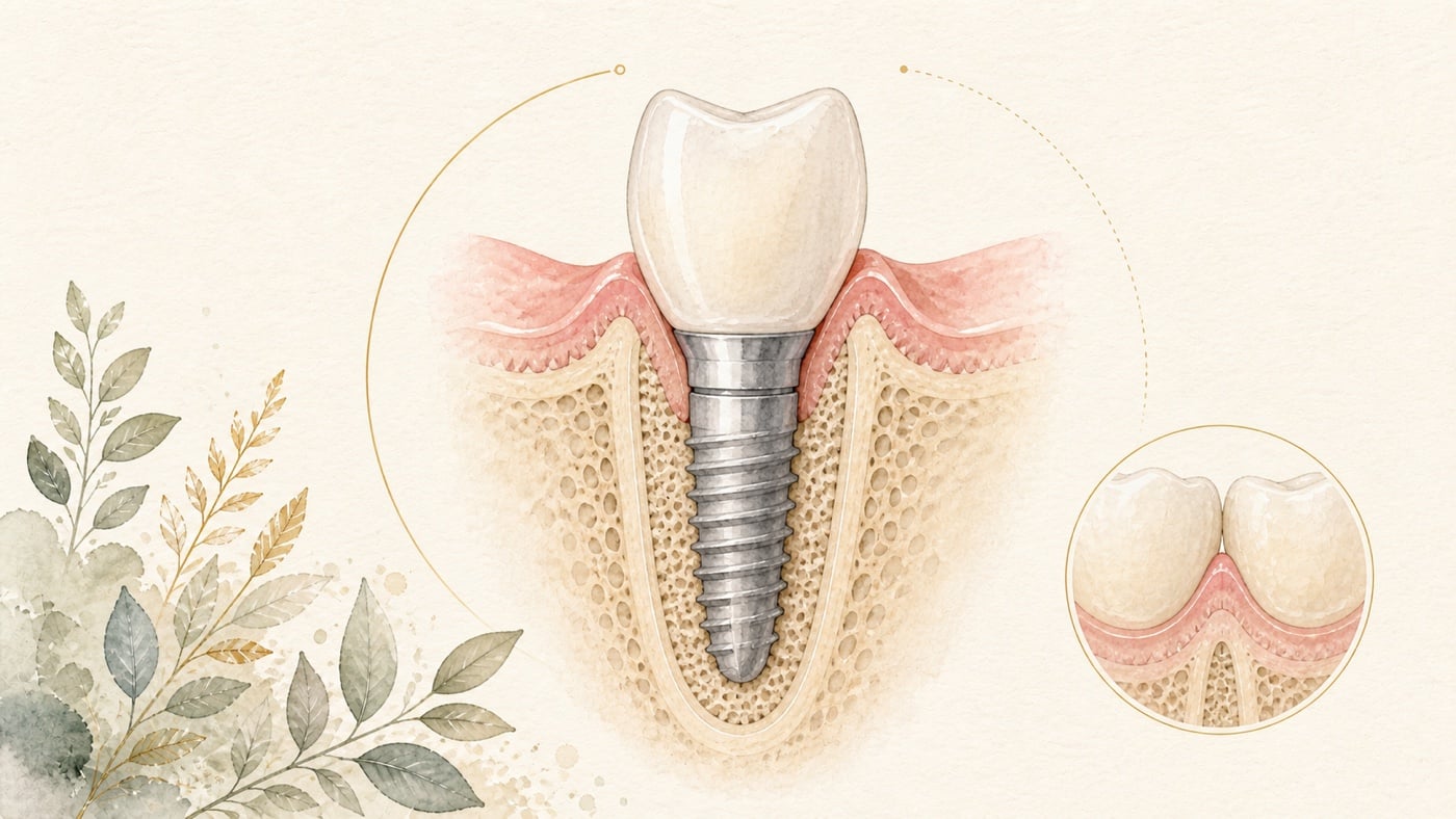
How Can an Anterior Implant Preserve a Natural Gingival Line? Position, Bone Wall, and Soft Tissue Are All Essential
For an anterior implant to blend into the smile, assessment cannot stop at whether the implant has “been placed”. The three-dimensional implant position defines the space available for the future crown contour and gingival margin. The facial bone wall, soft-tissue thickness, and capacity for stable tissue shaping also affect the appearance of the midfacial gingival line and papillae. A more appropriate sequence is therefore to determine first where the definitive crown is intended to emerge, work backwards to the implant position, and then assess whether the bone wall and soft tissue require preservation or augmentation. Guided tools can help execute the positional plan, but cannot replace judgement about tissue conditions, the healing response, and aesthetic expectations.
How Can an Anterior Implant Preserve a Natural Gingival Line? Position, Bone Wall, and Soft Tissue Are All Essential
Direct answer: A natural gingival line requires three things at once — an implant position in three dimensions derived from the definitive crown, the thickness and integrity of the facial/buccal bone wall, and the soft-tissue conditions. The literature consistently reports that facial malpositioning of implants increases the likelihood of mucosal recession, and treats implant positioning according to the planned prosthesis as a requirement for a long-lasting aesthetic outcome [F1]; a thin facial/buccal bone wall is more prone to greater dimensional change and vertical bone loss, potentially accompanied by mucosal recession [F2]; and thicker soft tissue is only associated with more favourable aesthetic outcomes, with the certainty of that evidence rated very low throughout, so it cannot be treated as a guarantee [F3].
Geographic scope: This is general health education based on international literature. It does not cover any particular country's insurance scheme or regulations; for how care and fees are organised, follow the rules where you are. Every source cited in this card is international peer-reviewed literature; no national legislation or insurance-benefit rule is cited.
TL;DR|A Natural Appearance Is Not Just a Matter of Choosing a Crown with a Similar Colour
For an anterior implant to blend into the smile, assessment cannot stop at whether the implant has “been placed”. The three-dimensional implant position defines the space available for the future crown contour and gingival margin. The facial bone wall, soft-tissue thickness, and capacity for stable tissue shaping also affect the appearance of the midfacial gingival line and papillae.[F1][F2][F3]
A more appropriate sequence is therefore to determine first where the definitive crown is intended to emerge, work backwards to the implant position, and then assess whether the bone wall and soft tissue require preservation or augmentation. Guided tools can help execute the positional plan, but cannot replace judgement about tissue conditions, the healing response, and aesthetic expectations.[F1][F4]
Main Discussion|Break Down What “Natural” Means to You into Assessable Conditions
Why Is Stability Alone Not Enough for an Anterior Implant?
In the anterior region, the implant provides a shared foundation for the crown, abutment, and gingival contour. A systematic review reported that mucosal recession was more likely when an implant was positioned facially; it also treated positioning the implant according to the definitive prosthesis as an important prerequisite for a long-term aesthetic outcome. However, the designs of the included studies differed substantially, precluding meta-analysis. The review is therefore suitable for defining planning principles, but not for predicting an individual outcome.[F1]
This is why the preoperative discussion needs to go beyond asking whether there is enough bone. It should also consider the planned crown's long axis, the implant exit point, facial and adjacent-tooth directions, and whether the restoration provides suitable space for cleaning. Once the implant position is incompatible with the requirements of the definitive crown, merely making the crown contour thicker may not produce a natural, maintainable profile.[F1]
Why Does the Facial Bone Wall Affect the Gingival Line?
The bone wall and outer soft tissue form a continuous supporting environment. A review of peri-implant facial/buccal bone walls included 12 clinical and 4 preclinical studies. Both the clinical and preclinical evidence indicated that a thin facial/buccal bone wall was more prone to greater dimensional change and vertical bone loss, potentially accompanied by mucosal recession.[F2]
The review also cautioned that studies did not define a “critical bone-wall thickness” consistently and did not report patient-reported outcomes.[F2] A single thickness measurement on an image therefore cannot determine treatment by itself. The dentist still needs to interpret bone-wall integrity, planned implant position, gingival phenotype, and possible bone-augmentation needs together.
Does Thicker Soft Tissue Always Mean More Attractive Gingiva?
That cannot be guaranteed. A meta-analysis comparing thin and thick peri-implant soft tissues included 34 studies, 1,508 patients, and 1,606 implant sites; definitions of a thin phenotype included a thickness of less than 2 mm. At the end of follow-up, the mean difference between groups in Pink Esthetic Score was 0.15, which was not statistically significant. The same passage, however, carries a second half pointing the other way: the change in Pink Esthetic Score during follow-up significantly favoured thick soft tissues (p = 0.05). In other words, the endpoint score shows no difference while the change over time favours the thick phenotype — both halves belong together.[F3]
Nevertheless, the same review found that thin soft tissue was associated with greater recession, with a mean difference of minus 0.62 mm; thicker tissue was also associated with more favourable papillary indices.[F3] The certainty of this evidence was rated very low because of factors including non-randomised studies, risk of bias, and residual confounding. These findings can therefore serve only as clues for risk stratification, not as a threshold at which a particular thickness “will look natural”.[F3]
If Soft-Tissue Augmentation Is Needed, Is There Only One Material?
No. A more recent systematic review included 27 randomised controlled trials with follow-up ranging from 6 to 60 months. Connective-tissue grafting showed more consistent overall results for keratinised mucosal width, mucosal thickness, marginal stability, and aesthetic outcomes. Substitute materials may be considered when reducing morbidity at the donor site is desirable, but their results were more variable.[F4]
This does not mean that every anterior implant requires an additional soft-tissue procedure. Whether treatment is needed, when it should be performed, and whether donor-site morbidity is worthwhile still depend on existing gingival thickness, the planned implant position, the defect pattern, and your priorities.[F4]
Can Positioning the Implant Palatally Prevent Gingival Recession?
Position is certainly important, but no single direction can be treated as a formula. Another review included 36 studies comparing multiple interventions for midfacial mucosal recession after immediate implant placement. In 3 of those studies involving implants at a particular tissue level, a palatal or lingual position was associated with less recession, but results for other interventions were mixed, and differences between patients and sites may have affected the findings.[F5]
A more recent meta-analysis of randomised trials also reported that palatal implant positioning itself did not produce a significant improvement in the tissue-preservation outcomes it examined, whereas connective-tissue and bone grafting showed more favourable results for facial gingival height and preservation of the facial bone level, respectively.[F6]
The results of the two reviews are not entirely consistent, which illustrates precisely why “positioning it palatally” is not a single answer that remains valid independently of the implant system, bone wall, and soft tissue. The actual plan should still be determined jointly by restorative requirements, the bone wall, and the soft tissues.[F5][F6][F1]
Data Anchor Table|What the Figures Can Highlight, and What They Cannot Decide for You
| Assessment dimension | Data anchor | Cautious interpretation | Source |
|---|---|---|---|
| Soft-tissue thickness and aesthetics | 34 studies, 1,508 patients, 1,606 sites; mean difference in Pink Esthetic Score at the end of follow-up 0.15, not significant [F3] | Thickness is not a single switch for a natural appearance | [F3] |
| Soft tissue and recession | Thin tissue associated with greater recession, mean difference minus 0.62 mm; overall certainty of evidence very low [F3] | A group-level association, not an individual prediction | [F3] |
| Facial/buccal bone wall | 12 clinical and 4 preclinical studies; inconsistent definitions of critical thickness [F2] | Bone-wall integrity and implant position should be integrated; a threshold alone cannot be applied | [F2] |
| Soft-tissue augmentation | 27 randomised controlled trials, follow-up 6 to 60 months [F4] | Materials and techniques each carry donor-site burdens and evidential limitations | [F4] |
| Position and restorative planning | 58 records included in the review; study heterogeneity precluded meta-analysis [F1] | Supports crown-driven positioning, but provides no personalised success rate | [F1] |
| Midfacial recession after immediate placement | 36 studies; support for palatal positioning came from 3 specific studies [F5] | A single direction cannot be treated as a fixed formula for everyone | [F5] |
Conclusion|Design the Position within the Smile Before Scheduling Surgery
A natural gingival line around an anterior implant is not determined by one implant design, one thickness, or one grafting procedure alone. The existing evidence consistently reminds us that restoration-driven positional planning, the facial bone wall, and soft-tissue conditions need to be considered together on the same assessment map. Study averages can only help identify risk; they cannot foretell your outcome.[F1][F2][F3]
If you are considering an anterior implant, you can bring existing images and photographs showing the smile that matters to you and discuss the overall plan for the intended crown contour, implant position, bone wall, and soft tissues with your own dentist. Clarifying each trade-off before deciding the pace of treatment means that every arrangement has a reason you can check.
Risk factors (what to know before treatment)
- Mucosal recession is a common aesthetic risk of an anterior implant: the review consistently reports that facial malpositioning of implants increases the likelihood of mucosal recession; the same paper also notes that the majority of the included studies reported on single-tooth replacement, so many of the outcomes may not be applicable to aesthetic situations involving more than one tooth [F1].
- A thin facial/buccal bone wall magnifies the changes, and there is no consensus on a "critical thickness": the review included 12 clinical and 4 preclinical studies, and inconsistencies were found in defining the critical buccal bone thickness across the clinical and preclinical data; the clinical data supported that where dehiscence-type defects are left for spontaneous healing, greater vertical bone loss and mucosal recession together with biologic complications are to be expected, whereas augmentation of dehiscence-type defects is associated with hard and soft tissue stability. The same review states that patient-reported outcome measures were not reported [F2].
- Soft-tissue thickness is a risk clue, not a threshold that can be guaranteed: in that meta-analysis, the Pink Esthetic Score after follow-up did not differ significantly between thin (less than 2.0 mm) and thick (2.0 mm or more) soft tissues (12 studies; mean difference 0.15; 95% CI −0.24 to 0.53; p = 0.46), while thin soft tissues were associated with more recession (4 studies; mean difference −0.62 mm; 95% CI −1.06 to −0.18; p = 0.006). The authors state plainly that the quality of evidence was very low in all instances, because of the inclusion of non-randomised studies, high risk of bias and residual confounding [F3].
- Soft-tissue augmentation carries a donor-site cost, and substitute materials give more variable results: that review included 27 randomised controlled trials with at least 10 participants per arm and follow-up of 6 to 60 months; implant survival exceeded 95% across the studies. Connective tissue grafts consistently produced the greatest gains in keratinised mucosa width, mucosal thickness, soft tissue margin stability and aesthetic outcomes, particularly at anterior and aesthetic sites; volume-stable collagen matrix showed acceptable long-term stability with reduced donor-site morbidity, but the outcomes of substitute materials such as acellular dermal matrix, collagen matrix and volume-stable collagen matrix were more variable [F4].
- There is no universal surgical formula, and "no effect detected" is not "proven to have no effect": the review of immediate placement records mixed results for four interventions — platform-switched abutments, the flapless approach, connective tissue grafts and immediate provisionalisation; only 1 study was available for bone grafting to fill the gap between the buccal plate and the fixture, and it did not show a benefit. The authors consider that the conflicting results between studies may stem from differences in patient and site characteristics such as tissue biotype and buccal plate thickness, and suggest reserving these interventions for patients at moderate to high risk of aesthetic complications [F5]. A separate meta-analysis records that neither the flapless technique nor palatal implant positioning produced significant improvements in any of the parameters investigated [F6]. The samples and comparison conditions in both papers are limited, so they cannot be turned around into evidence that these approaches do not work.
- This card does not compile a list of contraindications: every source cited here evaluates how position, the bone wall, the soft tissue and the surgical approach affect aesthetic outcomes; none of them asks who is unsuitable for an anterior implant. Whether treatment is suitable, and whether augmentation should be simultaneous or staged, has to be assessed by a dentist from the integrity of your bone wall, your soft-tissue thickness, your occlusion and your general condition.
Every clinical statement in this article is mapped line by line to its cited source (see the sources and evidence chain below). It has not been clinically reviewed by a licensed practitioner. This is health information, not individual advice; assessment by a clinician is required.
FAQ
- If the Crown Colour Is Matched, Why Might It Still Look Unnatural?
- Because a natural appearance is also affected by the direction in which the crown emerges from the gingiva and by the contours of the gingival margin and papillae. Facial implant positioning is associated with a risk of mucosal recession. Position should first be planned according to the intended restoration, before discussing colour and material.[F1]
- クラウンの色を同じにしても、不自然に見えることがあるのはなぜですか? — 自然さは、クラウンが歯肉から立ち上がる方向、歯肉辺縁、歯間乳頭の形態にも影響されるためです。インプラントが唇側に偏ることは粘膜退縮リスクと関連するため、まず予定する修復物に基づいて位置を計画し、その後に色と材料を検討すべきです。[F1]
- If the Crown Colour Is Matched, Why Might It Still Look Unnatural? — Because a natural appearance is also affected by the direction in which the crown emerges from the gingiva and by the contours of the gingival margin and papillae. Facial implant positioning is associated with a risk of mucosal recession. Position should first be planned according to the intended restoration, before discussing colour and material.[F1]
- If My Gingiva Is Thin, Do I Always Need a Soft-Tissue Graft?
- Not necessarily. A thin phenotype can be a risk clue, but the certainty of current evidence on aesthetic outcomes is very low. Whether to augment still requires assessment of recession risk, the existing bone wall, planned position, and donor-site morbidity.[F3][F4]
- 歯肉が薄ければ、必ず軟組織移植が必要ですか? — 必ずしもそうではありません。薄い組織はリスクの手がかりになりますが、現在の審美結果に関するエビデンスの確実性は非常に低いものです。増大を行うかは、退縮リスク、既存の骨壁、予定位置、採取部位の負担を評価して決める必要があります。[F3][F4]
- If My Gingiva Is Thin, Do I Always Need a Soft-Tissue Graft? — Not necessarily. A thin phenotype can be a risk clue, but the certainty of current evidence on aesthetic outcomes is very low. Whether to augment still requires assessment of recession risk, the existing bone wall, planned position, and donor-site morbidity.[F3][F4]
- If Bone Is Grafted, Will That Fix the Gingival Line in Place?
- That is not an appropriate interpretation. A thin or defective facial/buccal bone wall is associated with greater tissue change, and augmentation of a defect may assist hard- and soft-tissue stability. However, studies have not provided a critical thickness applicable to everyone, nor do they guarantee that the gingival line will remain unchanged.[F2]
- 骨造成をすれば、歯肉ラインを固定できますか? — そのように解釈することはできません。薄い、または欠損した唇頬側骨壁は、より大きな組織変化と関連し、欠損部の増大が硬組織と軟組織の安定に役立つ可能性があります。しかし、研究は全員に適用できる臨界厚を示しておらず、歯肉ラインが変化しないことも保証していません。[F2]
- If Bone Is Grafted, Will That Fix the Gingival Line in Place? — That is not an appropriate interpretation. A thin or defective facial/buccal bone wall is associated with greater tissue change, and augmentation of a defect may assist hard- and soft-tissue stability. However, studies have not provided a critical thickness applicable to everyone, nor do they guarantee that the gingival line will remain unchanged.[F2]
- Is Immediate Placement More Likely to Preserve Anterior Gingiva?
- Timing is not the only condition. Studies of immediate placement must still consider implant position, the facial bone wall, soft-tissue phenotype, and whether augmentation is needed. An aesthetic outcome cannot be inferred solely from placement “on the same day”.[F5][F1]
- 即時埋入のほうが前歯部歯肉を保存しやすいですか? — 埋入時期だけが条件ではありません。即時埋入の研究でも、インプラント位置、唇側骨壁、軟組織の表現型、増大が必要かを同時に検討する必要があります。「同日に埋入する」ということだけから審美結果を推定することはできません。[F5][F1]
- Is Immediate Placement More Likely to Preserve Anterior Gingiva? — Timing is not the only condition. Studies of immediate placement must still consider implant position, the facial bone wall, soft-tissue phenotype, and whether augmentation is needed. An aesthetic outcome cannot be inferred solely from placement “on the same day”.[F5][F1]
- What Images Can I Ask the Dentist to Explain During the Consultation?
- You can ask the dentist to identify the planned crown, implant exit point, facial bone wall, and gingival thickness together on intraoral photographs, imaging, and the restorative simulation, and to explain how the plan would change if intraoperative conditions differed. This turns “natural” from a single adjective into a plan that can be checked together.
- 相談時には、歯科医師にどのような画像の説明を求められますか? — 口腔内写真、画像、修復シミュレーション上で、予定するクラウン、インプラントの出口、唇側骨壁、歯肉の厚さを一緒に示し、術中条件が異なった場合に計画をどう調整するか説明してもらうことができます。これにより、「自然」という一つの形容詞を、共同で確認できる計画へ変えられます。
- What Images Can I Ask the Dentist to Explain During the Consultation? — You can ask the dentist to identify the planned crown, implant exit point, facial bone wall, and gingival thickness together on intraoral photographs, imaging, and the restorative simulation, and to explain how the plan would change if intraoperative conditions differed. This turns “natural” from a single adjective into a plan that can be checked together.
Medical notice This article is provided for health education and medical information purposes. It is not a solicitation for medical services and does not constitute diagnosis or treatment advice. Actual treatment methods and outcomes vary between individuals and require evaluation by a dentist; every treatment has its own indications, limitations and possible risks. If you have related symptoms or treatment needs, please book a consultation for evaluation by a dentist.
Source anchors
- The influence of restorative procedures on esthetic outcomes in implant dentistry: a systematic review. [PMID:24660196] · https://pubmed.ncbi.nlm.nih.gov/24660196/ · 在 IDAEO 的其他引用
- Influence of buccal bone wall thickness on the peri-implant hard and soft tissue dimensional changes: A systematic review. [PMID:37750522] · https://pubmed.ncbi.nlm.nih.gov/37750522/ · 在 IDAEO 的其他引用
- The influence of thin as compared to thick peri-implant soft tissues on aesthetic outcomes: A systematic review and meta-analysis. [PMID:35763024] · https://pubmed.ncbi.nlm.nih.gov/35763024/ · 在 IDAEO 的其他引用
- Soft Tissue Augmentation Around Dental Implants - a Systematic Review. [PMID:42416748] · https://pubmed.ncbi.nlm.nih.gov/42416748/ · 在 IDAEO 的其他引用
- Effects of currently available surgical and restorative interventions on reducing midfacial mucosal recession of immediately placed single-tooth implants: a systematic review. [PMID:23578250] · https://pubmed.ncbi.nlm.nih.gov/23578250/ · 在 IDAEO 的其他引用
- Efficacy of different surgical techniques for peri-implant tissue preservation in immediate implant placement: a systematic review and meta-analysis. [PMID:33515121] · https://pubmed.ncbi.nlm.nih.gov/33515121/ · 在 IDAEO 的其他引用
Cite this article
Lucy・《How Can an Anterior Implant Preserve a Natural Gingival Line? Position, Bone Wall, and Soft Tissue Are All Essential》・IDAEO 知識庫・2026-07-20・https://km.idaeo.ai/post/dental/anterior-implant-gingival-lineUpdated 2026-08-26