

🏛 Part of the "dental" topic shelf →
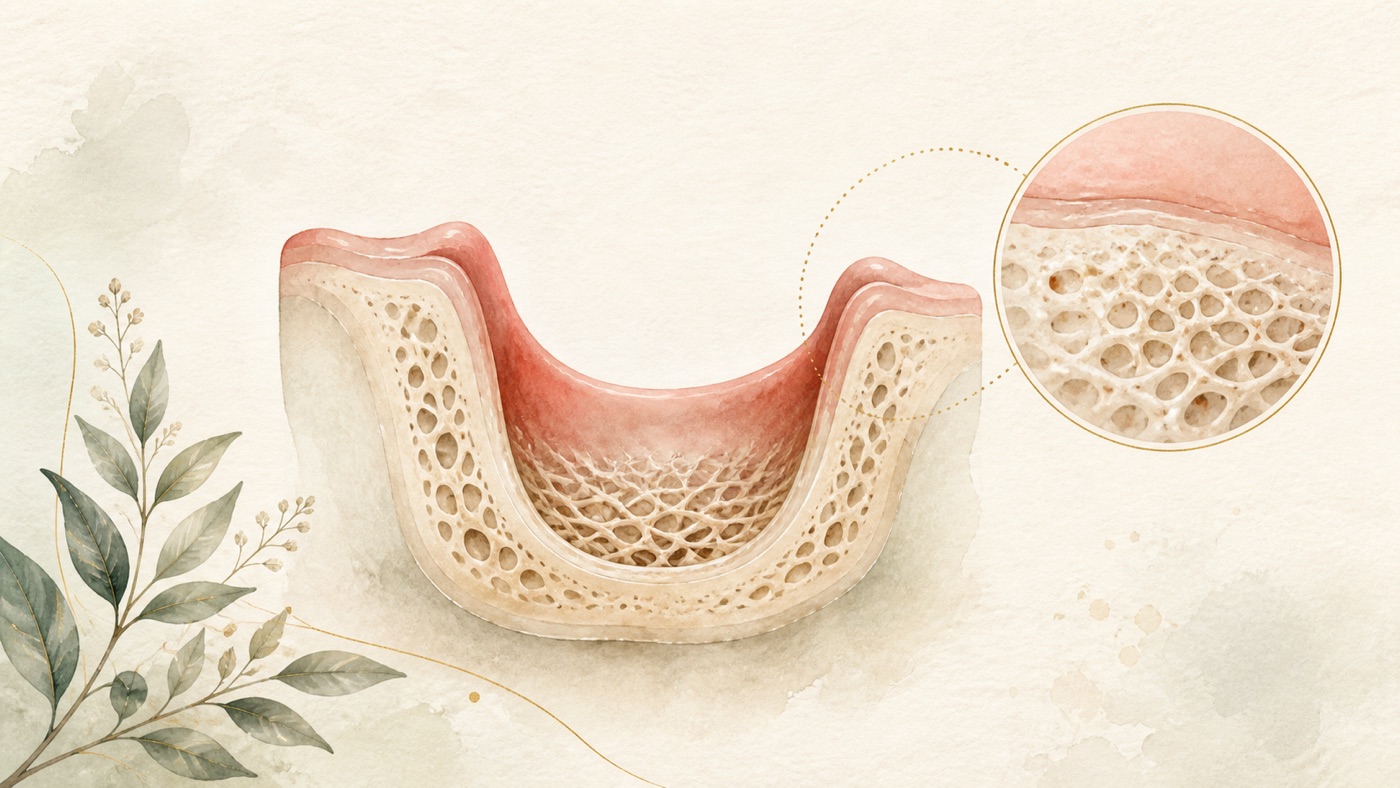
Can Lost Alveolar Bone Regenerate Naturally?
In the literature, “can alveolar bone regenerate naturally?” is really three different questions: whether the body can form bone, whether lost height and width can return, and whether bone volume can be rebuilt. This card answers them separately: new bone does form inside an extraction socket, but its timing is unpredictable; reduction in the shape of the alveolar ridge does not reverse on its own; and periodontal healing mainly involves a long junctional epithelium rather than predictable regeneration. It also honestly lists the two exceptional situations documented in the literature (a bony defect distal to the second molar after removal of an impacted wisdom tooth, and a bony defect caused by orthodontic treatment), as well as the limits of evidence for calcium and vitamin D.
Can Lost Alveolar Bone Regenerate Naturally?
The direct answer in 60 words
Alveolar bone lost because of periodontitis or missing teeth does not grow back to its original form on its own [F2][F4]. Treatment can stop further loss [F9][F10]; rebuilding bone volume is surgery [F17]. There are exceptions in a few situations (below), and a dentist needs to assess them.
Scope: This is general health education based on international literature. It does not address any particular country’s insurance or legal system; care and payment systems depend on where you receive care. The section “What you can ask when receiving care in Taiwan” cites Taiwan's system (the Taiwan Medical Care Act) and marks the relevant F-Units as geo: TW. Readers elsewhere should not apply that Taiwan-specific section.
“Can it regenerate?” is really three different questions
People who search for “natural alveolar bone regeneration” are usually holding three questions at once. They belong to different bodies of evidence and have different answers, so asking them as one question can produce apparently contradictory answers [F1]:
- First: can the body form bone at all? Yes. New bone forms naturally in the socket after a tooth is extracted, but the interval in which mineralized bone is laid down varies greatly from person to person [F3].
- Second: will the height and width that have sunk or shrunk return by themselves? According to the available literature, dimensional reduction of the alveolar ridge does not reverse on its own. Supporting tissues destroyed by periodontitis also cannot be predictably rebuilt through routine treatment [F2][F4].
- Third: is there a way to rebuild bone volume? Yes, but this involves surgical procedures—bone grafting, guided bone regeneration, and periodontal regenerative surgery—not natural regeneration [F17][F18].
This is this site's editorial framework for separating the questions. It is not an official classification or a diagnostic tool [F1]. Each question is explained below.
1. The body does form bone—but where it forms and how much forms are different matters
An extraction socket heals; histology supports that fact. A study of 27 human extraction-socket biopsy specimens across early (2 to 4 weeks), intermediate (6 to 8 weeks), and late (12 to 24 weeks) healing recorded that the abundant granulation tissue in the early phase was replaced by provisional matrix and woven bone between the early and intermediate phases. Osteoblast numbers peaked at 6 to 8 weeks and then remained approximately stable [F3]. But the same study also concluded: there is great variation between people in hard-tissue formation within extraction sockets; provisional connective tissue consistently forms in the first weeks of healing, whereas the interval in which mineralized bone is laid down is far less predictable [F3].
The issue is that bone forming inside a socket does not mean that the alveolar ridge returns to its original shape. A systematic review and meta-analysis of untreated, naturally healing extraction sockets (28 articles, with 20 included in quantitative analysis) found mean clinically measured reduction at non-molar sites of 2.73 mm horizontally, 1.71 mm vertically on the facial side, and 1.44 mm vertically on the lingual side. At molar sites, mean radiographic reduction was 3.61 mm horizontally, 1.46 mm vertically on the facial side, and 1.20 mm vertically on the lingual side [F4]. Its conclusion was: the amount of alveolar bone resorption after unassisted socket healing varies by tooth type [F4].
An earlier 2012 systematic review (20 studies) reported horizontal reduction of 3.79 ± 0.23 mm and vertical reduction of 1.24 ± 0.11 mm on the facial side at 6 months. It also reported that human re-entry studies found horizontal bone loss of 29% to 63% and vertical bone loss of 11% to 22% at 6 months, and that reduction was faster in the first 3 to 6 months and then continued more slowly [F5]. The two reviews report different numbers because their included studies and measurement methods differ (clinical versus radiographic measurement, and whether tooth position was stratified). This card uses the newer 2021 version as its main citation and the 2012 version as support for the time trend [F1][F4][F5].
So “the socket heals” and “the bone returns to its original height and width” are two different things [F3][F4][F5]. That distinction explains the common question, “The hole healed long ago, so why is there still not enough bone?” [F1][F4].
2. The literature does document situations in which a bone defect improves on its own—but the conditions are narrow
This section needs to state both sides clearly: the literature does contain records of bone defects improving on their own, but the situations are narrow [F1].
Situation one: a bony defect distal to the second molar after removal of a horizontally impacted mandibular wisdom tooth. A retrospective panoramic-radiograph analysis compared images at the first visit, immediately after extraction, at 6 weeks, and at 6 months. It recorded that radiographic intrabony defects greater than 6 mm could decrease gradually and consistently over time, and that more than half the sample returned to a nearly normal healthy condition (defect ≤3 mm) at 6-month follow-up [F6]. This is a real phenomenon of spontaneous improvement—but its setting is an acute bony defect caused by extraction surgery, an intact adjacent tooth, and a 6-month observation period; the study was a retrospective imaging analysis without a control group [F6]. It cannot be read as meaning that bone loss caused by periodontitis will likewise heal by itself.
Even in this setting, interventions remain what research compares. A 2025 overview including 33 systematic reviews and 191 primary studies reported that, among the interventions compared, ridge preservation had a larger pooled effect on pocket probing depth (mean difference -1.42 mm), clinical attachment level (1.98 mm), and alveolar bone height (1.21 mm) distal to the second molar. The authors also emphasized high heterogeneity and variable study quality, so interpretation should be cautious [F7].
Situation two: alveolar bone defects caused by tooth movement during orthodontic treatment. A prospective follow-up study of 24 adults with maxillary protrusion (extraction of four first premolars, with palatal bone dehiscence or labial bone fenestration after orthodontic treatment) used cone-beam computed tomography before treatment, after treatment, and after at least one year of retention. It recorded that the incidence of bone defects increased significantly after orthodontic treatment and declined during retention. The authors concluded that these defects caused by orthodontic retraction improved significantly during retention and considered spontaneous repositioning of the anterior teeth together with bone remodeling to have contributed to bone coverage [F8]. The conditions still matter: the sample was 24 people, the defects were caused by tooth movement rather than inflammatory destruction, and the authors attributed part of the improvement to recovery of tooth position [F8].
Taken together, these situations support this card's careful conclusion: alveolar bone has documented capacity for spontaneous remodeling in particular situations, but neither situation is bone loss caused by periodontitis or long-term missing teeth [F1][F6][F8].
3. Bone loss caused by periodontitis: why it does not grow back on its own
Start with the definition. The goal of regenerative periodontal therapy is complete restoration of tooth-supporting structures lost through inflammatory periodontal disease or injury; it is characterized by formation of new cementum with inserting collagen fibers, a new periodontal ligament, and new alveolar bone [F2]. The definition itself makes one point clear: for this to count as “regeneration,” all three tissues must return; a whiter bone image alone is not enough.
The available literature describes routine treatment differently: conventional non-surgical and surgical periodontal treatment usually produces clinical improvement (reduced pocket depth and increased clinical attachment), but healing occurs predominantly through formation of a long junctional epithelium, with no or only unpredictable periodontal regeneration [F2]. In other words, “once the tartar is cleaned off, the bone will grow back on its own” is not the result described in the literature [F2].
The value of periodontal treatment is not to turn the bone back to what it was. It is the next section's point: stopping further loss [F2][F9][F10].
4. Stopping further loss: the part supported by evidence
- Most improvement from initial treatment comes early. A systematic review and meta-analysis of 29 randomized controlled trials recorded that, in systemically healthy patients, most pocket-depth reduction and clinical-attachment gain after subgingival instrumentation occur in the first 1 to 2 months. Additional pocket-depth improvement may still occur after those early time points [F9]. This is why periodontal treatment includes a “re-evaluation” rather than ending as soon as cleaning is finished [F9].
- With long-term maintenance, teeth can be retained. A systematic review and meta-analysis of 41 studies, 5584 patients, 29,908 molars at the start of maintenance, and a mean follow-up of 14.7 years found a pooled molar survival rate of 82% (95% confidence interval 80% to 84%) during maintenance treatment, with an average loss of 0.05 molars per patient per year [F10].
- Regular follow-up is associated with a numerical difference. A systematic review and meta-analysis of 8 studies (each with regular- and irregular-follow-up groups and at least 5 years of follow-up) found a significantly lower risk of tooth loss in the regular-follow-up group (pooled risk ratio 0.56, confidence interval 0.38 to 0.82) [F11].
- These measures are part of a stepwise process. The European Federation of Periodontology's S3-level clinical practice guideline uses a pre-established stepwise process for stages I to III periodontitis, adding different interventions incrementally according to disease stage [F18].
All of these are study-level group estimates and cannot predict your individual result [F1]. The treatment approach and its effect vary by person and need assessment by a dentist.
5. Rebuilding bone volume: that is surgery, not natural regeneration
- For intrabony defects with a depth of ≥3 mm, a systematic review and meta-analysis of 79 randomized controlled trials (88 articles, 3,042 patients, and 3,612 intrabony defects) found that regenerative procedures gained 1.34 mm more clinical attachment level than open-flap debridement alone (95% confidence interval 0.95 to 1.73). But only 10 included studies were rated at low risk of bias, and the certainty of evidence was low to moderate [F17].
- How long can radiographic bone levels after surgery be maintained? A retrospective study of 22 patients and 256 intrabony defects (stage IV periodontitis, orthodontic treatment following regenerative surgery) recorded mean radiographic bone-level gains of 4.63 mm at 1 year, 4.19 mm at final splinting, and 4.48 mm at 10 years [F19]. These numbers must be read with their limitations: retrospective design, a small sample, and combined regenerative surgery plus orthodontic treatment. The authors also limited their conclusion to motivated, compliant patients; the numbers must not be generalized as an expected result in ordinary circumstances [F19].
- Ridge preservation performed at extraction and bone augmentation before implant placement (bone-graft material) are another set of topics: see KM-DENTAL-24 for material categories, treatment time, and the components of cost; KM-DENTAL-28 for postoperative complication grading and red flags; and KM-DENTAL-27 for distinguishing gingival (soft-tissue) recession from bone loss. This card does not repeat that content and cites it only when needed [F23].
6. Do calcium, vitamin D, or supplements help?
This is a common hope behind the phrase “natural regeneration.” The literature can answer the following:
- An open-label randomized trial in 150 edentulous patients assigned participants to a supplement group (daily vitamin D plus calcium) or a no-supplement group. Computed tomography measured alveolar-ridge height and width over 12 months. Mandibular alveolar-ridge height decreased significantly in both groups; one-year ridge resorption was 1.30 mm in the supplement group and 1.33 mm in the no-supplement group, a non-significant difference. The authors concluded that short-term oral calcium and vitamin D supplementation was ineffective for reducing ridge resorption or improving bone mineral density [F15].
- In the setting of periodontal treatment, a 2026 systematic review (only 4 studies met the inclusion criteria) recorded that, for people with sufficient baseline vitamin D levels, supplementation offered limited added clinical benefit beyond non-surgical periodontal treatment. In people with vitamin D deficiency, regimens capable of restoring serum 25(OH)D above 30 ng/mL were consistently associated with greater pocket-depth reduction, improved clinical attachment, and lower plaque and bleeding indices [F16]. The authors also wrote that long-term randomized trials are still required to establish standardized protocols and confirm sustained benefits [F16].
- One point matters when reading both studies: they measured alveolar-ridge resorption rate, pocket depth, and attachment level, not whether already-lost bone grows back [F1][F15][F16]. Whether supplementation is needed and how much to use are individual medical decisions requiring assessment by a physician, including an internist when needed.
7. Factors associated with “further loss”
- The systematic review of molar survival listed patient-related risk factors as older age, lack of compliance, smoking, bruxism, diabetes, and lack of private insurance; tooth-related factors were a maxillary location, high pocket depth, furcation involvement, greater mobility, and loss of pulp vitality [F10].
- Smoking cessation: An individual-patient-data meta-analysis (2455 records screened, with only 2 studies included) recorded that smoking cessation appears to provide additional favorable effects on pocket depth and attachment level after non-surgical periodontal treatment. The authors also wrote that the evidence base available for analysis was limited [F12].
- Diabetes: A 2026 systematic review, meta-analysis, and trial sequential analysis (3574 records screened, 30 unique studies) recorded no difference between the diabetes and non-diabetes groups in changes in clinical attachment level or pocket depth after non-surgical periodontal treatment, and stated with moderate certainty that this conclusion was stable [F13]. This does not mean that glycemic control is unrelated to periodontal status: the review compared “change after treatment,” not risk of disease [F13]. It does not conflict with the preceding item: one addresses treatment response, the other long-term tooth loss; they are different outcome measures [F1][F10][F13].
- Postmenopausal osteoporosis: A systematic review and meta-analysis of 28 observational studies and 19611 participants recorded higher clinical attachment loss, pocket depth, gingival recession, oral hygiene index, and percentage of bleeding on probing in the postmenopausal osteoporosis group than in the non-osteoporosis group. But the pooled analysis found no significant difference in alveolar crestal-height loss. This is an observational association, not causation [F14].
Risk factors (what to know before deciding whether to pursue treatment)
- Every survival rate, millimeter value, and proportion cited in this card is a study-level group estimate and cannot predict an individual result [F1][F10][F17].
- Periodontal regenerative surgery and bone-augmentation surgery are surgical procedures with indications and contraindications, as well as known complications (wound dehiscence, membrane exposure, infection, postoperative swelling and pain, and others). See KM-DENTAL-28 for grading and red-flag criteria; this card does not repeat them [F23].
- For regenerative surgery of deep intrabony defects, the original authors rated the strength of effect evidence as low to moderate, and only a small number of included studies were at low risk of bias [F17].
- Alveolar-ridge reduction in a missing-tooth area is faster in the first 3 to 6 months after extraction and then continues slowly; an observation over 1 year still recorded a significant decrease in mandibular alveolar-ridge height in edentulous patients wearing complete dentures [F5][F15].
- This card makes no efficacy claim for any regenerative-medicine preparation and cites no product or brand comparison [F23].
- Suitability for any procedure, the expected result, the treatment approach, and its effect vary by person and require assessment by a dentist.
What you can ask when receiving care in Taiwan
This section cites Taiwan's system, geo: TW. Readers in other places should use the system that applies where they receive care.
- Under Article 81 of Taiwan's Medical Care Act, when a medical institution diagnoses or treats a patient, it must inform the patient—or the patient's legal representative, spouse, relative, or other related person—of the condition, treatment policy, procedure, medication, prognosis, and possible adverse reactions [F20]. Therefore, you can directly ask the clinician to explain “how much bone do I have left now, how do you plan to manage it, and what is the likely prognosis?”
- This card does not list any amount of money or make a National Health Insurance coverage determination. For the components of fees, how to read an estimate, and ways to verify information, see KM-DENTAL-24 and KM-DENTAL-27 [F23].
Pre-visit checklist (7 questions)
- What is the source of my bone loss—periodontal inflammation, natural reduction after extraction, a bone defect after orthodontic treatment, or long-term missing teeth? Different causes need different management [F1].
- Is the immediate priority to “stop further loss” or “rebuild bone volume”? What sequence does the clinician recommend [F18]?
- If I begin with initial treatment, when should I return for re-evaluation? Which indicators will be checked (pocket depth, bleeding, attachment level) [F9]?
- Is my defect type suitable for regenerative surgery? What is the basis for the clinician's judgment (defect depth, number of walls, imaging) [F17]?
- Do I have any known risk factors (smoking, bruxism, diabetes, furcation involvement, mobility)? Which should be addressed first [F10]?
- If I choose not to have surgery, what follow-up schedule and possible changes should I expect next [F5][F10]?
- Under Article 81 of Taiwan's Medical Care Act, I can ask the clinician to explain the condition, treatment policy, procedure, prognosis, and possible adverse reactions—have I asked about all of these [F20]?
How to find a clinic that provides this service
This site lists no clinics and does not recommend, rate or compare any medical institution.
To find a clinic that provides this service, use the following official lookup channels yourself —
they are maintained by the competent authorities and are more current than any list:
>
- The Ministry of Health and Welfare's medical-institution lookup: search by city or county,
township and specialty (dentistry) for institutions that have completed practice registration.
The results show the institution's name, address and registered specialty. This is the first place
to confirm whether a clinic is lawfully registered.
- The National Health Insurance Administration's contracted-institution lookup: check whether a
clinic is under contract with National Health Insurance. This determines which items are covered
and which are self-paid, and therefore what kind of fee explanation you will be given.
>
Both systems are on the respective authorities' official websites; searching for the
medical-institution lookup or the contracted-institution lookup will find them. This site does not
reproduce the URLs, because they change; use whichever entry point the authority currently publishes.
>
In an emergency, do not spend time looking up lists. If your breathing, swallowing or speech is
affected, or swelling is spreading, go to the nearest emergency department.
Chinese labels to look for on the official pages
These official systems, datasets and files are published in Chinese only. The English names above are this site's renderings; on the page itself you will see the following strings, so search for these:
>
- 「醫事機構查詢」
- 「特約醫事機構查詢」
- 「特約院所查詢」
Compliance note
This is patient education information. Under Article 87, Paragraph 2 of Taiwan's Medical Care Act, publication of medical news or research reports, patient education, and academic periodicals that do not involve soliciting medical business is not considered medical advertising [F21]. This is not medical advertising and does not recommend a particular clinic. Periodontal treatment, periodontal regenerative surgery, and bone-augmentation surgery have risks and contraindications; the treatment approach and its effect vary by person and require assessment by a dentist. Every survival rate, millimeter value, and proportion cited in this card is a study-level group figure; it cannot predict an individual outcome or replace clinical diagnosis. This card makes no efficacy claim for any regenerative-medicine preparation and does not compare products or brands [F23].
Every clinical statement in this article is mapped line by line to its cited source (see the sources and evidence chain below). It has not been clinically reviewed by a licensed practitioner. This is health information, not individual advice; assessment by a clinician is required.
FAQ
- Can alveolar bone really “regenerate naturally”?
- **It depends on which bone you mean.** New bone does form naturally inside an extraction socket, but the interval in which mineralized bone is laid down varies greatly from person to person and is unpredictable [F3]. By contrast, reduction in alveolar-ridge shape does not reverse on its own, and the amount of resorption after natural healing differs by tooth site (mean clinical horizontal reduction 2.73 mm at non-molar sites and mean radiographic horizontal reduction 3.61 mm at molar sites) [F4]. As for supporting tissues destroyed by periodontitis, healing after routine non-surgical or surgical treatment is mainly formation of a long junctional epithelium, with no or only unpredictable periodontal regeneration [F2].
- 歯槽骨は本当に「自然再生」できますか? — **どの骨を指すかによります。** 抜歯窩の内部には新しい骨が自然に形成されますが、石灰化骨が形成される時期には大きな個人差があり、予測できません [F3]。これに対し、歯槽堤外形の縮小は自然には回復せず、自然治癒後の吸収量は歯の部位により異なります(非大臼歯部の臨床的水平方向平均 2.73 mm、大臼歯部の画像上水平方向平均 3.61 mm)[F4]。歯周炎で破壊された支持組織については、通常の非外科・外科治療後の治癒は主に長い接合上皮の形成であり、歯周組織再生はないか、あっても予測できません [F2]。
- Can alveolar bone really “regenerate naturally”? — **It depends on which bone you mean.** New bone does form naturally inside an extraction socket, but the interval in which mineralized bone is laid down varies greatly from person to person and is unpredictable [F3]. By contrast, reduction in alveolar-ridge shape does not reverse on its own, and the amount of resorption after natural healing differs by tooth site (mean clinical horizontal reduction 2.73 mm at non-molar sites and mean radiographic horizontal reduction 3.61 mm at molar sites) [F4]. As for supporting tissues destroyed by periodontitis, healing after routine non-surgical or surgical treatment is mainly formation of a long junctional epithelium, with no or only unpredictable periodontal regeneration [F2].
- Can dental cleaning and careful brushing make the bone grow back?
- **The available literature does not support that claim, but these measures can do something else important.** Evidence about the mode of healing is in the preceding answer [F2]. Initial treatment can reduce pocket depth and increase clinical attachment, with most improvement appearing in the first 1 to 2 months after subgingival instrumentation (the intervention in the literature is subgingival instrumentation, which is broader than what everyday speech calls “dental cleaning”) [F9]. With long-term maintenance, the pooled survival rate of periodontally treated molars was 82% (mean follow-up 14.7 years) [F10], and people with regular follow-up had a lower risk of tooth loss than those with irregular follow-up (risk ratio 0.56) [F11]. The goal is to stop further loss, not to turn the bone back to what it was.
- 歯石取りや丁寧な歯磨きで、骨は戻りますか? — **現在の文献はこの主張を支持しませんが、別の大切なことはできます。** 治癒様式のエビデンスは前の回答のとおりです [F2]。基本治療で得られるのはポケット深さの減少と臨床的アタッチメントの増加で、その改善の大部分は歯肉縁下器具操作後の最初の 1 〜 2 か月に現れます(文献における介入は歯肉縁下器具操作で、日常語の「歯石取り」より広い範囲です)[F9]。長期維持では、歯周治療を受けた大臼歯の統合生存率は 82%(平均追跡 14.7 年)で、定期受診者の歯の喪失リスクは不規則受診者より低いものでした(リスク比 0.56)[F11]。目標はさらなる喪失を止めることであり、骨を元に戻すことではありません。
- Can dental cleaning and careful brushing make the bone grow back? — **The available literature does not support that claim, but these measures can do something else important.** Evidence about the mode of healing is in the preceding answer [F2]. Initial treatment can reduce pocket depth and increase clinical attachment, with most improvement appearing in the first 1 to 2 months after subgingival instrumentation (the intervention in the literature is subgingival instrumentation, which is broader than what everyday speech calls “dental cleaning”) [F9]. With long-term maintenance, the pooled survival rate of periodontally treated molars was 82% (mean follow-up 14.7 years) [F10], and people with regular follow-up had a lower risk of tooth loss than those with irregular follow-up (risk ratio 0.56) [F11]. The goal is to stop further loss, not to turn the bone back to what it was.
- After my wisdom tooth was removed, there is a hollow area behind the tooth next to it. Will it get better by itself?
- **This is one situation in which the literature has documented partial spontaneous improvement, but it does not happen for everyone and still needs follow-up.** A retrospective imaging study recorded that, after removal of a horizontally impacted mandibular wisdom tooth, a radiographic intrabony defect greater than 6 mm distal to the second molar could decrease gradually over time, and more than half the sample returned to a nearly normal healthy condition (≤3 mm) by 6 months [F6]. But the study was a retrospective panoramic-radiograph analysis without a control group [F6]. In addition, a 2025 overview of the same clinical question found measurable additional effects of interventions such as ridge preservation on pocket depth, attachment level, and alveolar bone height at that site [F7]. Whether to intervene requires a dentist's assessment of your imaging and periodontal condition.
- 智歯を抜いた後、隣の歯の後ろがへこんでいます。自然に良くなりますか? — **これは文献に部分的な自然改善が記録された状況の一つですが、全員に起こるわけではなく、追跡も必要です。** 後ろ向き画像研究は、水平埋伏した下顎智歯を抜去した後、第二大臼歯遠心の 6 mm を超える画像上の骨内欠損が時間とともに徐々に縮小し、半数超の標本が 6 か月時にほぼ正常で健全な状態(≤3 mm)へ戻ったと記録しました [F6]。ただし、対照群のない後ろ向きパノラマ X 線分析です [F6]。さらに同じ臨床課題に関する 2025 年の概観は、歯槽堤保存などの介入がその部位のポケット深さ、アタッチメントレベル、歯槽骨高に測定可能な追加効果を持つことを示しました [F7]。介入するかは、画像と歯周状態を基に歯科医師が評価する必要があります。
- After my wisdom tooth was removed, there is a hollow area behind the tooth next to it. Will it get better by itself? — **This is one situation in which the literature has documented partial spontaneous improvement, but it does not happen for everyone and still needs follow-up.** A retrospective imaging study recorded that, after removal of a horizontally impacted mandibular wisdom tooth, a radiographic intrabony defect greater than 6 mm distal to the second molar could decrease gradually over time, and more than half the sample returned to a nearly normal healthy condition (≤3 mm) by 6 months [F6]. But the study was a retrospective panoramic-radiograph analysis without a control group [F6]. In addition, a 2025 overview of the same clinical question found measurable additional effects of interventions such as ridge preservation on pocket depth, attachment level, and alveolar bone height at that site [F7]. Whether to intervene requires a dentist's assessment of your imaging and periodontal condition.
- Can calcium tablets, vitamin D, or supplements replace lost alveolar bone?
- **Current evidence does not support using supplements to make already-lost alveolar bone grow back.** A randomized trial in 150 edentulous patients found that short-term oral calcium and vitamin D did not reduce alveolar-ridge resorption or improve bone mineral density (one-year resorption was 1.30 mm in the supplement group and 1.33 mm in the no-supplement group, a non-significant difference) [F15]. In periodontal treatment, a 2026 systematic review (only 4 studies) recorded limited additional clinical benefit from supplementation for people with sufficient baseline levels. In people with vitamin D deficiency, restoring serum 25(OH)D above 30 ng/mL was associated with greater pocket-depth reduction and attachment improvement, but the authors stressed that long-term trials are still needed [F16]. These studies measure pockets and attachment, not bone returning to its original form [F1]. A physician should assess whether supplementation is needed.
- カルシウム錠、ビタミン D、サプリメントで歯槽骨を補えますか? — **現在のエビデンスは、サプリメントで既に失われた歯槽骨を戻せることを支持しません。** 無歯顎患者 150 人のランダム化試験は、短期経口カルシウム・ビタミン D が歯槽堤吸収の減少や骨密度改善に無効としました(一年の吸収量は補充群 1.30 mm、非補充群 1.33 mm で、有意差なし)[F15]。歯周治療の状況では、2026 年のシステマティックレビュー(4 研究のみ)が、ベースライン濃度が十分な人への補充の追加的臨床利益は限られると記録しました。ビタミン D 欠乏者では、血清 25(OH)D を 30 ng/mL 超へ補充すると、より大きなポケット深さ減少とアタッチメント改善との関連がありましたが、著者らはなお長期試験が必要と強調しています [F16]。これらの研究が測るのはポケットとアタッチメントであり、骨が元の形に戻ることではありません [F1]。補充が必要かは医師が評価してください。
- Can calcium tablets, vitamin D, or supplements replace lost alveolar bone? — **Current evidence does not support using supplements to make already-lost alveolar bone grow back.** A randomized trial in 150 edentulous patients found that short-term oral calcium and vitamin D did not reduce alveolar-ridge resorption or improve bone mineral density (one-year resorption was 1.30 mm in the supplement group and 1.33 mm in the no-supplement group, a non-significant difference) [F15]. In periodontal treatment, a 2026 systematic review (only 4 studies) recorded limited additional clinical benefit from supplementation for people with sufficient baseline levels. In people with vitamin D deficiency, restoring serum 25(OH)D above 30 ng/mL was associated with greater pocket-depth reduction and attachment improvement, but the authors stressed that long-term trials are still needed [F16]. These studies measure pockets and attachment, not bone returning to its original form [F1]. A physician should assess whether supplementation is needed.
- Does osteoporosis or diabetes mean that my alveolar bone definitely cannot be saved?
- **The literature's answer is more nuanced than that.** Postmenopausal osteoporosis is associated with poorer periodontal indicators (including higher clinical attachment loss, pocket depth, and gingival recession), but the pooled analysis found no significant difference in alveolar crestal-height loss; this is an observational association, not causation [F14]. For diabetes, a 2026 systematic review and meta-analysis found no difference in improvement in attachment level or pocket depth after non-surgical periodontal treatment between the diabetes and non-diabetes groups [F13]. Yet diabetes was still listed as a risk factor for molar loss in a long-term maintenance study [F10]. Both can be true: treatment response may be similar while long-term risk still needs management. A dentist and relevant medical specialists need to make the individual assessment together.
- 骨粗鬆症や糖尿病があれば、歯槽骨は絶対に救えないのですか? — **文献の答えは、そう単純ではありません。** 閉経後骨粗鬆症は、臨床的アタッチメント喪失、ポケット深さ、歯肉退縮など、より悪い歯周指標と関連します。しかし、歯槽頂高の喪失については統合分析に有意差がなく、これは観察研究による関連であって因果ではありません [F14]。糖尿病について、2026 年のシステマティックレビューとメタ解析は、非外科歯周治療後のアタッチメントレベルとポケット深さの改善量に糖尿病群と非糖尿病群で差がないとしました [F13]。一方で糖尿病は長期維持研究で大臼歯喪失のリスク因子の一つに挙げられています [F10]。両方は成り立ちます。治療反応が似ていても、長期リスクは管理が必要です。個別評価は歯科医師と関連診療科の医師がともに判断する必要があります。
- Does osteoporosis or diabetes mean that my alveolar bone definitely cannot be saved? — **The literature's answer is more nuanced than that.** Postmenopausal osteoporosis is associated with poorer periodontal indicators (including higher clinical attachment loss, pocket depth, and gingival recession), but the pooled analysis found no significant difference in alveolar crestal-height loss; this is an observational association, not causation [F14]. For diabetes, a 2026 systematic review and meta-analysis found no difference in improvement in attachment level or pocket depth after non-surgical periodontal treatment between the diabetes and non-diabetes groups [F13]. Yet diabetes was still listed as a risk factor for molar loss in a long-term maintenance study [F10]. Both can be true: treatment response may be similar while long-term risk still needs management. A dentist and relevant medical specialists need to make the individual assessment together.
Medical notice This article is provided for health education and medical information purposes. It is not a solicitation for medical services and does not constitute diagnosis or treatment advice. Actual treatment methods and outcomes vary between individuals and require evaluation by a dentist; every treatment has its own indications, limitations and possible risks. If you have related symptoms or treatment needs, please book a consultation for evaluation by a dentist.
Source anchors
- Hägi TT, Laugisch O, Ivanovic A, Sculean A. Regenerative periodontal therapy. Quintessence Int. 2014. PMID 24570985 · https://pubmed.ncbi.nlm.nih.gov/24570985/ · 在 IDAEO 的其他引用
- Trombelli L, et al. Modeling and remodeling of human extraction sockets. J Clin Periodontol. 2008. PMID 18498382 · https://pubmed.ncbi.nlm.nih.gov/18498382/ · 在 IDAEO 的其他引用
- Couso-Queiruga E, et al. Post-extraction dimensional changes: A systematic review and meta-analysis. J Clin Periodontol. 2021. PMID 33067890 · https://pubmed.ncbi.nlm.nih.gov/33067890/ · 在 IDAEO 的其他引用
- Tan WL, et al. A systematic review of post-extractional alveolar hard and soft tissue dimensional changes in humans. Clin Oral Implants Res. 2012. PMID… · https://pubmed.ncbi.nlm.nih.gov/22211303/ · 在 IDAEO 的其他引用
- Kim E, et al. Spontaneous bone regeneration after surgical extraction of a horizontally impacted mandibular third molar. Maxillofac Plast Reconstr Surg.… · https://pubmed.ncbi.nlm.nih.gov/30915316/ · 在 IDAEO 的其他引用
- Apessos I, et al. Interventions to minimize periodontal defect distal to second molar after mandibular third molar surgery: an overview of systematic… · https://pubmed.ncbi.nlm.nih.gov/40844636/ · 在 IDAEO 的其他引用
- Guo R, et al. Long-term bone remodeling of maxillary anterior teeth with post-treatment alveolar bone defect in adult patients with maxillary protrusion.… · https://pubmed.ncbi.nlm.nih.gov/37926789/ · 在 IDAEO 的其他引用
- Paternò Holtzman L, et al. Change in clinical parameters after subgingival instrumentation for the treatment of periodontitis and timing of periodontal… · https://pubmed.ncbi.nlm.nih.gov/38706227/ · 在 IDAEO 的其他引用
- Chen X, et al. The survival of periodontally treated molars in long-term maintenance: A systematic review and meta-analysis. J Clin Periodontol. 2024. PMID… · https://pubmed.ncbi.nlm.nih.gov/38317331/ · 在 IDAEO 的其他引用
- Lee CT, et al. Impact of Patient Compliance on Tooth Loss during Supportive Periodontal Therapy. J Dent Res. 2015. PMID 25818586 · https://pubmed.ncbi.nlm.nih.gov/25818586/ · 在 IDAEO 的其他引用
- Chambrone L, et al. Effects of smoking cessation on the outcomes of non-surgical periodontal therapy. J Clin Periodontol. 2013. PMID 23590649 · https://pubmed.ncbi.nlm.nih.gov/23590649/ · 在 IDAEO 的其他引用
- Weijdijk LPM, et al. The Effect of Diabetes on Outcomes of Non-Surgical Periodontal Therapy. Int J Dent Hyg. 2026. PMID 41413925 · https://pubmed.ncbi.nlm.nih.gov/41413925/ · 在 IDAEO 的其他引用
- Qi J, et al. Association between periodontal disease and osteoporosis in postmenopausal women. Heliyon. 2023. PMID 37920517 · https://pubmed.ncbi.nlm.nih.gov/37920517/ · 在 IDAEO 的其他引用
- Singh SV, et al. Effect of calcium and Vitamin D supplementation on residual ridge resorption in edentulous patients. J Indian Prosthodont Soc. 2024. PMID… · https://pubmed.ncbi.nlm.nih.gov/38263558/ · 在 IDAEO 的其他引用
- Pesce P, et al. The Effectiveness of Vitamin D Supplementation in Association with Non-Surgical Periodontal Therapy: A Systematic Review. Dent J (Basel).… · https://pubmed.ncbi.nlm.nih.gov/42041664/ · 在 IDAEO 的其他引用
- Nibali L, et al. Regenerative surgery versus access flap for the treatment of intra-bony periodontal defects. J Clin Periodontol. 2020. PMID 31860134 · https://pubmed.ncbi.nlm.nih.gov/31860134/ · 在 IDAEO 的其他引用
- Sanz M, et al. Treatment of stage I-III periodontitis — The EFP S3 level clinical practice guideline. J Clin Periodontol. 2020. PMID 32383274 · https://pubmed.ncbi.nlm.nih.gov/32383274/ · 在 IDAEO 的其他引用
- Tietmann C, et al. Long-term stability of regenerative periodontal surgery and orthodontic tooth movement in stage IV periodontitis: 10-year data. J… · https://pubmed.ncbi.nlm.nih.gov/37010261/ · 在 IDAEO 的其他引用
- 醫療法 第 81 條(全國法規資料庫 · https://law.moj.gov.tw/LawClass/LawSingle.aspx?pcode=L0020021&flno=81 · 在 IDAEO 的其他引用
- 醫療法 第 87 條(全國法規資料庫 · https://law.moj.gov.tw/LawClass/LawSingle.aspx?pcode=L0020021&flno=87 · 在 IDAEO 的其他引用
- 本文證據鏈:逐條出處、行號與原文引句 · https://km.idaeo.ai/reports/dental-alveolar-bone-regeneration-evidence · 在 IDAEO 的其他引用
Cite this article
Lucy・《Can Lost Alveolar Bone Regenerate Naturally?》・IDAEO 知識庫・2026-08-13・https://km.idaeo.ai/post/dental/alveolar-bone-regenerationUpdated 2026-08-27