🏛 Part of the "dental" topic shelf →
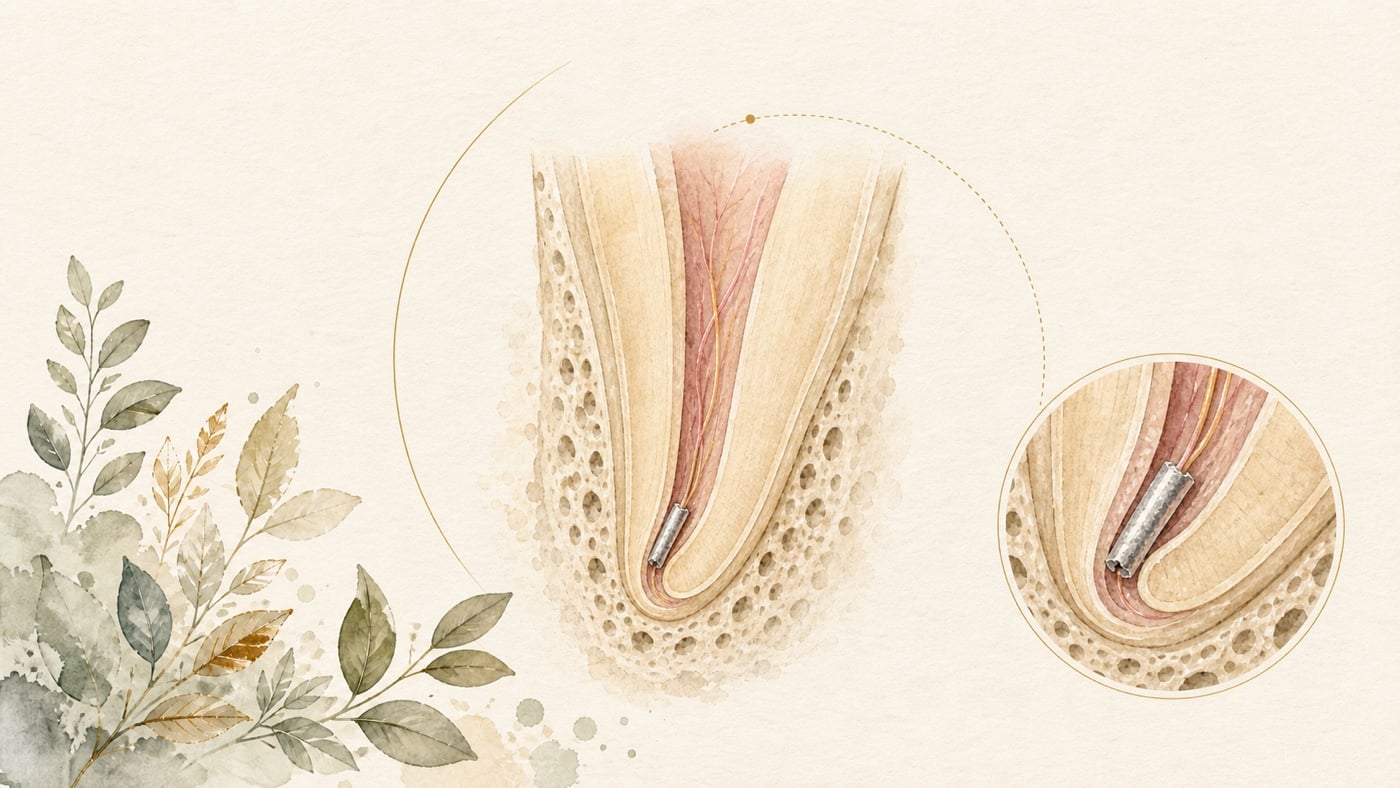
A Root Canal Instrument Has Broken Inside: What Should Guide the Next Step?
Separation of a root canal instrument is a technical event during treatment; it does not mean that the tooth has immediately failed. The questions that actually matter are where the fragment lies, whether it is before or beyond a curvature, whether it can be seen under microscopic visualisation, how far the canal had been cleaned before separation, and how much dentine retrieval would sacrifice. In a prospective clinical study, the overall completion rate for retrieval and bypassing combined was 70.5%; when the fragment was directly visible the study attempted retrieval and the completion rate was 85.3%, whereas for fragments that were not visible the study performed bypassing only, with no attempt at retrieval, and the completion rate was 47.7%. These are procedural rates from a particular team using a standardised protocol, not survival rates for an individual tooth. If forceful retrieval requires extensive removal of inner dentine, the original risk posed by a metal fragment may become a risk of perforation, ledging, or root weakening.
A Root Canal Instrument Has Broken Inside: What Should Guide the Next Step?
Direct answer: The next step depends on which part of the canal the fragment lies in, whether it is before or beyond a curvature, whether it is visible under microscopic visualisation, and how much dentine retrieval would sacrifice; the existing technical evidence is predominantly laboratory based and lacks well-designed clinical comparisons, so it does not support forceful retrieval in every case.[F3]
Geographic scope: Global. All cited evidence comes from international peer-reviewed literature; no country-specific reimbursement scheme or regulation is involved. The procedures, materials and fees actually available to you should follow your own health system and your dentist's assessment.
TL;DR|The Goal Is to Control Infection and Preserve Tooth Structure, Not Retrieval for Its Own Sake
Separation of a root canal instrument is a technical event during treatment; it does not mean that the tooth has immediately failed. The questions that actually matter are where the fragment lies, whether it is before or beyond a curvature, whether it can be seen under microscopic visualisation, how far the canal had been cleaned before separation, and how much dentine retrieval would sacrifice.
In a prospective clinical study, the overall completion rate for retrieval and bypassing combined was 70.5%; when the fragment was directly visible the study attempted retrieval and the completion rate was 85.3%, whereas for fragments that were not visible the study performed bypassing only, with no attempt at retrieval, and the completion rate was 47.7%.[F1] These are procedural rates from a particular team using a standardised protocol, not survival rates for an individual tooth. If forceful retrieval requires extensive removal of inner dentine, the original risk posed by a metal fragment may become a risk of perforation, ledging, or root weakening. The stopping point is therefore also part of treatment quality.
Main Discussion|Map the Fragment First, Then Compare Retrieval, Bypassing, and Retention
Why Do Instruments Separate? Understand the Event Before Assigning Blame
Nickel–titanium instruments may separate because of cyclic fatigue from repeated loading at a curvature, or torsional fatigue when the instrument tip binds while the shank continues to rotate. A systematic review included 12 studies and 939 separated instruments, reporting an overall incidence of 5%; across 10 studies, cyclic fatigue accounted for the predominant failure mode in a range of 62% to 92%.[F2]
This 5% is the overall estimate from the included studies, not a fixed incidence for every practice, instrument, or tooth. Canal curvature, instrument size, number of uses, method of movement, and the way a study identifies fracture surfaces can all affect the figure. Once separation has occurred, establishing the fragment's location and the remaining working space is more useful than pursuing the average incidence.
The First Map: Location, Curvature, and Visibility
Assessment commonly begins with periapical radiographs taken at different angles. If two-dimensional imaging cannot clarify the relationship between the fragment and the curvature, root thickness, or adjacent structures, limited-field CBCT may then be considered. A microscope provides illumination and magnification, but it cannot see through dentine; “not visible under the microscope” and “the fragment is absent” are two different things.
In a prospective study of 112 fragments, after straight-line access had been established, 68 were visible under the microscope and 44 were not. Retrieval was attempted for the 68 visible fragments using ultrasonic tips alone or combined with bypassing, whereas bypassing alone was performed for the 44 fragments that were not visible; the completion rates in the two groups were 85.3% and 47.7% respectively, a statistically significant difference.[F1] The 47.7% figure is therefore a completion rate for bypassing, and must not be read as meaning that a fragment which cannot be seen still has almost a one-in-two chance of being taken out. Visibility in fact reflects a combination of location, curvature, and the amount of dentine that would need to be removed, so it is a planning tool rather than a rigid threshold.
Retrieval: Under What Conditions Should It Be Discussed?
If the fragment lies in the coronal or middle third, conservative straight-line access can be established, and its coronal end can be exposed safely under microscopic visualisation, ultrasonic vibration, microtubes, or loop-type devices may be considered. A systematic review of fragments in the middle and apical thirds included only 14 in vitro studies. Ultrasonics were the most frequently studied method and produced generally better experimental results, but the more apically the fragment was located, the harder it was to retrieve, the more dentine was removed, and the lower the tooth's fracture resistance became.[F3]
“Better in vitro performance” cannot be translated directly into a first-line clinical choice, still less into one set of parameters for every canal. Cooling, ultrasonic power, duration of vibration, root concavities, and operator experience can all alter the thermal and structural cost.
Bypassing: Not a Second-Best Option, but Another Route to Infection Control
Bypassing means using a fine instrument to establish a path alongside the fragment so that the canal space beyond it can be cleaned, irrigated, and filled. When the fragment is difficult to retrieve but a path beside it can still be explored safely, bypassing may offer a better balance than continuing to remove dentine. Whether bypassing can be completed also depends on fragment length, canal curvature, and whether the fragment is locked in a constriction.
The review considers bypassing an option when retrieval is not feasible, but it also notes that the existing evidence is predominantly laboratory based and that well-designed clinical comparisons remain lacking.[F3] The dentist should therefore explain the intended duration of the attempt, the point at which it will stop, and whether acceptable cleaning and sealing can be completed coronal to the fragment if bypassing is unsuccessful.
Retaining the Fragment and Monitoring It: When Might This Be Safer Than Forceful Retrieval?
If the fragment is at the apical end, beyond a pronounced curvature, or would require excessive dentine loss to expose, retaining it while cleaning and sealing the accessible portion may be a reasonable route. In that situation, the extent of cleaning already completed before separation, whether the original pulpal condition was infected or non-infected, the presence of an apical lesion, and the quality of the coronal seal should all be included in the risk discussion.
A retrospective study at a teaching clinic identified 108 instrument separations among 3,150 treatments, an incidence of 3.4%; 53.7% were in mandibular molars and 42.6% in maxillary molars, and the level at which the fragment was located was significantly associated with subsequent management.[F4] This is the distribution of events at a single institution and does not represent the long-term prognosis after retaining a fragment. It supports the proposition that location changes management, not that a fragment in a particular tooth position must be retrieved.
Beyond the Technical Completion Rate, Consider the Cost to Tooth Structure
In a retrospective study of 123 cases of ultrasonic retrieval, 97 instruments were successfully retrieved (78.9%). The fragment's position relative to the canal curvature, the angle of access, and the angle of curvature all affected the result; the chance of failure was higher with an access angle > 20 degrees and a Schneider curvature angle > 40 degrees.[F5]
In the same study, only 26.3% of canals met all technical standards for obturation after retrieval, while 64.2% showed altered taper, all accompanied by ledge formation.[F5] This reminds us that procedurally “getting it out” does not mean that both the biological and structural outcomes are ideal. The preoperative plan should include acceptable dentine loss and stopping criteria within its definition of success.
Data Anchors|Procedural Rates Cannot Be Separated from Anatomical Conditions
| Evidence question | Data anchor | Cautious interpretation | Source |
|---|---|---|---|
| Instrument separation pattern | 12 studies, 939 instruments; overall incidence 5%; cyclic fatigue accounted for 62%–92% | Influenced by instruments and study criteria; not a fixed practice rate | [F2] |
| Visibility under microscopic visualisation | 112 instruments; overall 70.5%, visible group (retrieval attempted) 85.3%, non-visible group (bypassing only) 47.7% | The two groups underwent different procedures; these are procedural completion rates, not long-term tooth survival | [F1] |
| In vitro technical review | 14 studies; ultrasonics studied most frequently | More apical locations carry less favourable costs in tooth removal and fracture resistance; clinical comparisons are lacking | [F3] |
| Event distribution at a teaching clinic | 108 of 3,150 treatments, incidence 3.4% | Single-institution retrospective data; supports stratification by location but does not provide prognosis | [F4] |
| Ultrasonic retrieval cases | 97 retrieved among 123 cases (78.9%); access and curvature angles affected outcomes | Only 26.3% of fillings met all technical standards; the structural cost must also be considered | [F5] |
Risk factors: what to know before treatment
- When retrieval is more appropriate to discuss: retrieval is better placed for discussion when the fragment lies in the coronal or middle third, conservative straight-line access can be established, and its coronal end can be exposed safely under microscopic visualisation; the more apically the fragment lies, the harder it is to retrieve, the more dentine is removed, and the lower the tooth's fracture resistance becomes.[F3]
- The cost that retrieval may carry: a retrospective study of ultrasonic retrieval showed that obturation after retrieval did not necessarily meet all technical standards, with altered taper and ledge formation observed; in that study the highest chance of removal failure was associated with a canal access angle greater than 20 degrees and a Schneider angle greater than 40 degrees.[F5]
- Limits of the evidence, and why forceful retrieval is not appropriate in every case: the technical review of fragments in the middle and apical thirds included in vitro studies only and lacks well-designed clinical comparisons, so it cannot be extrapolated directly to the long-term outcome of an individual tooth.[F3] Retrospective data from a single teaching institution showed that the level at which the fragment lies was significantly associated with subsequent management, but that is a distribution of events and does not provide prognosis.[F4]
Conclusion|Reframe “Retrieving the Fragment” as “Preserving Infection Control and Tooth Safety”
After instrument separation, a sound decision begins with location, curvature, visibility, infection status, and remaining tooth structure. Retrieval, bypassing, and retention with monitoring each have appropriate indications. Existing technical reviews are predominantly in vitro, and clinical rates also depend heavily on anatomy and team experience; they do not support forceful retrieval in every case.[F3][F5]
If you have radiographs from before and after treatment or referral records, bring them with you to your dental appointment so that they can inform the assessment. During assessment, ask the dentist to identify the fragment's level, its relationship to the curvature, the planned extent of exposure, alternative routes for infection control, and the stopping point. This allows every step to be judged by how well it preserves tooth structure, rather than by whether the fragment leaves the canal.
Every clinical statement in this article is mapped line by line to its cited source (see the sources and evidence chain below). It has not been clinically reviewed by a licensed practitioner. This is health information, not individual advice; assessment by a clinician is required.
FAQ
- Will a Fragment Left Inside Keep Rusting or Moving?
- Root canal instruments are usually made of stainless steel or nickel–titanium alloy. The central question in deciding whether treatment is needed is not imagined “rust”, but whether the fragment obstructs infection control, whether its position is stable, and whether the surrounding tooth structure can withstand the retrieval procedure.
- 中に残った破折片は、錆び続けたり移動したりしますか? — 根管器具は通常、ステンレス鋼またはニッケルチタン合金です。処置が必要かを判断する要点は、想像される「錆」ではなく、破折片が感染制御を妨げるか、その位置が安定しているか、周囲の歯質が除去手技に耐えられるかです。
- Will a Fragment Left Inside Keep Rusting or Moving? — Root canal instruments are usually made of stainless steel or nickel–titanium alloy. The central question in deciding whether treatment is needed is not imagined “rust”, but whether the fragment obstructs infection control, whether its position is stable, and whether the surrounding tooth structure can withstand the retrieval procedure.
- Does Having a Microscope Mean That It Can Always Be Retrieved?
- No. A microscope improves illumination and the field of view, but it cannot see through dentine beyond a curvature. The prospective study also showed a lower rate of retrieval or bypassing for fragments that were not visible.[F1]
- 顕微鏡があれば必ず取り出せますか? — いいえ。顕微鏡は照明と視野を改善しますが、湾曲の先にある象牙質を透視することはできません。前向き研究でも、視認できない破折片は除去またはバイパス形成の割合が低いことが示されています。[F1]
- Does Having a Microscope Mean That It Can Always Be Retrieved? — No. A microscope improves illumination and the field of view, but it cannot see through dentine beyond a curvature. The prospective study also showed a lower rate of retrieval or bypassing for fragments that were not visible.[F1]
- If It Cannot Be Retrieved, Does the Tooth Have to Be Extracted?
- Not necessarily. Depending on infection status, fragment location, and accessible space, options may include bypassing; cleaning and sealing coronal to the fragment with follow-up; or, in appropriate cases, assessment for apical surgery. Extraction is generally reserved for situations in which the tooth is unrestorable or the cause cannot be controlled.
- 取り出せなければ、抜歯が必要ですか? — 必ずしもそうではありません。感染状態、破折片の位置、到達可能な空間に応じて、バイパス形成、破折片の歯冠側で清掃と封鎖を完了して経過観察する方法、または適切な症例で根尖手術を評価する方法を検討できます。抜歯は通常、歯を修復できない場合や原因を制御できない状況で選択されます。
- If It Cannot Be Retrieved, Does the Tooth Have to Be Extracted? — Not necessarily. Depending on infection status, fragment location, and accessible space, options may include bypassing; cleaning and sealing coronal to the fragment with follow-up; or, in appropriate cases, assessment for apical surgery. Extraction is generally reserved for situations in which the tooth is unrestorable or the cause cannot be controlled.
- Why Might the Dentist Stop After Trying for a While?
- Continuing ultrasonic treatment accumulates heat and dentine loss; this cost is generally higher the more apically the fragment lies.[F3] Stopping at a predetermined boundary is intended to avoid turning a manageable obstruction into a perforation or a weakened root.
- 歯科医師が一定時間試みた後に中止するのはなぜですか? — 超音波処置を続けると、熱と象牙質の喪失が蓄積します。破折片が根尖側にあるほど、この代償は一般に大きくなります。[F3] あらかじめ定めた中止基準で止めるのは、管理可能な障害を穿孔や歯根の脆弱化に変えないためです。
- Why Might the Dentist Stop After Trying for a While? — Continuing ultrasonic treatment accumulates heat and dentine loss; this cost is generally higher the more apically the fragment lies.[F3] Stopping at a predetermined boundary is intended to avoid turning a manageable obstruction into a perforation or a weakened root.
Medical notice This article is provided for health education and medical information purposes. It is not a solicitation for medical services and does not constitute diagnosis or treatment advice. Actual treatment methods and outcomes vary between individuals and require evaluation by a dentist; every treatment has its own indications, limitations and possible risks. If you have related symptoms or treatment needs, please book a consultation for evaluation by a dentist.
Source anchors
- Success rates for removing or bypassing fractured instruments: a prospective clinical study. [PMID:22414826] · https://pubmed.ncbi.nlm.nih.gov/22414826/ · 在 IDAEO 的其他引用
- Incidence of Different Types of Intracanal Fracture of Nickel-Titanium Rotary Instruments: A Systematic Review. [PMID:34267014] · https://pubmed.ncbi.nlm.nih.gov/34267014/ · 在 IDAEO 的其他引用
- Techniques to address fractured instruments in the middle or apical third of the root canal in human permanent teeth: a systematic review of the in vitro studies. [PMID:34698940] · https://pubmed.ncbi.nlm.nih.gov/34698940/ · 在 IDAEO 的其他引用
- Assessment of Incidence, Management and Contributory Factors of Root Canal Instrument Separation in an Endodontics Post-Graduate Program: A Retrospective Clinical Study. [PMID:38317030] · https://pubmed.ncbi.nlm.nih.gov/38317030/ · 在 IDAEO 的其他引用
- Factors affecting ultrasonic removal of separated endodontic instruments: A retrospective clinical study. [PMID:29532817] · https://pubmed.ncbi.nlm.nih.gov/29532817/ · 在 IDAEO 的其他引用
Cite this article
Lucy・《A Root Canal Instrument Has Broken Inside: What Should Guide the Next Step?》・IDAEO 知識庫・2026-07-20・https://km.idaeo.ai/post/dental/separated-instrument-decisionUpdated 2026-08-26