

🏛 Part of the "dental" topic shelf →
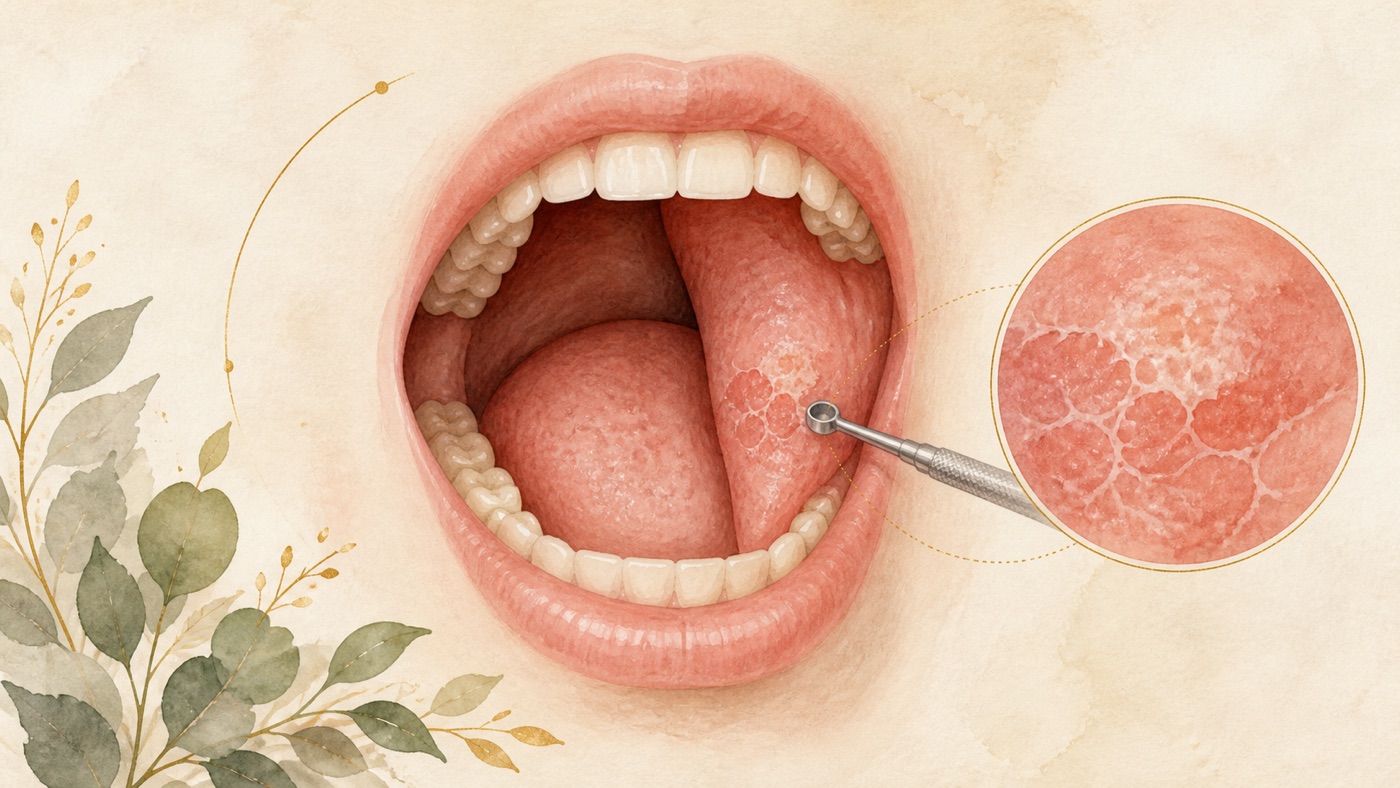
A complete guide to the oral mucosa and oral cancer screening: a domain map from the classification of ulcers, through the risk of potentially malignant disorders, to the limits of the screening evidence
This is the map-layer article for the field of oral medicine; it does not answer any single question. It covers: the two-axis reading framework for lesions of the oral mucosa (those that resolve by themselves / those that do not), what recurrent aphthous stomatitis looks like in the literature and its nature as a diagnosis of exclusion, the international classification system for oral potentially malignant disorders (OPMDs) and why the malignant transformation rates reported for the subtypes do not line up with one another, where leukoplakia and erythroplakia sit on the risk axis, the quantified risk evidence for tobacco, alcohol and betel quid, the difference in strength of evidence and the harms across the three layers of screening (routine screening of asymptomatic adults / high-risk groups / people who already have a lesion), why adjunctive tests cannot replace a biopsy, the source of the two-week rule and how it should be read, a conceptual framework for the care pathway, and the logic of what costs are made of. Every question-level specific issue is summarised in one sentence and pointed to its corresponding canonical card.
A complete guide to the oral mucosa and oral cancer screening: a domain map from the classification of ulcers, through the risk of potentially malignant disorders, to the limits of the screening evidence
TL;DR
Recurrent aphthous stomatitis is described in the literature as a common clinical disease of the oral mucosa [Fn102]; but an ulcer that has not healed after more than two weeks, or a red or white patch, has to be examined by a dentist [Fn20], and confirmation rests on biopsy [Fn17].
(49 characters in the zh-Hant original)
Introduction
This article is general oral-health education based on international literature. It does not address any country's insurance or regulations; consult local rules for care pathways and costs.
This article deliberately does not answer “what is this hole in my mouth”. That kind of judgement can only be made by the dentist or doctor who has examined that lesion in person. What it handles is the domain-level gap: which classes the professional community divides mucosal lesions into, what the concept of a precancerous lesion actually refers to, why the frequently quoted risk figures differ so widely from one another, into how many separate questions “oral screening” is cut on the evidence, and where the red-flag lists come from and how much weight they should be read with.
One thing has to be stated first, because it governs how every later section should be read: in this field, clinical appearance cannot replace a pathological diagnosis. Suspected oral cancer or its precursor lesions always require a tissue biopsy for confirmation [Fn17]; and a Cochrane systematic review (the current version being the 2021 update), having compared the various adjunctive tests, concluded that no adjunctive test can be recommended as a replacement for the current standard of surgical biopsy and histological assessment [Fn62]. So what this article provides is a map for reading and a set of evidence boundaries, not a self-diagnosis tool.
1. First establish two axes: time and appearance
There are many kinds of lesion on the oral mucosa, but the things a patient can observe for themselves — and that are genuinely used as criteria in the literature — come down to two.
1-1 The time axis: two weeks
The American Dental Association's consumer education page sets “lasting longer than two weeks” as the threshold for contacting a dentist; its own wording is that you should call your dentist right away if any of these issues last longer than two weeks [Fn20]. The oral cancer education page of the National Institute of Dental and Craniofacial Research (NIDCR) uses the same time threshold: if you have any of these symptoms for more than two weeks, see a dentist or a doctor [Fn118].
What makes this “two-week rule” important at domain level is not that two weeks carries any biological meaning, but that two independent institutions use the same duration as the prompt threshold for “this ought to be examined”.
But read it correctly: two weeks is a persistence threshold for a warning sign, not an observation window that assures safety. What both sources give is “see someone if it lasts longer than two weeks” [Fn20][Fn118], not “you may wait it out within two weeks”; not one of the sources cited in this article has validated that “two weeks” safely separates the lesions that will resolve by themselves from those that will not. Anyone whose lesion is new, is getting worse, looks suspicious, or comes with other warning signs may be examined earlier; and there is a further class of emergency signals to which the two-week threshold does not apply — see the opening of section 6. (This sentence and 6-0 are both an editorial safety reservation of this article, not a criterion taken from any single source; it only widens when to seek care and never narrows it, and it contains no criterion for judging a cause. It is booked as F30.)
Note that both of these sources are institutional patient education pages, not the text of a clinical guideline; this article cites only their warning-sign lists and time thresholds, and derives no diagnosis or management from them.
1-2 The appearance axis: white and red
The screening summary of the National Cancer Institute explains that a dentist or medical doctor may check the oral cavity during a routine check-up [Fn7], and that this examination includes looking for lesions, including areas of leukoplakia (an abnormal white patch of cells) and erythroplakia (an abnormal red patch of cells) [Fn8]; the same page states plainly that leukoplakia and erythroplakia lesions on the mucous membranes may become cancerous [Fn9].
Note that the mood of that sentence is “may”, not “is”. Leukoplakia and erythroplakia are written into the items examined at screening because they belong to the OPMD category discussed in the next section, not because they are equivalent to cancer.
1-3 Neither of these two axes is a diagnosis
The primary screening test for oral cancer set out by the US Preventive Services Task Force is a systematic clinical examination, including inspection and palpation of the oral cavity [Fn16]; and suspected oral cancer or its precursors detected on examination require confirmation by tissue biopsy [Fn17]. Put another way: time and appearance are the signals institutional lists use to prompt “this needs to be examined by a professional” [Fn20][Fn118]; determining “what it is” rests on a systematic clinical examination [Fn16] and, where necessary, a tissue biopsy [Fn17] — not on these two axes.
2. Recurrent aphthous stomatitis: common, cause unknown, and a diagnosis of exclusion
This is where misunderstanding clusters most densely in this domain. The literature describes recurrent aphthous stomatitis as a common clinical disease of the oral mucosa [Fn102], but “common” does not mean “most mouth ulcers are this” — the sources cited in this article do not report the proportion of oral ulcers attributable to each cause, and this article therefore makes no claim about proportions.
2-1 What it looks like in the literature
A clinical review describes recurrent aphthous stomatitis (RAS) as a common clinical disease of the oral mucosa [Fn102]; its prevalence in the general population is reported to vary between 5% and 25%, with its peak appearance in the second decade of life [Fn103]. The ulcers are described as round or oval, with well-defined erythematous margins and a shallow ulcerated centre covered with a gray or yellowish fibrinous pseudomembrane [Fn105]. Another narrative review describes them in the same terms: round or oval, covered by a gray-white fibrin layer and encircled by an erythematous ring [Fn81].
The prevalence is reported as a range as wide as 5%–25% [Fn103]; that source gives the range itself and does not state what produces its width. Whenever you meet a single figure, ask first by what criterion it was measured.
2-2 Cause: no settled conclusion at present
Two independent sources agree on this point: one review writes that so far, the etiopathogenesis is not clear [Fn104]; and the opening of a systematic review and meta-analysis likewise writes that the etiology of RAS remains unclear and its management is challenging [Fn41].
What has been quantified is “association”, not “cause”. The meta-analytic results compiled in one review show that people with recurrent aphthous stomatitis have a higher frequency of nutritional deficiencies, including vitamin B12 (OR 3.75, 95% CI 2.38–5.94), folic acid (OR 7.55, 95% CI 3.91–14.60) and ferritin (OR 2.62, 95% CI 1.69–4.06) [Fn82]; the same review also records a meta-analysis of 21 case-control studies showing that celiac disease is associated with a higher incidence of recurrent aphthous stomatitis (25% vs 11%; OR 3.79, 95% CI 2.67–5.39; P < .001) [Fn83].
⚠ These are associations from case-control designs, not causation, and they constitute no supplement or dietary advice. This article gives no nutritional-supplement or medication instruction of any kind.
2-3 It is a diagnosis that stands only after other conditions have been ruled out
This is the key sentence of this section at domain level. The literature states it explicitly: faced with ulcers that appear periodically on the oral mucosa, the first thing to do is to make a correct differential diagnosis, rule out associated systemic diseases and assess treatable causes before reaching the diagnosis of RAS [Fn106].
That is to say, “recurrent mouth ulcers” is not a label anyone can attach to themselves. Within the professional process it is an end point, not a starting point.
2-4 The evidence spectrum for management (containing no drug name, dose or method of use)
| Evidence source | Scale | Summary of conclusion |
|---|---|---|
| Network meta-analysis of topical medications (2023) | 43 RCTs, 3,067 participants, comparing 20 topical medications [Fn85] | On the outcome of pain reduction, that network meta-analysis failed to show any statistically significant difference between the topical treatments, or even against placebo [Fn86]; low to moderate quality evidence showed no superiority of any topical treatment over others as regards pain reduction [Fn87] |
| Systematic review and meta-analysis of one single topical medication (2026) | 5 RCTs included in the review, only 3 entering the meta-analysis (2 excluded for insufficient sample size) [Fn42] | The quantitative evidence base is extremely thin. The main text, the gist column of the footnotes, the F-Units and the FAQ of this article all omit that drug's name, route of administration, frequency of use and efficacy figures (a prescription drug name combined with an efficacy claim must not be presented to the public); the source list at the end retains that publication's original bibliographic title — bibliographic information must not be altered. The scope of and reason for the withdrawal are in the self-check record, section 11 |
| The management ladder compiled in a review [Fn84] | — | Oral conditions that do not improve with first-line treatment may require treatment with systemic medications [Fn84] |
The key information in this table is not which medication comes out ahead, but how thin the quantitative evidence in this field is. The analysis with the larger scale compared 20 topical medications [Fn85], yet its conclusion was that on the pain outcome no statistically significant difference could be seen between them, or even against placebo [Fn86], and that this rests on low to moderate quality evidence [Fn87]; while the meta-analysis conducted separately on one of those medications admitted only 3 studies into the quantitative pooling, with a further 2 excluded for insufficient sample size [Fn42]. This is precisely why section 11 of this article lists it as a weak point.
And at the other end of the spectrum, the review literature puts it very directly: at present, there is no curative treatment [Fn107]. The target of management is the symptom side, not eradication of the disease.
⚠ This section is a compilation of conclusions from the literature, not treatment advice. This section (and the whole main text, the gist column of the footnotes, the F-Units and the FAQ) lists no named drug, dose, route of administration or method of use; the source list at the end retains the original bibliographic titles of the publications, which is required for traceability and constitutes no medication instruction. Whether management is needed, and which management, has to be assessed by a dentist or doctor.
Adjacent specific questions (each has its own canonical card; this article does not expand on them)
- The differential sources of painful, swollen gums and the red flags for seeking care — see canonical card KM-DENTAL-05 (in production).
- The positioning and evidence for each ingredient class of mouthwash — see canonical card KM-DENTAL-06 (in production).
3. OPMD: what the category of oral potentially malignant disorders is saying
What the phrase “early signs of oral cancer” corresponds to professionally is not one sign, but a whole disease category.
3-1 The authoritative source of the classification
The consensus report produced by the expert seminar convened by the WHO Collaborating Centre for Oral Cancer deals with exactly this nomenclature and classification [Fn46]. That consensus defines them thus: oral potentially malignant disorders (OPMDs) are associated with an increased risk of occurrence of cancers of the lip or oral cavity [Fn46]; and it lists the disorders covered, including leukoplakia, erythroplakia, proliferative verrucous leukoplakia, oral lichen planus, oral submucous fibrosis, palatal lesions in reverse smokers, lupus erythematosus, epidermolysis bullosa, and dyskeratosis congenita [Fn47]. That update also records that there is sufficient evidence for an increased risk of oral cancer among patients diagnosed with certain further conditions, which were consequently added to the OPMD list [Fn48].
This list is the professional version of “early signs of oral cancer”. It is not a list of symptoms but a list of diseases; being on it does not mean a lesion will become cancer — it means that this class of lesion carries a higher risk than ordinary mucosa.
3-2 How common these disorders are
A systematic review and meta-analysis (in which only studies where a clinical assessment and histopathological confirmation were performed were included [Fn114]) found the overall prevalence of OPMD to be 4.47% (95% CI 2.43–7.08) [Fn111]; the most prevalent among them were oral submucous fibrosis (4.96%, 95% CI 2.28–8.62) and leukoplakia (4.11%, 95% CI 1.98–6.97) [Fn112]. The same analysis shows a marked geographical difference: 10.54% (95% CI 4.60–18.55) in Asian populations and 3.93% (95% CI 2.43–5.77) in South American/Caribbean populations [Fn113].
3-3 Malignant transformation rates: first the figures, then why the figures do not line up
This is the passage in this field that is most easily misread, so it is handled on its own.
A systematic review and meta-analysis that included 92 studies [Fn54] reports an overall malignant transformation (MT) rate across all OPMD groups of 7.9% (99% CI 4.9%–11.5%) [Fn50]; by subtype the rates were lichen planus 1.4% (99% CI 0.9%–1.9%), leukoplakia 9.5 (99% CI 5.9%–14.00%), oral lichenoid lesions 3.8% (99% CI 1.6%–7.00%), oral submucous fibrosis 5.2% (99% CI 2.9%–8.00%), erythroplakia 33.1% (99% CI 13.6%–56.1%) and proliferative verrucous leukoplakia 49.5% (99% CI 26.7%–72.4%) [Fn51]. (The leukoplakia entry is written in the source as “LE 9.5”, without a percent sign; its 99% CI is 5.9%–14.00%.) Converting by the mean follow-up period, the same study also gives annual transformation rates: lichen planus 0.28%, oral lichenoid lesions 0.57%, leukoplakia 1.56%, proliferative verrucous leukoplakia 9.3% and oral submucous fibrosis 0.98% [Fn53].
The same thing, measured by different studies, yields different figures. Three comparable examples:
| Disorder | Source A | Source B | Origin of the difference |
|---|---|---|---|
| Oral lichen planus | 1.4% (99% CI 0.9%–1.9%) [Fn51] | 1.14% (95% CI 0.84–1.49), with 82 studies and 26,742 patients included [Fn55][Fn58] | Different inclusion criteria, different CI width setting, different follow-up periods |
| Oral submucous fibrosis | 5.2% (99% CI 2.9%–8.00%) [Fn51] | 4%–7% (global expert consensus) [Fn44] | One is a meta-analytic point estimate, the other a consensus range |
And on that lichen planus figure, the original study attaches a key footnote of its own: the authors hold that the malignant transformation rates of OLP, OLLs and lichenoid reactions are underestimated, due essentially to restrictive diagnostic criteria, inadequate follow-up periods and/or low quality of studies [Fn57].
A portable way of reading this: whenever you meet a malignant transformation rate, ask three things first — which version of the diagnostic criteria was used, how long the follow-up ran, and what confidence level the CI uses [Fn51][Fn55][Fn57]. Inclusion criteria, follow-up duration and study quality can all affect the estimate [Fn57], so figures from different studies should not be treated as contradictory without examining those definitions.
3-4 One of the risk axes that has been quantified: the grade of dysplasia, not the colour
The meta-analysis just cited compared grades of dysplasia: moderate/severe dysplasia is meaningfully associated with a much greater risk of MT compared to mild dysplasia, with an odds ratio of 2.4 (95% CI 1.5–3.8) [Fn52]; on annual transformation rates, mild dysplasia was 1.7% and severe dysplasia 3.57% (reported directly by the source) [Fn53].
This does not mean the other factors are unimportant. The same analysis itself reports transformation rates by lesion subtype that differ enormously [Fn51], and another analysis quantifies factors such as site, lesion form, tobacco and alcohol [Fn56]. This article claims one thing only: the grade of dysplasia is one of the important risk axes that has been quantified, and it is not determined by colour or appearance.
The grade of dysplasia can only be determined by histopathology, not by appearance — that is the concrete reason behind the sentence at the head of this article that appearance cannot replace pathology, and it is why a suspicious lesion needs a biopsy for confirmation [Fn17][Fn62].
3-5 Two supplementary facts about oral lichen planus
A systematic review and meta-analysis reports a global pooled prevalence of oral lichen planus of 1.01%, with a marked geographical difference [Fn59]; from the age of 40 years, the prevalence increases significantly and progressively (OR 3.43, 95% CI 2.48–4.73, p < .001) [Fn60].
Another observation with real bearing on the question of who should be looking at these lesions is this: studies performed by oral medicine / oral pathology specialists report a significantly higher prevalence (1.80%) than dentists (0.61%) and dermatologists (0.33%) [Fn61]. This is an observed association, not a rating of competence, but it shows that the proportion in which one and the same lesion is recognised is not the same across professional training backgrounds.
As for the factors associated with malignant transformation of oral lichen planus, the meta-analysis in that field lists: tongue localization (RR 1.82, 95% CI 1.21–2.74, p = 0.004), presence of atrophic-erosive lesions (RR 4.09, 95% CI 2.40–6.98, p < 0.001), tobacco use (RR 1.98, 95% CI 1.28–3.05, p = 0.002) and alcohol consumption (RR 2.28, 95% CI 1.14–4.56, p = 0.02) [Fn56].
3-6 What happens after leukoplakia has been treated
A Cochrane systematic review included 14 studies with 909 participants [Fn78], and its conclusion is written without any ambiguity: we do not currently have evidence of a treatment that is effective for preventing the development of oral cancer [Fn76]. The same review also records that certain drug treatments may be effective in healing oral lesions, but that relapses and adverse effects are common [Fn77] (this article does not list the ingredient names, and does not advise anyone to obtain or use any ingredient on their own; whether management is needed, and which management, has to be assessed by a dentist or doctor). The review further notes that surgical treatment (including laser and cryotherapy) has never been assessed in a randomised trial that included a no treatment or placebo comparison group [Fn79]. It also explains that most leukoplakias are asymptomatic; therefore, the primary objective of treatment should be to prevent onset of cancer [Fn80].
Beyond that, a meta-analysis of oral leukoplakia with epithelial dysplasia reports a pooled overall response rate of 0.85 (95% CI 0.74–0.93) for 5-ALA photodynamic therapy [Fn45] — but the evidence level of that analysis was graded as very low, and its outcome is a lesion response rate, not cancer prevention. This is a procedure carried out by professionals; this article lists it only in order to show the state of the evidence in that field, and it cannot be used as a basis for choosing or requesting any therapy.
What the two of them mean together is this: leukoplakia can be managed, but “once it has been managed it will not turn into cancer” is not supported by the current evidence [Fn76]. As to what should happen next, what the consensus report in this field offers is a research agenda, not a follow-up frequency — it calls for multi-center longitudinal studies with uniform diagnostic criteria to improve the identification and cancer risk stratification of patients with OPMDs [Fn49]. This article derives from it no recall interval, follow-up method or care routine.
⚠ This section is a compilation of conclusions from the literature, not treatment advice. Actual treatment approaches and outcomes vary from person to person and must be assessed by a dentist.
4. Risk factors: which exposures have been quantified
4-0 First, put the scale in its correct place
Before talking about multiples of risk, one has to know the baseline scale of the disease, otherwise any multiple will be read as heavier than it is. The World Health Organization's oral health fact sheet records that oral cancers (lip, other parts of the oral cavity and the oropharynx combined) rank as the 13th most common cancer worldwide [Fn1], and that the global occurrence of lip and oral cavity cancer is estimated to be 389,846 new cases and 188,438 deaths in 2022 [Fn2].
How to read it: the “risk is such-and-such times higher” entries in the table below are relative risks — relative changes on top of that baseline scale, not an individual's probability of developing the disease. The lower the baseline scale, the smaller the absolute increment of risk to which the same multiple corresponds.
4-1 The qualitative lists at institutional level
The WHO oral health fact sheet states plainly that tobacco, alcohol and areca nut (betel quid) use are among the leading causes of oral cancer [Fn3]. The risk-assessment passage of the US Preventive Services Task Force lists them in more detail: the primary risk factors for oral cancer are tobacco and alcohol use, and additional risk factors include male sex, older age, use of betel quid, ultraviolet light exposure, infection with Candida or bacterial flora, and a compromised immune system [Fn15].
The epidemiological distribution is consistent with this: oral cancer is more common in men and in older people, more deadly in men compared to women, and it varies strongly by socio-economic circumstances [Fn4]; the American Dental Association's education page likewise records that males tend to get oral cancer more often than females, and that older people are generally at higher risk [Fn27].
4-2 The effect sizes that have been quantified
| Exposure | Quantified result | Nature of the study and its limits |
|---|---|---|
| Heavy drinking | Relative risk 5.13 for oral and pharyngeal cancer, compared with nondrinkers and occasional drinkers [Fn89] | A dose-response meta-analysis of 572 studies including 486,538 cancer cases [Fn90]; for those neoplasms there was a clear dose-risk relationship [Fn91] |
| Chewing tobacco | Pooled odds ratio 4.7 (3.1–7.1) [Fn92] | A systematic review and meta-analysis in South Asia; that paper records that over 90% of the global smokeless tobacco use burden is in South Asia [Fn93] — population-restricted, not to be extrapolated as a universal value |
| Betel quid with tobacco (paan) | Pooled odds ratio 7.1 (4.5–11.1) [Fn92] | As above |
| Betel-quid addictive use disorder (severe) | Risk of oral cancer 42.3 (mild 8.5, moderate 8.2) [Fn39] | A systematic review and meta-analysis for the South, Southeast and East Asian region; regional data |
| Betel quid without tobacco (restricted to people who neither smoke nor drink) | Oral leukoplakia OR 22.2 (95% CI 11.3–43.7), oral submucous fibrosis OR 56.2 (95% CI 21.8–144.8), erythroplakia OR 29.0 (95% CI 5.63–149.5) [Fn100] | A single large case-control study; the outcome is precancerous lesions and not cancer; dose-response relationships for frequency and duration were also observed [Fn101] |
One further point about betel quid needs to be flagged on its own: the IARC monographs have classified chewing betel quid without tobacco as a human carcinogen [Fn99]. Which means that “it is fine as long as no tobacco is added” does not stand up on the evidence.
4-3 The position of HPV has to be stated clearly
What the WHO fact sheet says is: in North America and Europe, human papillomavirus infections are responsible for a growing percentage of oral cancers among young people [Fn5]. The wording of the US Preventive Services Task Force marks the anatomical site more precisely: sexually transmitted oral HPV infection is recognised as a risk factor for the growing incidence of oropharyngeal cancer, another subgroup of head and neck cancer [Fn15].
The difference between those two sentences matters: the evidence of association for HPV points mainly at the oropharynx (the tonsils and the base of the tongue), not at the oral mucosa that is the main subject of this article. Conflating the two throws the risk judgement off.
⚠ The figures in this section are population-level associations and risk estimates; they cannot be used to calculate any individual's probability of developing disease. Most of them come from case-control or observational studies in particular regions; where the population differs, the values cannot be carried across from one to another.
5. Screening: three different questions, with completely different strengths of evidence
“What is oral screening doing” is bound to produce answers that look contradictory unless the question is taken apart first. This section takes it apart into three layers.
5-1 Is routine screening of asymptomatic adults worth it
The recommendation statement of the US Preventive Services Task Force is a Grade I (insufficient evidence) conclusion: the current evidence is insufficient to assess the balance of benefits and harms of screening for oral cancer in asymptomatic adults [Fn13]; and that Task Force records that it found inadequate evidence on the diagnostic accuracy, benefits, and harms of screening for oral cancer [Fn18]. The statement was issued in 2013, and the same page separately records that literature scans conducted in July 2023 in the MEDLINE database and the Cochrane Library showed a lack of new evidence to support an updated systematic review on the topic at this time [Fn14] — which is to say, this is a conclusion that was re-examined and left standing, not a forgotten old document.
The wording of the National Cancer Institute points the same way: there are no standard or routine screening tests for oral cavity and nasopharyngeal cancers [Fn6].
How that sentence is read decides whether you misunderstand the whole domain. “The evidence is insufficient to assess the balance of benefits and harms” is not the same as “screening is useless”, and it is not the same as “do not see a dentist”. What it says is: when routine screening is treated as a public-health intervention applied to all asymptomatic adults, the existing research is still not enough to judge whether the benefits it brings outweigh the harms. It does not negate the other two questions discussed in 5-2 and 5-3 below.
5-2 What the evidence looks like for visual screening of high-risk groups
At this layer there is actual randomised trial data, and its conclusion points in a different direction from 5-1 [Fn13][Fn69][Fn115].
A cluster-randomised controlled trial carried out in Kerala, India produced this result: 205 oral cancer cases and 77 oral cancer deaths were recorded in the intervention group compared with 158 cases and 87 deaths in the control group, with a mortality rate ratio of 0.79 (95% CI 0.51–1.22) [Fn72] — overall this did not reach statistical significance. But among people who used tobacco or alcohol, or both, 70 oral cancer deaths took place in the intervention group compared with 85 in controls, a mortality rate ratio of 0.66 (95% CI 0.45–0.95) [Fn73], and the trial therefore drew the conclusion that oral visual screening can reduce mortality in high-risk individuals [Fn75].
The Cochrane systematic review of this material sets out the limits of the same data more completely. Note the version: its current version is the 2013 update, and what it includes is the 15-year follow-up data of that same trial (13 clusters, 191,873 participants) [Fn115]. The results of that update are: there was no statistically significant difference in the oral cancer mortality rates for the screened group and the control group (15.4 per 100,000 person-years versus 17.1 per 100,000 person-years, RR 0.88, 95% CI 0.69–1.12) [Fn68]; among high-risk individuals who used tobacco or alcohol or both, a 24% reduction in mortality was reported between the screening group and the control group (30 per 100,000 person-years versus 39.0 per 100,000), and that difference was statistically significant (RR 0.76, 95% CI 0.60–0.97) [Fn69]. The same update separately records that a statistically significant reduction in the number of individuals diagnosed with stage III or worse oral cancer was found for those in the screening group (RR 0.81, 95% CI 0.70–0.93) [Fn116]. That review states at the same time that the evidence is limited to one study, which has a high risk of bias and did not account for the effect of cluster randomisation in the analysis [Fn70].
There is a portable piece of methodology here. Reviews of this kind get updated, and after an update both the follow-up period and the effect size may change — both of the Cochrane reviews cited in this article have updated versions: the current version of the screening review extends the follow-up period to 15 years [Fn115], and the current version of the diagnostic accuracy review states of itself that it is an update of a Cochrane Review first published in 2015 [Fn117]. Before citing a systematic review, confirm whether what you have is the current version; this article uses the current version throughout and does not use effect sizes from versions that have been superseded.
Another implementation figure that often gets overlooked: of the 5,145 individuals in that trial who screened positive, only 3,218 (63%) complied with referral [Fn74]. Screening is not itself the end point; whether the benefit can be realised turns on whether people go on to be seen.
5-3 What is actually done at screening, and is it accurate
The action side is straightforward: the primary screening test is a systematic clinical examination, including inspection and palpation of the oral cavity [Fn16]; what the examination covers includes looking for lesions, including areas of leukoplakia and erythroplakia [Fn8].
On accuracy there are three sets of figures that can be set side by side, and they do not measure the same thing:
| Figure | What it measures | Nature of the source |
|---|---|---|
| Sensitivity 74%, specificity 94% [Fn28] | Oral visual examination by frontline health workers after training | A systematic review and meta-analysis including 15 papers; that paper also warns that considerable heterogeneity across studies warrants cautious interpretation [Fn29] |
| Pooled sensitivity 88.8% (95% CI 71.6–96.1), pooled specificity 91.9% (95% CI 78.3–97.3) [Fn97] | Conventional oral examination performed by frontline health workers | A systematic review and meta-analysis with only 5 papers included, and 4 of those completed before the year 2000 [Fn98] |
| Sensitivity 0.87, specificity 0.81 [Fn38] | Artificial-intelligence image interpretation of OPMDs and oral cancer | A systematic review and meta-analysis |
These three rows cannot be compared with one another: the people performing the examination differ (trained health workers / AI), what is being interpreted differs, and the span of years is wide. What they jointly support amounts to just one sentence: visual examination has a degree of discriminatory ability, but not one of them performs anywhere near the level at which a diagnosis could be settled on that basis.
5-4 Adjunctive tools: they can add, they cannot replace
The direction of the literature on this point is highly consistent.
- The current version of the Cochrane diagnostic accuracy review is the 2021 update (which states of itself that it is an update of a Cochrane Review first published in 2015 [Fn117]); this update included 63 studies (79 datasets) evaluating 7,942 lesions [Fn67], and its pooled results are: vital staining sensitivity 0.86 (95% CI 0.79–0.90), specificity 0.68 (0.58–0.77), the sensitivity being low-certainty evidence and the specificity very low-certainty evidence [Fn63]; oral cytology sensitivity 0.90 (0.82–0.94), specificity 0.94 (0.88–0.97), both of them moderate-certainty evidence [Fn64]; light-based detection sensitivity 0.87 (0.78–0.93), specificity 0.50 (0.32–0.68), the sensitivity low-certainty and the specificity very low-certainty evidence [Fn65]. The authors' conclusion in that review is that at present none of the adjunctive tests can be recommended as a replacement for the currently used standard of a surgical biopsy and histological assessment [Fn62]; on the bias side it records that two studies were classed as being at low risk of bias across all domains, and 33 studies were at low concern for applicability across the three domains [Fn66].
- Another systematic review (which expressly excluded studies not reporting diagnostic accuracy using biopsy results as the gold standard [Fn33]) screened 137 articles down to 17 papers included in the review, comparing five classes of visual aid [Fn30]; its conclusion paragraph states plainly that none of the investigated visual aids seem suited for screening purposes or use by the general dentist [Fn31], and that high study heterogeneity and lack of randomized clinical trials limit the conclusions of that review [Fn32]. (This article does not relay the individual accuracy figures of the named brands of instrument in that review — statements about the performance of named medical devices are governed by medical regulation and must not go beyond the approved labelling, and that review has itself declared that its conclusions are limited by high heterogeneity; this article takes only the part of it that can support “cannot replace biopsy”. What the preceding Cochrane review reports is pooled accuracy at the level of *classes of test* [Fn63][Fn64][Fn65], involving no brand, and it is therefore retained.)
- The Cochrane screening review (2013 update) likewise records that there was no evidence to support the use of adjunctive technologies like toluidine blue, brush biopsy or fluorescence imaging as a screening tool to reduce oral cancer mortality [Fn71].
- Viewed as a technology spectrum, the screening tools that have been studied in low-resource settings include oral visual examinations, toluidine blue staining, autofluorescence, chemiluminescence and mobile health (mHealth) applications [Fn37] — there are many tools, but the three conclusions above do not change because the number of tools has grown.
The portable conclusion: the current evidence does not support any adjunctive test replacing biopsy and histological assessment [Fn62], nor does it support using the visual aids that were investigated for screening purposes or by the general dentist [Fn31]. This article does not describe from this any other role these tools may play in the clinical process — the sources cited have not assessed those uses.
5-5 The harms side of screening (this passage usually gets skipped)
The National Cancer Institute writes the risks of screening very explicitly, and this article relays all three of them: finding these cancers may not improve health or help someone live longer; false-negative test results can occur [Fn12]; and false-positive results can occur as well. The definition of overdiagnosis among them is written particularly clearly: some cancers never cause symptoms or become life-threatening, but if found by a screening test, the cancer may be treated, and finding these cancers is called overdiagnosis [Fn11].
The same page carries one further fact, placed alongside that one and pointing the opposite way: more than half of oral cancers have already spread to lymph nodes or other areas by the time they are found [Fn10].
Both of these things are true at the same time, and they do not conflict. One speaks of the harm screening may bring; the other speaks of when detection actually happens in the absence of screening. All the domain layer can honestly offer is “this is a trade-off in which both benefit and harm exist”, and not a one-sided conclusion in either direction.
6. The institutional warning-sign lists, the time threshold, and the weight they should carry
This article compiles no symptom-grading table of its own and does not rank symptoms by severity — the full framework for symptom grading and red flags for seeking care is a domain of its own, taken up by P13 (symptom triage and guide to seeking care). This section does three things: first it writes a safety floor to which the two-week threshold does not apply; second it sets the two institutional lists side by side exactly as they stand (neither of them is a complete list); and third it explains how much weight they ought to be read with.
6-0 First, a safety floor that does not wait two weeks
⚠ The following situations are not subject to the “lasting more than two weeks” threshold: difficulty breathing, or difficulty swallowing that comes on suddenly or is worsening rapidly, especially when accompanied by swelling of the mouth, tongue or throat, a feeling of tightness in the throat, or a change in the voice. In situations of this kind emergency medical assessment should be sought immediately (through whatever emergency medical channel applies where you are), without waiting two weeks and without booking only an ordinary dental appointment.
This passage is an editorial reservation of a safety floor, not a criterion taken from any single source. The reason for keeping it is verifiable: both of the institutional patient-education lists cited in this article place difficulty swallowing under the two-week threshold [Fn25][Fn120], and neither sets any separate exception for “sudden in onset, rapidly worsening, or with the airway involved”; this article will not on that account leave readers with the impression that such situations can wait. All that is written here is that these ought to be assessed by a professional immediately; it writes no criterion for judging a cause, and it does not replace the full triage in P13.
6-1 The two institutional lists (neither is a complete list)
⚠ First be clear about what these two lists are: they are warning-sign prompts that institutions set out for patients, not the complete set of the symptoms of oral cancer, and not a checklist anyone can interpret for themselves. A symptom that is not listed does not mean it can be ignored; and meeting the list and going to be examined is very far from meaning that it is cancer — in one single-hospital referral sample most were not cancer (the limits of that denominator are in 6-3). The lists have only one use: to prompt “if these appear, have them examined by a professional”.
The warning signs listed on the consumer education page of the American Dental Association, for which you should call your dentist right away if any of these issues last longer than two weeks [Fn20], are:
- A sore or irritation that doesn't go away [Fn21]
- Red or white patches on your gums, tongue or lining of the mouth [Fn22]
- Pain, tenderness or numbness in your mouth or lips [Fn23]
- A lump, thickening, rough spot, crust or small rough area [Fn24]
- Difficulty chewing, swallowing, speaking or moving your tongue or jaw [Fn25]
- A change in the way your teeth fit together when you close your mouth [Fn26]
The list of the National Institute of Dental and Craniofacial Research (NIDCR) adopts the same time threshold [Fn118], but sets out several items the previous list does not — and that is in itself direct evidence that “a single list is not the complete set”:
- Persistent sore throat, a feeling that something is caught in your throat, or hoarseness or loss of your voice [Fn119]
- A lump in the neck [Fn120]
- Swelling of your jaw that causes dentures to fit poorly or become uncomfortable [Fn121]
- Pain or bleeding in the mouth, numbness in your tongue or other areas of your mouth, ear pain [Fn122]
The correct way to read the two lists side by side: even institutions do not draw up the same list, which means no single one of them should be treated as a tool for ruling things out on the basis that “it isn't listed, so it's fine”. Any of the items above, or any oral abnormality that is not on the lists but is persistent, recurrent or changing, is a reason to have it examined by a professional, and not a basis for deciding for yourself what it is.
The same ADA page also records that dentists are often the first health professionals to notice the signs of oral cancer [Fn19].
6-2 Why “putting it off” is unfavourable on the evidence
The result of one meta-analysis is this: the fixed-effects pooled relative risk of advanced stages of oropharyngeal cancer when diagnostic delay is present was 1.32 (95% CI 1.07–1.62) [Fn94]; the association was stronger when the analysis was restricted to oral cancer (pooled RR 1.47, 95% CI 1.09–1.99), and when delay was longer than 1 month (pooled RR 1.69, 95% CI 1.26–2.77) [Fn95]. The authors of that paper state at the same time that new prospective studies with strict methodology are needed to shed more light on this association [Fn96].
Note that this outcome is “stage at the time of diagnosis”, not mortality. This article derives no claim about survival from it.
6-3 But meeting a warning sign is not the same as cancer
This passage exists so as not to manufacture anxiety. A retrospective analysis at a single district general hospital recorded 883 referrals for suspected head and neck cancer received through the urgent referral route [Fn108]; the overall prevalence of cancer was 6.2% [Fn109], and that study also records that most referrals received were for commonly occurring benign conditions [Fn110].
How to read it: in that single hospital's urgent referral sample, most were not cancer [Fn109][Fn110]. Note that this denominator is “people who have already been referred”, not “everyone in whom a warning sign appears”; the two are not interchangeable, and no individual's probability can be calculated from it. The function of a warning-sign list is to prompt “please have this examined by a professional”, not to “determine what it is” — which is also why the suffix of every item above is contacting a dentist or a doctor, rather than any diagnostic conclusion.
⚠ This section is a relay of existing institutional lists together with an explanation of their evidential context; it does not constitute a diagnosis. That study is service data from a single district hospital, and this article describes no country's referral system on the basis of it.
Adjacent specific questions (each has its own canonical card; this article does not expand on them)
- How to read toothache in the moment and when to seek care — see canonical card KM-DENTAL-33 (in production); the full framework for symptom grading is in domain article P13.
7. A conceptual framework for the care pathway (involving no country's system)
The answer to “which specialty should I see” varies with the division of medical labour where you are, and this article describes no country's specialty structure, referral rules or reimbursement system. Local systems and costs are covered in the corresponding canonical card (TW) and domain article P12. This section offers only three concepts that can be supported in the international literature.
First, the entry point is usually on the dental side. Dentists are often the first health professionals to notice the signs of oral cancer [Fn19]; a dentist or medical doctor may check the oral cavity during a routine check-up [Fn7].
Second, the rate at which things are recognised is bound up with professional training background. The prevalence study of lichen planus mentioned earlier shows that the figures reported by oral medicine / oral pathology specialists are significantly higher than those reported by dentists and by dermatologists [Fn61]; and another review states outright that none of the visual aids investigated seem suited for screening purposes or use by the general dentist [Fn31]. Put together, those two points show that the assessment of mucosal lesions involves a division of labour at specialty level.
Third, the end point is pathology, not an impression formed in the clinic. Suspected oral cancer or its precursor lesions detected on examination require confirmation by tissue biopsy [Fn17], and no adjunctive test can replace biopsy and histological assessment [Fn62].
As for how the pathway itself ought to be institutionalised, international consensus is still forming: one multinational Delphi study produced seven recommendations that achieved consensus, addressing key domains of oral cavity cancer control including primary prevention [Fn43]. This article cites it in order to show that “the referral pathway is itself a subject still being studied”, and not as an operational guide for any region.
8. Self-examination at home: what it can do and what it cannot
One systematic review included 11 studies, with sample sizes ranging from 53 to 34,766 individuals [Fn36]; the steps of mouth self-examination (MSE) that it compiled generally included visual inspection of the oral mucosa and neck palpation, the frequency of performance differing between studies [Fn34].
But the conclusion of that same review is a limitation rather than a recommendation: the lack of standardized procedures and assessment impairs reproducibility and recommendations [Fn35].
This article therefore provides no instruction in self-examination steps. What the domain layer can honestly give amounts to two sentences: first, the valid output of self-observation is “whether to go and have this examined by a professional” [Fn20][Fn118], not “working out what this is”; second, no self-examination changes the fact that confirmation requires a biopsy [Fn17][Fn62].
9. What costs are made of (containing no monetary figures)
This article provides no price, fee or reimbursement information. This section explains only which structural factors drive costs.
- The diagnostic pathway itself contains a pathology procedure: a suspected lesion requires confirmation by tissue biopsy [Fn17], and no adjunctive test can replace biopsy and histological assessment [Fn62]. That means histological interpretation is a necessary procedure, not an optional extra.
- Adding adjunctive tools does not reduce the need for a biopsy: visual aids have been expressly assessed as not suited for screening purposes or for use by the general dentist [Fn31], and there is no evidence to support using adjunctive technologies as a screening tool to reduce mortality [Fn71]. An increase in the number of tools is therefore usually additive, not substitutive.
- The evidence gap on the management side of leukoplakia itself drives up the volume of care: there is at present no evidence of a treatment for leukoplakia that is effective for preventing the development of oral cancer [Fn76], and relapses and adverse effects are common [Fn77]; while what the consensus in that field calls for is multi-center longitudinal studies with uniform diagnostic criteria to improve the identification and cancer risk stratification of patients with OPMDs [Fn49] — which is to say, even “how it ought to be followed up” is still being researched. This article derives no follow-up interval from that.
- Risk stratification changes the density of care: the risk of malignant transformation differs meaningfully with the grade of dysplasia (odds ratio 2.4 for moderate/severe compared with mild) [Fn52]; where the risk stratification differs, the number of follow-ups and interventions naturally differs too.
- The evidence for managing recurrent aphthous stomatitis is limited: topical treatments showed no statistically significant difference on the pain outcome against each other or against placebo [Fn86][Fn87], and there is no treatment that can stop the disease from occurring again [Fn107] — which means management is often repeated symptom management rather than a single closed episode.
10. Risk factors (indications / side effects / contraindications and limits)
Indications (the circumstances in which these assessments and interventions are indicated)
- Where an oral lesion persists for more than two weeks (an ulcer, a red or white patch, a lump, numbness, a change in swallowing or in the bite, and so on), what the institutional patient-education page recommends is to contact a dentist [Fn20][Fn21][Fn22][Fn23][Fn24][Fn25][Fn26]; the other institutional list adds, under the same threshold, persistent sore throat or change of voice, a lump in the neck, swelling of the jaw, bleeding inside the mouth and ear pain [Fn118][Fn119][Fn120][Fn121][Fn122]. Neither of them is a complete list, and the emergency safety floor to which the two-week threshold does not apply is in 6-0.
- Where suspected oral cancer or a precursor lesion is detected on examination, confirmation by tissue biopsy is required [Fn17].
- Before a diagnosis of RAS is reached for recurrent oral ulceration, a correct differential diagnosis has to be made first, associated systemic diseases ruled out and treatable causes assessed [Fn106].
- Most leukoplakias are asymptomatic, and the primary objective of treatment is therefore positioned as preventing the onset of cancer [Fn80].
Possible side effects and adverse outcomes
- The harms of screening itself: false-negative test results can occur [Fn12]; overdiagnosis is an expressly listed risk — some cancers never cause symptoms or become life-threatening, but if found by a screening test the cancer may be treated [Fn11].
- Drug treatment of leukoplakia: certain drug treatments may be effective in healing oral lesions, but relapses and adverse effects are common [Fn77] — this article does not list their ingredient names, and it gives no instruction for obtaining or using anything on one's own.
- Management is not the same as being able to prevent cancer: there is at present no evidence that any treatment for leukoplakia is effective for preventing the development of oral cancer [Fn76]; surgical treatment has not been assessed in an RCT that included a no treatment or placebo comparison [Fn79].
- The ceiling on what management of RAS achieves: topical treatments showed no statistically significant difference on the pain outcome [Fn86], and there is no treatment that can stop the disease from occurring again [Fn107]; oral conditions that do not improve with first-line treatment may require treatment with systemic medications [Fn84] — the latter falls within the scope of a doctor's prescription, and this article gives no medication instruction of any kind.
Contraindications and limits of application (the boundaries of the evidence cited in this article)
- The ceiling on the level of evidence: routine screening of asymptomatic adults is a Grade I statement (insufficient evidence) [Fn13][Fn18]; the mortality evidence for high-risk groups is limited to a single study, which has a high risk of bias and did not account for the effect of cluster randomisation in the analysis [Fn70]; in the 2021 update of the Cochrane diagnostic accuracy review, only 2 studies were classed as being at low risk of bias across all domains [Fn66], and the certainty of the individual pooled estimates ranges from moderate to very low [Fn63][Fn64][Fn65]; the review of visual aids is limited by high heterogeneity and by the lack of randomised trials [Fn32]; the level of evidence for photodynamic therapy of leukoplakia with dysplasia was rated very low [Fn45 — for the corresponding passage see the notes to the footnote master table].
- Restriction to a source version: both Cochrane citations in this article use the current updated version — the screening review uses the 2013 update (15-year follow-up) [Fn115], and the diagnostic accuracy review uses the 2021 update (which states of itself that it is an update of the 2015 version) [Fn117]; effect sizes from superseded older versions must not be used alongside those in this article or compared with them.
- Population restrictions cannot be extrapolated: the odds ratios for smokeless tobacco and for betel quid with tobacco come from a South Asian population [Fn92], a region that carries over 90% of the global smokeless tobacco use burden [Fn93]; the risk figures for betel-quid addictive use disorder come from the South, Southeast and East Asian region [Fn39]; and the population and exposure structure of the Kerala trial are likewise those of one particular region [Fn72].
- Outcome measures are not interchangeable: the outcome of the meta-analysis on diagnostic delay is “stage at the time of diagnosis” and not mortality [Fn94][Fn95]; the outcome of the case-control study on betel quid without tobacco is “precancerous lesions” and not cancer [Fn100].
- The figures are not comparable with one another: the malignant transformation rates for OPMDs use two kinds of confidence interval, 99% and 95%, across different reviews, with different inclusion criteria and follow-up periods [Fn51][Fn55][Fn58]; and the transformation rate for lichen planus is marked by the original authors as an underestimate [Fn57].
- The age limits of the examination methods: the pooled accuracy of conventional oral examination by frontline workers rests on only 5 papers included, 4 of which were completed before the year 2000 [Fn98].
- Anatomical sites must not be conflated: the association for HPV in the literature points mainly at oropharyngeal cancer [Fn15], which is a different subgroup from the lesions of the oral mucosa that form the main subject of this article.
- This article contains no medication, dose, supplement or procedural instruction: every citation touching on drugs and ingredients is there only to present the conclusions of research and their limits.
- This article compiles no symptom-grading table and does not rank symptoms by severity: the warning-sign lists are a relay of the original text of two institutions [Fn20]–[Fn26], [Fn118]–[Fn122], and neither of them is the complete set of the symptoms of oral cancer; the criteria for grading belong to the P13 domain. This article separately keeps, in 6-0, one emergency safety floor to which the two-week threshold does not apply; that passage is expressly marked as an editorial reservation, not a criterion taken from any single source, its content points only to “be assessed by a professional immediately”, and it contains no criterion for judging a cause, nor does it name any country's emergency service or route.
⚠ This section is a disclosure of medical risk and constitutes no individual treatment advice. Actual treatment methods and their effects vary from person to person and have to be assessed by a dentist.
Downlinks: related canonical cards and domain articles
This article is the map layer of the domain. This domain has no question-level card mapped to it in the 76-node coverage matrix; what follows is the internal citation chain of adjacent cards and domain articles, each of which is answered by its own document, with only a one-sentence summary here and no repetition of their content.
| Card / domain article | One-sentence summary |
|---|---|
| KM-DENTAL-05 (in production) | Painful, swollen gums are a symptom and not a diagnosis; the differential of their sources and the red flags for immediate care. |
| KM-DENTAL-06 (in production) | The positioning, evidence and costs of the four main ingredient classes of mouthwash, and why it is an adjunct rather than a replacement. |
| KM-DENTAL-33 (in production) | How to read a toothache in the moment and when to seek care. |
| Domain article P06 (extraction and oral surgery) | The complete domain map of oral surgical procedures (including how to read post-operative wounds and healing). |
| Domain article P11 (scaling and everyday oral care) | The full framework for the prevention side and regular check-ups. |
| Domain article P13 (symptom triage and guide to seeking care) | The criteria for symptom grading, the full red-flag framework and the care pathway — this article compiles no symptom-grading table of its own. |
| Domain article P17 (halitosis and dry mouth) | The classification and differential of the causes of dry mouth and of changes in the mucosal environment (adjacent to the mucosal subject of this article). |
| Domain article P19 (systemic disease at the interface with dentistry) | How systemic diseases present in the mouth and their reciprocal effects (downstream of “ruling out systemic disease” in section 2-3 of this article). |
| Domain article P12 (the complete guide to costs and insurance systems) | A typology of reimbursement models across countries and the universal logic of what costs are made of; local systems and costs are taken up there throughout. |
Every clinical statement in this article is mapped line by line to its cited source (see the sources and evidence chain below). It has not been clinically reviewed by a licensed practitioner. This is health information, not individual advice; assessment by a clinician is required.
FAQ
- Q1. My mouth ulcer keeps not getting better — when should I have it looked at?
- **Both institutional patient-education pages give the same time threshold, two weeks: if a sore or irritation that doesn't go away, or a similar issue, lasts longer than two weeks, you should call your dentist right away [Fn20][Fn21]; the other one puts it as, if you have any of these symptoms for more than two weeks, see a dentist or a doctor [Fn118]. But two weeks is a persistence threshold for a warning sign, not an observation window that assures safety**—anyone whose lesion is new, is getting worse, or comes with other warning signs may be examined earlier; and **difficulty breathing, or difficulty swallowing that comes on suddenly or is worsening rapidly (especially with swelling of the mouth, tongue or throat, tightness in the throat or a change in the voice), is not subject to the two-week threshold and calls for emergency medical assessment immediately**. (Both of those two items in this sentence are an **editorial reservation of a safety floor by this article, not a criterion taken from any single source**; they are booked as F30 and explained in 6-0.) What needs to be known alongside this is why there is a threshold at all — where diagnostic delay is present, the pooled relative risk of oral cancer presenting at an advanced stage at diagnosis is 1.47 (95% CI 1.09–1.99), and 1.69 (95% CI 1.26–2.77) where the delay is longer than 1 month [Fn95]. But please remember this at the same time: in the urgent referral sample of one single district hospital, the overall prevalence of cancer was 6.2% [Fn109], and most were commonly occurring benign conditions [Fn110] — that denominator is “people who have already been referred”, not “everyone in whom a warning sign appears”. **The function of a warning-sign threshold is to prompt that this should be examined by a professional, not to determine what it is.** The time threshold in this passage comes from patient-education pages rather than the text of a clinical guideline, and this article describes no country's referral system.
- Q1. 口内炎がずっと治りません。いつ受診すべきですか? — **二つの機関の衛生教育ページが示す時間の閾値はいずれも 2 週間です:消えない痛みや刺激感などの状況が 2 週間を超えて持続する場合は、ただちに歯科医師に連絡すべきです [Fn20][Fn21];もう一方の書き方は、2 週間を超えて持続する場合は歯科医師または医師の診察を受けてください、というものです [Fn118]。しかし 2 週間は持続性のレッドフラッグの閾値であって、安全を保証する観察期間ではありません**——新たに生じたもの、悪化しつつあるもの、または他のレッドフラッグを伴うものは、より早く診察を受けることができます。**呼吸困難、または突然生じて急速に悪化する嚥下困難(とくに口・舌・のどの腫れ、のどの締めつけ感や声の変化を伴うもの)は 2 週間の閾値が当てはまらず、ただちに救急の医学的な評価を求めるべきです**。(この文のこの二つの項目はいずれも本記事の**安全のための最低線としての編集上の留保であり、単一の文献から取った判断基準ではありません**。台帳上は F30、説明は 6-0 をご覧ください。)あわせて知っておく必要があるのは、なぜ閾値があるのかということです——診断の遅れが存在する場合、口腔がんが診断時に進行した病期で現れる統合された相対危険は 1.47(95% CI 1.09–1.99)、遅れが 1 か月を超える場合は 1.69(95% CI 1.26–2.77)です [Fn95]。しかし同時に覚えておいてください:ある単一の地域の病院の緊急の紹介の標本では、全体のがんの有病率は 6.2% であり [Fn109]、多くはよく起こる良性の病態でした [Fn110]——この分母は「すでに紹介された人」であって、「レッドフラッグが現れたすべての人」ではありません。**レッドフラッグの閾値の機能は専門家の診察を受けるべきことを促すことであって、それが何であるかを判定することではありません。** 本段落の時間の閾値は患者向けの衛生教育ページに由来するものであって臨床ガイドラインの原文ではなく、また本記事はいかなる国の紹介の制度も記述しません。
- Q1. My mouth ulcer keeps not getting better — when should I have it looked at? — **Both institutional patient-education pages give the same time threshold, two weeks: if a sore or irritation that doesn't go away, or a similar issue, lasts longer than two weeks, you should call your dentist right away [Fn20][Fn21]; the other one puts it as, if you have any of these symptoms for more than two weeks, see a dentist or a doctor [Fn118]. But two weeks is a persistence threshold for a warning sign, not an observation window that assures safety**—anyone whose lesion is new, is getting worse, or comes with other warning signs may be examined earlier; and **difficulty breathing, or difficulty swallowing that comes on suddenly or is worsening rapidly (especially with swelling of the mouth, tongue or throat, tightness in the throat or a change in the voice), is not subject to the two-week threshold and calls for emergency medical assessment immediately**. (Both of those two items in this sentence are an **editorial reservation of a safety floor by this article, not a criterion taken from any single source**; they are booked as F30 and explained in 6-0.) What needs to be known alongside this is why there is a threshold at all — where diagnostic delay is present, the pooled relative risk of oral cancer presenting at an advanced stage at diagnosis is 1.47 (95% CI 1.09–1.99), and 1.69 (95% CI 1.26–2.77) where the delay is longer than 1 month [Fn95]. But please remember this at the same time: in the urgent referral sample of one single district hospital, the overall prevalence of cancer was 6.2% [Fn109], and most were commonly occurring benign conditions [Fn110] — that denominator is “people who have already been referred”, not “everyone in whom a warning sign appears”. **The function of a warning-sign threshold is to prompt that this should be examined by a professional, not to determine what it is.** The time threshold in this passage comes from patient-education pages rather than the text of a clinical guideline, and this article describes no country's referral system.
- Q2. Is oral leukoplakia cancer?
- **Leukoplakia is not the same as cancer, but neither can dysplasia or cancer be ruled out on appearance alone — it takes a clinical examination and, where necessary, determination by tissue biopsy [Fn16][Fn17].** It belongs to a risk category that is kept under watch: leukoplakia is one of the oral potentially malignant disorders listed in the consensus of the WHO Collaborating Centre for Oral Cancer [Fn47], and the definition of an OPMD is precisely that it is “associated with an increased risk of occurrence of cancers of the lip or oral cavity” [Fn46]. Quantitatively, a meta-analysis that included 92 studies reports a malignant transformation rate for leukoplakia of 9.5 (99% CI 5.9%–14.00%) [Fn51][Fn54], with an annual transformation rate of 1.56% [Fn53]. The important risk axis that has been quantified is the degree of dysplasia, not the colour: compared with mild dysplasia, moderate/severe dysplasia carries an odds ratio for the risk of malignant transformation of 2.4 (95% CI 1.5–3.8) [Fn52], and dysplasia can only be determined by histopathology, which is why a suspected lesion requires confirmation by biopsy [Fn17][Fn62]. Note also that “leukoplakia” as a patient uses the word is often only a description of appearance, and before examination it is not equivalent to the leukoplakia that is clinically and pathologically confirmed in the literature (the inclusion condition of that class of study includes histopathological confirmation [Fn114]). The state of the management side has to be stated honestly too: there is at present no evidence that any treatment for leukoplakia is effective for preventing the development of oral cancer [Fn76].
- Q2. 口腔白板症はがんですか? — **白板症はがんと同じではありませんが、見た目だけで異形成やがんを除外することもできません——臨床診察が必要で、必要な場合は組織の生検によって判定します [Fn16][Fn17]。** それは管理の対象とされるリスクのカテゴリーに属します:白板症は世界保健機関の口腔がん協力センターの合意が挙げる口腔潜在的悪性疾患の一つであり [Fn47]、OPMD の定義そのものが「口唇または口腔のがんの発生リスクの上昇と関連する」というものです [Fn46]。定量的には、92 件の研究を組み入れたメタアナリシスは白板症の悪性転化率を 9.5(99% CI 5.9%–14.00%)と報告しており [Fn51][Fn54]、年あたりの転化率は 1.56% です [Fn53]。定量化された重要なリスクの軸は色ではなく異形成の程度です:中等度・高度の異形成は軽度の異形成と比べて悪性転化のリスクのオッズ比が 2.4(95% CI 1.5–3.8)であり [Fn52]、そして異形成は病理組織によってしか判定できないため、疑わしい病変には生検による確認が必要です [Fn17][Fn62]。またご注意ください、患者が言う「白板症」はしばしば見た目についての記述にすぎず、診察を受ける前の段階では、文献において臨床と病理によって確認された白板症と同じではありません(この種の研究の組み入れの条件には病理組織学的な確認が含まれます [Fn114])。管理の側の現状も誠実に説明する必要があります:現時点で、いかなる白板症の治療も口腔がんの発生を有効に予防できるというエビデンスはありません [Fn76]。
- Q2. Is oral leukoplakia cancer? — **Leukoplakia is not the same as cancer, but neither can dysplasia or cancer be ruled out on appearance alone — it takes a clinical examination and, where necessary, determination by tissue biopsy [Fn16][Fn17].** It belongs to a risk category that is kept under watch: leukoplakia is one of the oral potentially malignant disorders listed in the consensus of the WHO Collaborating Centre for Oral Cancer [Fn47], and the definition of an OPMD is precisely that it is “associated with an increased risk of occurrence of cancers of the lip or oral cavity” [Fn46]. Quantitatively, a meta-analysis that included 92 studies reports a malignant transformation rate for leukoplakia of 9.5 (99% CI 5.9%–14.00%) [Fn51][Fn54], with an annual transformation rate of 1.56% [Fn53]. The important risk axis that has been quantified is the degree of dysplasia, not the colour: compared with mild dysplasia, moderate/severe dysplasia carries an odds ratio for the risk of malignant transformation of 2.4 (95% CI 1.5–3.8) [Fn52], and dysplasia can only be determined by histopathology, which is why a suspected lesion requires confirmation by biopsy [Fn17][Fn62]. Note also that “leukoplakia” as a patient uses the word is often only a description of appearance, and before examination it is not equivalent to the leukoplakia that is clinically and pathologically confirmed in the literature (the inclusion condition of that class of study includes histopathological confirmation [Fn114]). The state of the management side has to be stated honestly too: there is at present no evidence that any treatment for leukoplakia is effective for preventing the development of oral cancer [Fn76].
- Q3. What are the early signs of oral cancer?
- What corresponds to this professionally is not a single sign but a whole list of disorders (oral potentially malignant disorders, defined as being “associated with an increased risk of occurrence of cancers of the lip or oral cavity” [Fn46]) together with the warning-sign lists institutions set out for patients (the threshold being to seek care if it lasts longer than two weeks [Fn20][Fn118]). ⚠ Neither of the two warning-sign lists below is a complete set, and something not listed does not mean it can be ignored; the correct use is “if the following appear, have them examined by a professional”, not “if the following appear it means there may be cancer”.** On the disorder side, the consensus of the WHO Collaborating Centre for Oral Cancer lists leukoplakia, erythroplakia, proliferative verrucous leukoplakia, oral lichen planus, oral submucous fibrosis, palatal lesions in reverse smokers, lupus erythematosus, epidermolysis bullosa and dyskeratosis congenita [Fn47]. On the symptom side, one institutional education page lists a sore that doesn't go away [Fn21], red or white patches [Fn22], pain or numbness [Fn23], a lump or rough spot [Fn24], difficulty chewing and swallowing [Fn25], and a change in the bite [Fn26]; the other institutional list adds, under the same threshold, persistent sore throat or a feeling that something is caught in the throat, hoarseness or loss of voice [Fn119], a lump in the neck [Fn120], swelling of the jaw that makes dentures fit poorly [Fn121], pain or bleeding in the mouth, numbness in the tongue or other areas of the mouth, and ear pain [Fn122] — **what the two institutions list is not identical, and that is exactly what shows that no single list can be treated as a complete set.** In addition, **difficulty breathing, or difficulty swallowing that comes on suddenly or is worsening rapidly, is not subject to the two-week threshold and calls for emergency medical assessment immediately** (an editorial reservation of a safety floor by this article, not a criterion taken from any single source; see 6-0). On the risk-factor side, the primary ones are tobacco and alcohol, with the further inclusion of male sex, older age, betel quid, ultraviolet light exposure, infection with Candida or bacterial flora, and a compromised immune system [Fn15]. **These are risk indicators at population level and cannot be used to judge one's own case.
- Q3. 口腔がんの前兆にはどのようなものがありますか? — 専門的に対応するのは単一の徴候ではなく、疾患のリスト全体(口腔潜在的悪性疾患。その定義は「口唇または口腔のがんの発生リスクの上昇と関連する」です [Fn46])と、機関が患者に向けて挙げたレッドフラッグのリスト(閾値は 2 週間を超えて持続する場合は受診すること [Fn20][Fn118])です。⚠ 以下の二つのレッドフラッグのリストはどちらも完全な集合ではなく、挙げられていないものが無視してよいことを意味しません。正しい使い方は「以下の状況が現れたら専門家の診察を受けてください」であって、「以下の状況が現れたらがんの可能性があることを意味する」ではありません。** 疾患の面では、世界保健機関の口腔がん協力センターの合意は白板症、紅板症、増殖性疣贅性白板症、口腔扁平苔癬、口腔粘膜下線維症、逆喫煙者の口蓋病変、エリテマトーデス、表皮水疱症、先天性角化不全症を挙げています [Fn47]。同じ更新はまた、いくつかの追加の集団について口腔がんリスクの上昇を支持する十分な証拠が既にあり、OPMD リストに新たに追加されたことも記載しています [Fn48]。症状の面では、ある機関の衛生教育ページが消えない痛み [Fn21]、赤い斑や白い斑 [Fn22]、痛みまたはしびれ [Fn23]、しこりやざらつき [Fn24]、咀嚼や嚥下の困難 [Fn25]、噛み合わせの変化 [Fn26] を挙げています。別の機関のリストは同じ閾値のもとで、持続するのどの痛みやのどの異物感、声のかすれや声が出なくなること [Fn119]、首のしこり [Fn120]、顎が腫れて義歯が合わなくなること [Fn121]、口の中の痛みまたは出血、舌や口の他の部位のしびれ、耳の痛み [Fn122] を別に挙げています——**二つの機関が挙げた内容は一致しておらず、これはまさにどの単一のリストも完全な集合として扱えないことを示しています。** また、**呼吸困難または突然生じて急速に悪化する嚥下困難は 2 週間の閾値が当てはまらず、ただちに救急の医学的な評価を求めるべきです**(本記事の安全のための最低線としての編集上の留保であり、単一の文献から取った判断基準ではありません。6-0 をご覧ください)。リスク因子の面では、主にタバコとアルコールであり、その他に男性であること、高齢、ビンロウ、紫外線曝露、カンジダまたは細菌叢の感染、免疫機能の低下が含まれます [Fn15]。**これらは集団レベルのリスクの指標であり、自己判断に用いることはできません。
- Q3. What are the early signs of oral cancer? — What corresponds to this professionally is not a single sign but a whole list of disorders (oral potentially malignant disorders, defined as being “associated with an increased risk of occurrence of cancers of the lip or oral cavity” [Fn46]) together with the warning-sign lists institutions set out for patients (the threshold being to seek care if it lasts longer than two weeks [Fn20][Fn118]). ⚠ Neither of the two warning-sign lists below is a complete set, and something not listed does not mean it can be ignored; the correct use is “if the following appear, have them examined by a professional”, not “if the following appear it means there may be cancer”.** On the disorder side, the consensus of the WHO Collaborating Centre for Oral Cancer lists leukoplakia, erythroplakia, proliferative verrucous leukoplakia, oral lichen planus, oral submucous fibrosis, palatal lesions in reverse smokers, lupus erythematosus, epidermolysis bullosa and dyskeratosis congenita [Fn47]. On the symptom side, one institutional education page lists a sore that doesn't go away [Fn21], red or white patches [Fn22], pain or numbness [Fn23], a lump or rough spot [Fn24], difficulty chewing and swallowing [Fn25], and a change in the bite [Fn26]; the other institutional list adds, under the same threshold, persistent sore throat or a feeling that something is caught in the throat, hoarseness or loss of voice [Fn119], a lump in the neck [Fn120], swelling of the jaw that makes dentures fit poorly [Fn121], pain or bleeding in the mouth, numbness in the tongue or other areas of the mouth, and ear pain [Fn122] — **what the two institutions list is not identical, and that is exactly what shows that no single list can be treated as a complete set.** In addition, **difficulty breathing, or difficulty swallowing that comes on suddenly or is worsening rapidly, is not subject to the two-week threshold and calls for emergency medical assessment immediately** (an editorial reservation of a safety floor by this article, not a criterion taken from any single source; see 6-0). On the risk-factor side, the primary ones are tobacco and alcohol, with the further inclusion of male sex, older age, betel quid, ultraviolet light exposure, infection with Candida or bacterial flora, and a compromised immune system [Fn15]. **These are risk indicators at population level and cannot be used to judge one's own case.
- Q4. Does a sore on the tongue matter?
- **That depends on which of the two axes it meets, not on the fact that it is on the tongue: on the time axis, the threshold on the institutional education pages is that care should be sought once it lasts longer than two weeks [Fn20][Fn118]; but two weeks is a warning-sign threshold, not an observation window that assures safety, and anyone whose lesion is new or is getting worse may be examined earlier (that sentence is an **editorial reservation of a safety floor** by this article, booked as F30).** As to the site itself, the literature does record that tongue localization is one of the factors associated with malignant transformation of oral lichen planus (RR 1.82, 95% CI 1.21–2.74, p = 0.004) [Fn56] — but note that this figure is restricted to the population of patients already diagnosed with lichen planus, and **it must not be read as “any sore on the tongue carries a higher risk”**. On the other hand, recurrent aphthous stomatitis is one of the differentials for recurrent oral ulceration, and the literature describes it as a common clinical disease of the oral mucosa [Fn102], with a prevalence in the general population reported as 5%–25% [Fn103]; but RAS is a diagnosis that stands only once other causes have been ruled out [Fn106], and this article makes no claim that it accounts for the majority of recurrent ulcers. The correct next step is therefore to have a clinical examination, rather than to extend the period of watching it yourself.
- Q4. 舌にできた傷は大丈夫でしょうか? — **これはそれがどの軸に合致するかによるものであって、舌にできているかどうかによるものではありません:時間の軸では、機関の衛生教育ページの閾値は 2 週間を超えて持続すれば受診すべきというものです [Fn20][Fn118]。しかし 2 週間はレッドフラッグの閾値であって、安全を保証する観察期間ではなく、新たに生じたものや悪化しつつあるものはより早く診察を受けることができます(この文は本記事の**安全のための最低線としての編集上の留保**であり、台帳上は F30 に記載しています)。** 部位そのものについて言えば、文献は確かに舌の部位が口腔扁平苔癬の悪性転化に関連する因子の一つであることを記録しています(RR 1.82、95% CI 1.21–2.74、p = 0.004)[Fn56]——しかしご注意ください、このデータはすでに扁平苔癬と診断された患者の集団に限定されており、**「舌にできたどのような傷でもリスクが高い」と読むことはできません**。他方で、再発性アフタ性口内炎は繰り返す口腔潰瘍の鑑別の一つであり、文献はそれを口腔粘膜のよくある臨床疾患と記述しており [Fn102]、一般集団での有病率の報告は 5%–25% です [Fn103]。しかし RAS は他の病因を除外したあとで初めて成立する診断であり [Fn106]、本記事はそれが繰り返す潰瘍のうち多数を占めるとは主張しません。したがって正しい次の一歩は臨床診察を受けることであって、自己観察を延長することではありません。
- Q4. Does a sore on the tongue matter? — **That depends on which of the two axes it meets, not on the fact that it is on the tongue: on the time axis, the threshold on the institutional education pages is that care should be sought once it lasts longer than two weeks [Fn20][Fn118]; but two weeks is a warning-sign threshold, not an observation window that assures safety, and anyone whose lesion is new or is getting worse may be examined earlier (that sentence is an **editorial reservation of a safety floor** by this article, booked as F30).** As to the site itself, the literature does record that tongue localization is one of the factors associated with malignant transformation of oral lichen planus (RR 1.82, 95% CI 1.21–2.74, p = 0.004) [Fn56] — but note that this figure is restricted to the population of patients already diagnosed with lichen planus, and **it must not be read as “any sore on the tongue carries a higher risk”**. On the other hand, recurrent aphthous stomatitis is one of the differentials for recurrent oral ulceration, and the literature describes it as a common clinical disease of the oral mucosa [Fn102], with a prevalence in the general population reported as 5%–25% [Fn103]; but RAS is a diagnosis that stands only once other causes have been ruled out [Fn106], and this article makes no claim that it accounts for the majority of recurrent ulcers. The correct next step is therefore to have a clinical examination, rather than to extend the period of watching it yourself.
- Q5. What does oral screening actually do? Does having it done mean things will be found early?
- **On the action side it is a systematic clinical examination, including inspection and palpation of the oral cavity [Fn16], and what it covers includes looking for lesions such as leukoplakia and erythroplakia [Fn8]; but “having it done means it will be found early and I will benefit from that” has to be looked at on the evidence in three layers.** The first is routine screening of asymptomatic adults, where the conclusion of the US Preventive Services Task Force is that the current evidence is insufficient to assess the balance of benefits and harms [Fn13], and where the literature scan in 2023 showed a lack of new evidence to support an update [Fn14]; the National Cancer Institute likewise records that there are at present no standard or routine screening tests [Fn6]. The second is high-risk groups — among people who used tobacco or alcohol, the 9-year report of that cluster-randomised trial showed a mortality rate ratio of 0.66 (95% CI 0.45–0.95) [Fn73]; the current Cochrane version (the 2013 update, with 15-year follow-up [Fn115]) records a 24% reduction in mortality (RR 0.76, 95% CI 0.60–0.97) [Fn69], though the evidence is limited to a single study, at high risk of bias and without accounting for the effect of cluster randomisation [Fn70]. The third is accuracy: the sensitivity and specificity of oral visual examination were 74% / 94% [Fn28] in one study and 88.8% / 91.9% [Fn97] in another, and no adjunctive test can replace biopsy and histological assessment [Fn62]. The harms of screening have to be set out alongside these: false negatives [Fn12] and overdiagnosis [Fn11] are both listed risks.
- Q5. 口腔のスクリーニングは結局何をしているのですか?受ければ早期に発見できますか? — **動作の面は口腔の視診と触診を含む系統的な臨床診察であり [Fn16]、診察の内容には白板症や紅板症などの病変を探すことが含まれます [Fn8]。しかし「受ければ早期に発見でき、それによって利益を得られる」はエビデンスの上では三つの層に分けて見る必要があります。** 一つ目は無症状の成人への通常のスクリーニングで、米国予防医学専門委員会の結論は現在のエビデンスは利益と害のバランスを評価するには不十分だというものであり [Fn13]、しかも 2023 年の文献のスキャンは更新を支持する新しいエビデンスの欠如を示しています [Fn14]。米国国立がん研究所も、現時点で標準的あるいは通常のスクリーニング検査はないと記載しています [Fn6]。二つ目は高リスク集団です——タバコまたはアルコールを使用する人では、当該のクラスター無作為化試験の 9 年の報告は死亡率比が 0.66(95% CI 0.45–0.95)であることを示しています [Fn73]。Cochrane の現行版(2013 年更新版、15 年の追跡 [Fn115])は 24% の死亡率の低下と記載していますが(RR 0.76、95% CI 0.60–0.97)[Fn69]、エビデンスは単一の研究に限られ、バイアスのリスクが高く、クラスター無作為化の効果を考慮していません [Fn70]。三つ目は精度です:口腔の視診の感度と特異度は研究によってそれぞれ 74%/94% [Fn28] と 88.8%/91.9% [Fn97] であり、そしていかなる補助的な検査も生検と病理組織学的な判読に取って代わることはできません [Fn62]。スクリーニングの害も併記する必要があります:偽陰性 [Fn12] と過剰診断 [Fn11] はいずれも挙げられているリスクです。
- Q5. What does oral screening actually do? Does having it done mean things will be found early? — **On the action side it is a systematic clinical examination, including inspection and palpation of the oral cavity [Fn16], and what it covers includes looking for lesions such as leukoplakia and erythroplakia [Fn8]; but “having it done means it will be found early and I will benefit from that” has to be looked at on the evidence in three layers.** The first is routine screening of asymptomatic adults, where the conclusion of the US Preventive Services Task Force is that the current evidence is insufficient to assess the balance of benefits and harms [Fn13], and where the literature scan in 2023 showed a lack of new evidence to support an update [Fn14]; the National Cancer Institute likewise records that there are at present no standard or routine screening tests [Fn6]. The second is high-risk groups — among people who used tobacco or alcohol, the 9-year report of that cluster-randomised trial showed a mortality rate ratio of 0.66 (95% CI 0.45–0.95) [Fn73]; the current Cochrane version (the 2013 update, with 15-year follow-up [Fn115]) records a 24% reduction in mortality (RR 0.76, 95% CI 0.60–0.97) [Fn69], though the evidence is limited to a single study, at high risk of bias and without accounting for the effect of cluster randomisation [Fn70]. The third is accuracy: the sensitivity and specificity of oral visual examination were 74% / 94% [Fn28] in one study and 88.8% / 91.9% [Fn97] in another, and no adjunctive test can replace biopsy and histological assessment [Fn62]. The harms of screening have to be set out alongside these: false negatives [Fn12] and overdiagnosis [Fn11] are both listed risks.
Medical notice This article is provided for health education and medical information purposes. It is not a solicitation for medical services and does not constitute diagnosis or treatment advice. Actual treatment methods and outcomes vary between individuals and require evaluation by a dentist; every treatment has its own indications, limitations and possible risks. If you have related symptoms or treatment needs, please book a consultation for evaluation by a dentist.
Source anchors
- #O1 | official_statement | Oral health(Fact sheet) | World Health Organization, 頁面時間戳 17 March 2025 · https://www.who.int/news-room/fact-sheets/detail/oral-health · 在 IDAEO 的其他引用
- #O2 | official_statement | Oral Cavity and Nasopharyngeal Cancers Screening (PDQ®)–Patient Version | National Cancer Institute (NCI/NIH), Updated May 8, 2025 · https://www.cancer.gov/types/head-and-neck/patient/oral-screening-pdq · 在 IDAEO 的其他引用
- #G1 | clinical_guideline | Oral Cancer: Screening(Final Recommendation Statement, Grade I) | US Preventive Services Task Force, November 15, 2013(2023-07… · https://www.uspreventiveservicestaskforce.org/uspstf/recommendation/oral-cancer-screening · 在 IDAEO 的其他引用
- #G2 | official_patient_education | Oral Cancer(消費者衛教頁) | American Dental Association/MouthHealthy.org · https://www.mouthhealthy.org/all-topics-a-z/oral-cancer · 在 IDAEO 的其他引用
- #R1 | peer_reviewed | Effectiveness of oral cancer training programs for frontline health workers: A systematic review and meta-Analysis | J Cancer Policy… · https://pubmed.ncbi.nlm.nih.gov/42019578/ · 在 IDAEO 的其他引用
- #R2 | peer_reviewed | Beyond Visual Inspection: A Systematic Review of Adjunctive Aids for the Early Detection of Oral Squamous Cell Carcinoma | J Clin Med… · https://pubmed.ncbi.nlm.nih.gov/41899069/ · 在 IDAEO 的其他引用
- #R3 | peer_reviewed | Reported mouth self-examination methods for oral cancer screening: a systematic review | Oral Surg Oral Med Oral Pathol Oral Radiol… · https://pubmed.ncbi.nlm.nih.gov/41352997/ · 在 IDAEO 的其他引用
- #R4 | peer_reviewed | Screening tools and strategies for early detection of oral cancer and potentially malignant disorders in rural and low-resource… · https://pubmed.ncbi.nlm.nih.gov/41285247/ · 在 IDAEO 的其他引用
- #R5 | peer_reviewed | Diagnostic performance of artificial intelligence in detecting oral potentially malignant disorders and oral cancer using medical… · https://pubmed.ncbi.nlm.nih.gov/39568787/ · 在 IDAEO 的其他引用
- #R6 | peer_reviewed | Betel-quid addictive use disorders and Oral potentially malignant disorders and Oral cancer in south, southeast, and East Asia: A… · https://pubmed.ncbi.nlm.nih.gov/39164987/ · 在 IDAEO 的其他引用
- #R7 | peer_reviewed | A systematic review and meta-analysis of single-application topical doxycycline for recurrent aphthous stomatitis | Saudi Dent J… · https://pubmed.ncbi.nlm.nih.gov/42034874/ · 在 IDAEO 的其他引用
- #R8 | peer_reviewed | Implementation-focused consensus recommendations for oral cavity cancer prevention and early detection in Latin America and the… · https://pubmed.ncbi.nlm.nih.gov/42378752/ · 在 IDAEO 的其他引用
- #R9 | peer_reviewed | Global expert consensus on oral submucous fibrosis: Standardizing care, surveillance, and prevention strategies | J Oral Maxillofac… · https://pubmed.ncbi.nlm.nih.gov/42064137/ · 在 IDAEO 的其他引用
- #R10 | peer_reviewed | Efficacy of 5-ALA Photodynamic Therapy in Dysplastic Oral Leukoplakia: Systematic Review and Meta-Analysis | Pharmaceutics… · https://pubmed.ncbi.nlm.nih.gov/41754995/ · 在 IDAEO 的其他引用
- #O3 | official_statement | Oral Cancer(含 Symptoms 清單) | National Institute of Dental and Craniofacial Research (NIDCR/NIH), 頁面標示 Last Reviewed November… · https://www.nidcr.nih.gov/health-info/oral-cancer · 在 IDAEO 的其他引用
- #W1 | peer_reviewed | Oral potentially malignant disorders: A consensus report from an international seminar on nomenclature and classification, convened by… · https://pubmed.ncbi.nlm.nih.gov/33128420/ · 在 IDAEO 的其他引用
- #W2 | peer_reviewed | Potentially malignant disorders of the oral cavity and oral dysplasia: A systematic review and meta-analysis of malignant… · https://pubmed.ncbi.nlm.nih.gov/31803979/ · 在 IDAEO 的其他引用
- #W3 | peer_reviewed | Malignant transformation risk of oral lichen planus: A systematic review and comprehensive meta-analysis | Oral Oncol, 2019;96:121-130… · https://pubmed.ncbi.nlm.nih.gov/31422203/ · 在 IDAEO 的其他引用
- #W4 | peer_reviewed | Worldwide prevalence of oral lichen planus: A systematic review and meta-analysis | Oral Dis, 2021;27(4):813-828 | PMID 32144836 · · https://pubmed.ncbi.nlm.nih.gov/32144836/ · 在 IDAEO 的其他引用
- #W5 | peer_reviewed | Diagnostic tests for oral cancer and potentially malignant disorders in patients presenting with clinically evident lesions(Cochrane… · https://pubmed.ncbi.nlm.nih.gov/34282854/ · 在 IDAEO 的其他引用
- #W6 | peer_reviewed | Screening programmes for the early detection and prevention of oral cancer(Cochrane,2013 更新版;PubMed 紀錄自載 `UpdateOf` 指向其 2010 舊版) |… · https://pubmed.ncbi.nlm.nih.gov/24254989/ · 在 IDAEO 的其他引用
- #W7 | peer_reviewed | Effect of screening on oral cancer mortality in Kerala, India: a cluster-randomised controlled trial | Lancet, 2005;365(9475):1927-1933… · https://pubmed.ncbi.nlm.nih.gov/15936419/ · 在 IDAEO 的其他引用
- #W8 | peer_reviewed | Interventions for treating oral leukoplakia to prevent oral cancer(Cochrane) | Cochrane Database Syst Rev, 2016;7(7):CD001829 | PMID… · https://pubmed.ncbi.nlm.nih.gov/27471845/ · 在 IDAEO 的其他引用
- #W9 | peer_reviewed | Common Oral Conditions: A Review | JAMA, 2024;331(12):1045-1054 | PMID 38530258 · · https://pubmed.ncbi.nlm.nih.gov/38530258/ · 在 IDAEO 的其他引用
- #W10 | peer_reviewed | Topical medications for the treatment of recurrent aphthous stomatitis: A network meta-analysis | J Oral Pathol Med… · https://pubmed.ncbi.nlm.nih.gov/37753744/ · 在 IDAEO 的其他引用
- #W11 | peer_reviewed | Alcohol consumption and site-specific cancer risk: a comprehensive dose-response meta-analysis | Br J Cancer, 2015;112(3):580-593 |… · https://pubmed.ncbi.nlm.nih.gov/25422909/ · 在 IDAEO 的其他引用
- #W12 | peer_reviewed | Smokeless tobacco and oral cancer in South Asia: a systematic review with meta-analysis | J Cancer Epidemiol, 2014;2014:394696 | PMID… · https://pubmed.ncbi.nlm.nih.gov/25097551/ · 在 IDAEO 的其他引用
- #W13 | peer_reviewed | Is diagnostic delay related to advanced-stage oral cancer? A meta-analysis | Eur J Oral Sci, 2009;117(5):541-546 | PMID 19758250 · · https://pubmed.ncbi.nlm.nih.gov/19758250/ · 在 IDAEO 的其他引用
- #W14 | peer_reviewed | Diagnostic Accuracy of Screening of Lip and Oral Cavity Cancers or Potentially Malignant Disorders (PMD) by Frontline Workers: A… · https://pubmed.ncbi.nlm.nih.gov/36579978/ · 在 IDAEO 的其他引用
- #W15 | peer_reviewed | Betel quid without tobacco as a risk factor for oral precancers | Oral Oncol, 2004;40(7):697-704 | PMID 15172639 · · https://pubmed.ncbi.nlm.nih.gov/15172639/ · 在 IDAEO 的其他引用
- #W16 | peer_reviewed | Recurrent aphthous stomatitis | Med Clin (Barc), 2023;161(6):251-259 | PMID 37357066 · · https://pubmed.ncbi.nlm.nih.gov/37357066/ · 在 IDAEO 的其他引用
- #W17 | peer_reviewed | Appropriateness of two-week wait head and neck cancer referrals to a district general hospital | Br Dent J, 2023 | PMID 37723310 · · https://pubmed.ncbi.nlm.nih.gov/37723310/ · 在 IDAEO 的其他引用
- #W18 | peer_reviewed | Prevalence of oral potentially malignant disorders: A systematic review and meta-analysis | J Oral Pathol Med, 2018;47(7):633-640 |… · https://pubmed.ncbi.nlm.nih.gov/29738071/ · 在 IDAEO 的其他引用
- 本文證據鏈:逐條出處、行號與原文引句 · https://km.idaeo.ai/reports/dental-pillar-oral-medicine-evidence · 在 IDAEO 的其他引用
Cite this article
Lucy・《A complete guide to the oral mucosa and oral cancer screening: a domain map from the classification of ulcers, through the risk of potentially malignant disorders, to the limits of the screening evidence》・IDAEO 知識庫・2026-08-13・https://km.idaeo.ai/post/dental/pillar-oral-medicineUpdated 2026-08-27