

🏛 Part of the "dental" topic shelf →
Can Periodontal Bone Defects Regenerate?
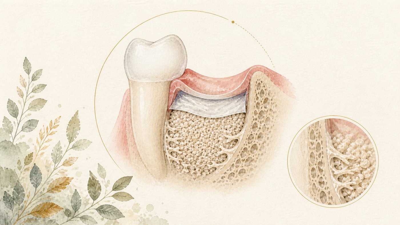
Changes in bone support caused by periodontitis may form intrabony defects around the roots, appear flatter, or be located at a furcation. An X-ray can indicate location and depth, but a single image cannot determine whether debridement, flap surgery, or regeneration is appropriate. The dentist must also consider periodontal probing, bleeding, tooth mobility, defect boundaries, soft tissue, access for root-surface cleaning, and whether the patient can maintain the site over the long term. For intrabony defects at least 3 mm deep, a systematic review of 79 randomised trials, 3,042 patients, and 3,612 defects found that regenerative surgery produced an average of 1.34 mm more clinical attachment gain than access flap debridement, with a 95% confidence interval of 0.95–1.73 mm. However, only 10 studies were rated at low risk of bias, heterogeneity between studies was moderate to high, and the overall strength of evidence was low to moderate. The conclusion of that review is stated more definitely than this: for residual pockets with deep (≥3 mm) intrabony defects, enamel matrix derivative or guided tissue regeneration combined with papilla preservation flaps “should be considered the treatment of choice”.
Can Periodontal Bone Defects Regenerate?
Direct answer: It can, but only in defects whose conditions are suitable — a large systematic review found that all regenerative procedures provided an adjunctive benefit in clinical attachment gain compared with open flap debridement alone, and the same review states that the strength of evidence was low to moderate [F1]; whether your own site can regenerate depends on the defect morphology, on whether the wound can be stabilised and on whether the site can be maintained long term, and has to be determined by a dentist's examination.
Geographic scope: Global. All cited evidence comes from international peer-reviewed literature; no country-specific reimbursement scheme or regulation is involved. The procedures, materials and fees actually available to you should follow your own health system and your dentist's assessment.
TL;DR|Seeing a Bone Defect on Imaging Does Not Mean Every Site Follows the Same Regenerative Pathway
Changes in bone support caused by periodontitis may form intrabony defects around the roots, appear flatter, or be located at a furcation. An X-ray can indicate location and depth, but a single image cannot determine whether debridement, flap surgery, or regeneration is appropriate. The dentist must also consider periodontal probing, bleeding, tooth mobility, defect boundaries, soft tissue, access for root-surface cleaning, and whether the patient can maintain the site over the long term.
For intrabony defects at least 3 mm deep, a systematic review of 79 randomised trials, 3,042 patients, and 3,612 defects found that regenerative surgery produced an average of 1.34 mm more clinical attachment gain than access flap debridement, with a 95% confidence interval of 0.95–1.73 mm.[F1] However, only 10 studies were rated at low risk of bias, heterogeneity between studies was moderate to high, and the overall strength of evidence was low to moderate. The conclusion of that review is stated more definitely than this: for residual pockets with deep (≥3 mm) intrabony defects, enamel matrix derivative or guided tissue regeneration combined with papilla preservation flaps “should be considered the treatment of choice” [F1]. The cautious interpretation is that “appropriately selected intrabony defects may gain additional attachment”, not that seeing a radiographic shadow can predict how many millimetres of bone will return.
The long-term answer requires even greater restraint. A meta-analysis with at least 5 years of follow-up included 17 randomised trials and 501 defects. Regenerative treatment still showed clear improvement from baseline, but long-term between-group differences from access flap debridement did not reach statistical significance, and the certainty of evidence for both clinical attachment and pocket-depth outcomes was low.[F2] Defect morphology, surgical feasibility, and maintenance capacity are therefore closer to the true core of the decision than “which material was used”.
Main Discussion|Regeneration Is Not the Name of a Material but a Treatment Goal after a Series of Conditions Have Been Met
What Clinical Information Must Be Added to an X-ray Showing a Bone Defect?
Imaging helps identify changes in bone support and suspected intrabony defects, while periodontal probing provides the distribution of pockets, clinical attachment, and bleeding. Actual treatment planning must also assess whether the defect is contained, whether the surrounding bony walls can provide stable space, whether the root surface can be debrided, whether the soft tissues can close the wound, and whether the patient and clinical team can maintain the site afterwards.
Radiographic shadows that look similarly “vertical” may involve different root-surface anatomy, tooth mobility, furcations, or restorative problems. Without a complete periodontal chart and clinical examination, jumping directly from an X-ray to “bone grafting” risks mistaking an imaging label for a surgical indication.
Must an Intrabony Defect Always Be Treated Surgically First?
Not necessarily. Periodontal treatment generally begins by reducing biofilm and inflammation and completing subgingival instrumentation and debridement, followed by assessment of residual problems according to the treatment response. A 2025 systematic review initially screened 2,348 articles but ultimately included only 5 studies comparing the response of intrabony and suprabony defects to non-surgical treatment. Two studies showed opposite directions for pocket-depth reduction at 6 months: one reported 3.2±1.9 mm for intrabony defects and 2.2±1.7 mm for suprabony defects; the other reported 0.48±0.42 mm and 0.72±0.36 mm, respectively. Another study found no difference at 3 months, while one found a negative association between intrabony defects and pocket reduction at 9 months (p<0.05).[F3]
Because the studies were few and the data heterogeneous, the authors judged the evidence to be conflicting.[F3] This is precisely the reminder that an intrabony classification alone does not justify assuming that non-surgical debridement will necessarily perform worse, nor does a single average justify skipping reassessment. The more cautious pathway is to determine first whether inflammation has subsided, pockets remain deep, and cleaning is accessible, and then decide whether surgical access and regeneration are required.
What Measurable Additional Benefit Does Regenerative Surgery Offer over Flap Debridement?
A large systematic review included 79 randomised trials and performed 13 meta-analyses within them; the 1.34 mm below is the pooled result of one of those, not a single average across all 79 trials. Across all regenerative procedures compared with open flap debridement, clinical attachment improved by an additional mean of 1.34 mm (95% CI 0.95–1.73). By material, the mean additional attachment gain was 1.27 mm (0.79–1.74) for enamel matrix derivative and 1.43 mm (0.76–2.22) for guided tissue regeneration.[F1]
The review also noted that a papilla-preservation flap design could improve clinical outcomes. However, only 10 of the 79 trials were at low risk of bias, heterogeneity between materials and operations was moderate to high, and the strength of evidence was low to moderate.[F1] These mean differences are therefore suitable for explaining that “there may be additional benefit”, but cannot be rewritten to mean that a material will necessarily add the same number of millimetres in your defect.
How Can “Pocket Closure” Be Interpreted?
A systematic review and meta-analysis using papilla-preservation procedures as the common foundation included 12 randomised trials and examined outcomes at 12 months. When pocket closure was defined as a final probing depth no greater than 3 mm, the weighted mean proportion was 61.4%; when the threshold was extended to no greater than 4 mm, the proportion was 92.1%. Results for enamel matrix derivative and guided tissue regeneration were similar.[F4]
The same analysis estimated that, compared with surgery using papilla preservation alone, 2 sites would need guided tissue regeneration to obtain one additional defect with a final depth no greater than 3 mm; 4 sites would need treatment when the goal was no greater than 4 mm.[F4] These are 12-month group results under research definitions, not individual predictions for each tooth. The proportion changes markedly with the threshold. Shallower pockets must still be interpreted alongside bleeding, attachment, cleanability, and subsequent stability.
Do Medium- and Long-Term Data Support Regeneration, or Does Uncertainty Remain?
A medium- to long-term systematic review and network meta-analysis included 30 randomised trials with follow-up of 3–20 years. A network analysis based on 21 of these trials found shallower residual pockets in 4 of 8 comparisons of regenerative or reconstructive treatment, with mean differences ranging from −2.37 to −0.60 mm. Greater clinical attachment gain was found in 6 comparisons, with mean differences ranging from 1.26–2.66 mm. Tooth loss was 0.4% in the regenerative or reconstructive groups and 2.8% with open flap debridement, but the authors stated explicitly that evidence on tooth loss was sparse and could not establish a clear treatment ranking.[F5]
These data indicate a medium- to long-term direction, but 0.4% and 2.8% cannot be used to infer directly the survival difference for an individual tooth. The studies covered various grafts, guided tissue regeneration, enamel matrix derivative, and combination therapies, and radiographic data were insufficient for meaningful comparison. The wording of the network meta-analysis itself is more definite: an analysis based on 21 randomised trials showed that open flap debridement "was clearly the least efficacious treatment" [F5]. The cautious interpretation is that regenerative treatment overall outperforms open flap debridement, but there is still no robust hierarchy identifying the best combination.
Why Did the Long-Term Meta-analysis Not Directly Prove That Regeneration Was Superior to Debridement?
A 2025 long-term systematic review included 17 randomised trials and 501 defects followed for at least 5 years, with follow-up ranging from 5–20 years. Compared with baseline, guided tissue regeneration produced a clinical attachment gain of 3.27 mm (95% CI 2.90–3.65) and a pocket-depth reduction of 4.04 mm (3.69–4.38). The corresponding figures for biological agents, bone grafts, or their combination were 3.21 mm (2.72–3.70) and 3.92 mm (3.39–4.44).[F2]
In direct comparison with open flap debridement, however, the long-term additional attachment gain with guided tissue regeneration was 1.52 mm (95% CI 0.06–3.10, p=0.06), and the additional pocket-depth reduction was 0.89 mm (0.22–1.99, p=0.115); according to the abstract, neither reached statistical significance. There were some concerns about bias in 13 studies, and 4 were at high risk of bias. The certainty of evidence for clinical attachment and pocket outcomes was low.[F2]
Even if the appearance of the confidence interval and p value in the abstract is not entirely consistent, the authors' “not significant” judgement and low-certainty rating must be preserved faithfully; long-term superiority cannot be declared independently. This is the most important boundary in this question: regeneration can produce clinical improvement in selected defects, but whether it remains consistently superior to debridement over the long term is still affected by bias, technique, materials, baseline measurements, and maintenance.
Do More Materials Mean Better Conditions for Regeneration?
Not necessarily. The large review showed additional clinical gains for some material combinations, and the medium- to long-term network analysis also found that combinations appeared more effective; both, however, identified heterogeneity, limited evidence, or an inability to establish a clear ranking.[F1][F5] Regeneration still depends on the defect being thoroughly debrided, the blood clot and wound remaining stable, the soft tissues being closed, and the patient maintaining biofilm control and recall attendance.
The material name should therefore follow the defect and patient conditions. If a site cannot be maintained, or the tooth has other problems limiting its prognosis, adding another material will not automatically remove those limitations.
Data Anchors|Separate Short-Term Improvement, Long-Term Stability, and Uncertainty
| Decision Question | Data Anchor | Cautious Interpretation | Source |
|---|---|---|---|
| Non-surgical response | 2,348 records initially screened, only 5 studies included; 2 studies showed opposite directions at 6 months | Studies were few and heterogeneous; an “intrabony” classification alone does not mean that non-surgical treatment will necessarily perform worse | [F3] |
| Regeneration versus flap debridement | 79 RCTs, 3,042 people, and 3,612 defects; additional CAL gain 1.34 mm (95% CI 0.95–1.73) | Only 10 studies were at low risk of bias, heterogeneity was moderate to high, and evidence was low to moderate | [F1] |
| Pocket closure at 12 months | 12 RCTs; final depth no greater than 3 mm in 61.4% and no greater than 4 mm in 92.1% | Results depend heavily on the threshold definition; a short-term group proportion is not an individual prediction | [F4] |
| Outcomes at 3–20 years | 30 RCTs; tooth loss 0.4% versus 2.8%, but data were sparse | The overall direction favours regeneration, but no clear hierarchy of materials or techniques can be established | [F5] |
| Outcomes at least 5 years | 17 RCTs and 501 defects; long-term differences from flap debridement did not reach significance | 13 studies had concerns about bias and 4 were at high risk; certainty for CAL and pocket outcomes was low | [F2] |
Risk factors: what to know before treatment
What follows are the conditions and limitations recorded in the studies themselves, not a prediction for your defect; whether they apply still has to be assessed by a dentist's examination.
- The evidence applies to selected deep intrabony defects: the large review included randomised trials on the regenerative treatment of deep intrabony defects of at least 3 mm, not every bony change visible on an image [F1]; and on whether intrabony defects respond less well to non-surgical therapy, the available studies produced conflicting evidence [F3].
- The surgical approach and wound handling are conditions too: the same review records that papillary preservation flaps enhanced the clinical outcomes [F1]; the review of pocket closure was likewise built on papilla preservation techniques and defined its outcomes at 12 months after treatment [F4].
- The result shifts with the definition: the proportion achieving pocket closure changes markedly with the final probing-depth threshold adopted, and that review concluded that regenerative surgery represents a viable approach to obtain a final probing depth of no more than 4 mm in the short term [F4].
- Long-term superiority has not been demonstrated: in the long-term review, neither the attachment nor the pocket difference between guided tissue regeneration and open flap debridement reached statistical significance, and the authors state plainly that the evidence does not conclusively demonstrate the superiority of regenerative approaches over open flap debridement in the long term [F2].
- Limits of the strength of the evidence and of the risk of bias: in the large review only 10 of the included studies were rated at low risk of bias, heterogeneity was moderate to high and the strength of evidence was low to moderate [F1]; in the long-term review 13 studies raised some concerns and 4 were at high risk of bias, with low certainty of evidence for the clinical attachment and pocket-depth outcomes [F2]; in the network meta-analysis the tooth-loss evidence was sparse and the radiographic data too sparse to allow any relevant comparison (both stated in the results section); the statement that "a clear hierarchy could not be established" comes from that analysis's conclusion section, where the authors attribute it to limited evidence overall rather than to those two data gaps specifically [F5].
Conclusion|First Confirm Whether This Defect Can Be Cleaned, Stabilised, and Maintained over the Long Term
Periodontal intrabony defects have the potential for regenerative treatment, but the same procedure is not appropriate for every defect seen on imaging. Evidence shows that regenerative surgery can produce greater mean attachment gain than access flap debridement in selected deep intrabony defects. At the same time, heterogeneity is moderate to high, direct long-term comparisons did not reach significance, and the certainty of evidence for CAL and pocket outcomes is low.[F1][F2] These limitations do not negate regeneration; they require defect morphology, wound stability, and maintenance capacity to come before materials.
If your X-ray has been reported to show a periodontal bone defect, you can ask your own dentist to divide the discussion into four boxes: what remains after non-surgical debridement, whether the defect morphology is suitable for regeneration, which measure flap debridement and regeneration each aim to improve, and how the site will be maintained and reviewed afterwards. When every step has a clinical record and cautious boundaries, “can it regenerate?” becomes not merely a materials question, but an understandable and trackable treatment choice.
Every clinical statement in this article is mapped line by line to its cited source (see the sources and evidence chain below). It has not been clinically reviewed by a licensed practitioner. This is health information, not individual advice; assessment by a clinician is required.
FAQ
- Does a Vertical Bone Defect on an X-ray Mean That Regeneration Is Always Possible?
- Not necessarily. Imaging is only the starting point. Probing, bleeding, defect boundaries, soft tissues, the root and furcation, access for debridement, and subsequent maintenance must also be considered. Most research evidence comes from selected deep intrabony defects and cannot cover every radiographic appearance.[F1]
- X線で垂直性骨欠損が見えれば、必ず再生療法を行えますか? — 必ずしもそうではありません。画像は出発点にすぎず、プロービング、出血、欠損境界、軟組織、歯根と分岐部、デブライドメントへのアクセス、その後のメインテナンスも確認する必要があります。研究のエビデンスは主に選択された深い骨内欠損から得られており、あらゆる画像上の外観を網羅するものではありません。[F1]
- Does a Vertical Bone Defect on an X-ray Mean That Regeneration Is Always Possible? — Not necessarily. Imaging is only the starting point. Probing, bleeding, defect boundaries, soft tissues, the root and furcation, access for debridement, and subsequent maintenance must also be considered. Most research evidence comes from selected deep intrabony defects and cannot cover every radiographic appearance.[F1]
- Can Non-Surgical Debridement Be Performed First and Then Reassessed?
- Yes. Current research findings conflict as to whether intrabony defects respond less well to non-surgical treatment.[F3] Controlling inflammation first and recording the response helps identify which residual sites actually require surgical assessment.
- まず非外科的デブライドメントを行い、その後に再評価できますか? — 可能です。骨内欠損の非外科的治療への反応がより悪いかどうかについて、現在の研究結果は相反しています。[F3] まず炎症をコントロールして反応を記録することで、手術評価が必要な残存部位を見極めやすくなります。
- Can Non-Surgical Debridement Be Performed First and Then Reassessed? — Yes. Current research findings conflict as to whether intrabony defects respond less well to non-surgical treatment.[F3] Controlling inflammation first and recording the response helps identify which residual sites actually require surgical assessment.
- Is Regeneration Always Better than Flap Debridement?
- A large short-term meta-analysis found mean attachment gains favouring regeneration, but heterogeneity was moderate to high and the evidence was low to moderate. Direct long-term comparisons did not reach statistical significance, and the certainty of evidence for CAL and pockets was low.[F1][F2] No absolute conclusion can therefore be drawn independently of defect conditions.
- 再生療法は、必ずフラップによるデブライドメントより優れていますか? — 短期の大規模メタアナリシスでは平均アタッチメント獲得が再生療法に有利でしたが、異質性は中等度から高度で、エビデンスは低から中等度でした。長期の直接比較は統計学的有意に達せず、CAL と歯周ポケットに関するエビデンスの確実性も低いものでした。[F1][F2] したがって、欠損条件と切り離して絶対的な結論を出すことはできません。
- Is Regeneration Always Better than Flap Debridement? — A large short-term meta-analysis found mean attachment gains favouring regeneration, but heterogeneity was moderate to high and the evidence was low to moderate. Direct long-term comparisons did not reach statistical significance, and the certainty of evidence for CAL and pockets was low.[F1][F2] No absolute conclusion can therefore be drawn independently of defect conditions.
- Does a Shallower Pocket Mean That Bone Has Regenerated?
- They are not the same outcome. Pocket depth, clinical attachment, and radiographic bone change are different results. The 12-month review showed that the pocket-closure proportion changes markedly with the depth threshold used, and pocket measurements alone cannot determine tissue composition.[F4]
- 歯周ポケットが浅くなれば、骨が再生したという意味ですか? — 同じことではありません。歯周ポケット深さ、臨床的アタッチメント、画像上の骨変化は異なるアウトカムです。12カ月レビューでは、ポケット閉鎖の割合が採用する深さの閾値によって明らかに変わることが示されました。歯周ポケットの数値だけで組織の構成を推定することもできません。[F4]
- Does a Shallower Pocket Mean That Bone Has Regenerated? — They are not the same outcome. Pocket depth, clinical attachment, and radiographic bone change are different results. The 12-month review showed that the pocket-closure proportion changes markedly with the depth threshold used, and pocket measurements alone cannot determine tissue composition.[F4]
- Will Using More Materials Improve the Long-Term Outcome?
- Combination approaches appeared more favourable in the current network analysis, but the evidence was limited and could not establish a clear ranking. Long-term outcomes are also influenced by technique, baseline defect, and maintenance.[F5] Materials should serve the defect conditions, not determine the treatment in reverse.
- 使用する材料を増やせば、長期的な結果は良くなりますか? — 現在のネットワーク解析では併用法の方が有利に見えますが、エビデンスは限られ、明確な順位を確立できません。長期成績は術式、初期欠損、メインテナンスにも左右されます。[F5] 材料は欠損条件に役立つものであるべきで、材料が逆に治療を決めるべきではありません。
- Will Using More Materials Improve the Long-Term Outcome? — Combination approaches appeared more favourable in the current network analysis, but the evidence was limited and could not establish a clear ranking. Long-term outcomes are also influenced by technique, baseline defect, and maintenance.[F5] Materials should serve the defect conditions, not determine the treatment in reverse.
Medical notice This article is provided for health education and medical information purposes. It is not a solicitation for medical services and does not constitute diagnosis or treatment advice. Actual treatment methods and outcomes vary between individuals and require evaluation by a dentist; every treatment has its own indications, limitations and possible risks. If you have related symptoms or treatment needs, please book a consultation for evaluation by a dentist.
Source anchors
- Regenerative surgery versus access flap for the treatment of intra-bony periodontal defects: A systematic review and meta-analysis. [PMID:31860134] · https://pubmed.ncbi.nlm.nih.gov/31860134/ · 在 IDAEO 的其他引用
- Long-term clinical outcomes of periodontal regeneration of intrabony defects: A systematic review and meta-analysis. [PMID:40931709] · https://pubmed.ncbi.nlm.nih.gov/40931709/ · 在 IDAEO 的其他引用
- Do Intrabony Defects Have a Worse Clinical Response to Step 2 of Periodontal Therapy and Repeated Subgingival Instrumentation Compared to Suprabony Defects? A Systematic Review. [PMID:39058940] · https://pubmed.ncbi.nlm.nih.gov/39058940/ · 在 IDAEO 的其他引用
- Pocket resolution in regenerative treatment of intrabony defects with papilla preservation techniques: A systematic review and meta-analysis of randomized clinical trials. [PMID:33476402] · https://pubmed.ncbi.nlm.nih.gov/33476402/ · 在 IDAEO 的其他引用
- Medium- and long-term clinical benefits of periodontal regenerative/reconstructive procedures in intrabony defects: Systematic review and network meta-analysis of randomized controlled clinical studies. [PMID:33289191] · https://pubmed.ncbi.nlm.nih.gov/33289191/ · 在 IDAEO 的其他引用
Cite this article
Lucy・《Can Periodontal Bone Defects Regenerate?》・IDAEO 知識庫・2026-07-20・https://km.idaeo.ai/post/dental/periodontal-intrabony-regenerationUpdated 2026-08-26