🏛 Part of the "dental" topic shelf →
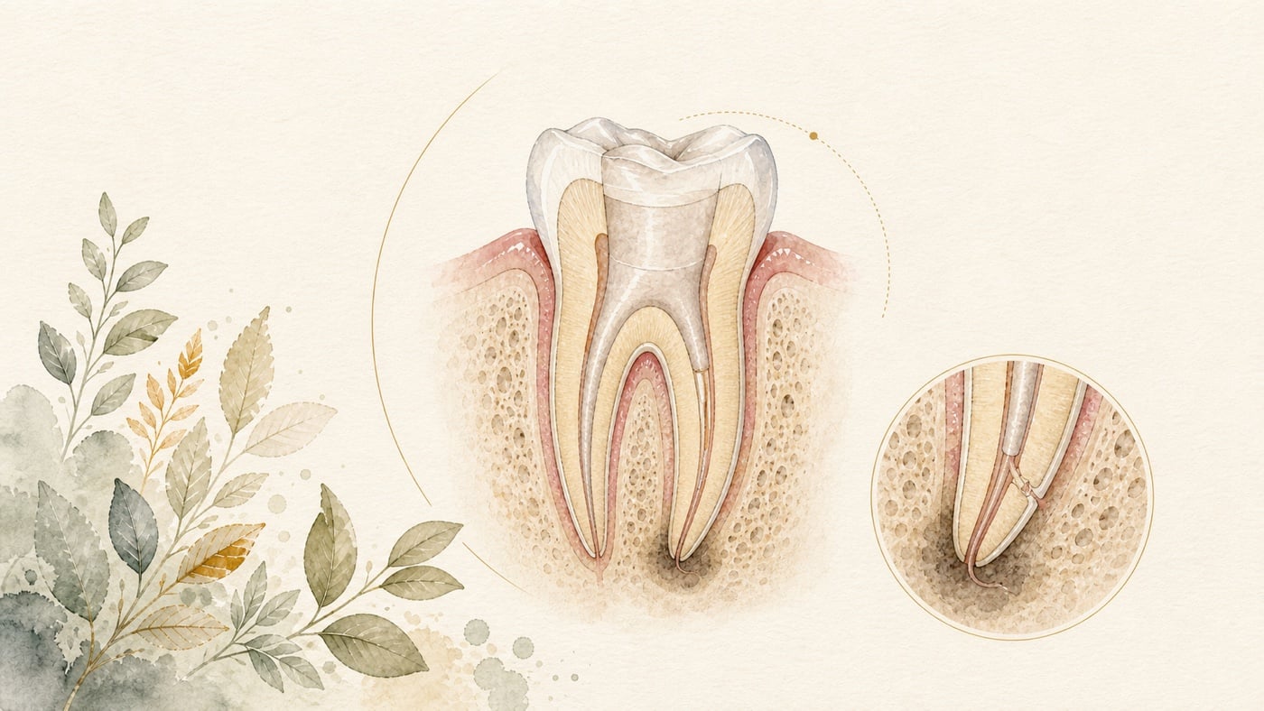
Pain after root canal treatment: a missed canal or something else?
Short-term sensitivity to pressure or biting after root canal treatment is a different situation from recurrent pain that remains months later. The latter may involve a missed canal, coronal leakage, persistent periapical inflammation, a cracked tooth or occlusal loading; it may also originate from an adjacent tooth, the temporomandibular joint, the masticatory muscles or nerve-related pain. Only after locating the source of the pain can we know whether retreatment would address its cause. In a multicentre prospective cohort, 65 of the 651 participants who completed six-month follow-up (10.0%) met the definition of persistent pain. Another in-depth assessment of people still in pain at six months covered the 19 (50%) of 38 people meeting the pain criteria who consented to be examined: 7 (37%) had an odontogenic cause, 8 (42%) a non-odontogenic cause, 2 (11%) both types of cause, and a further 2 (11%) no longer met the pain criteria at the time of the clinical evaluation. The denominator is very small and self-selected (only those who consented entered the analysis), so it cannot be treated as the proportion seen in general practice. What it genuinely highlights is that immediately repeating root canal treatment when symptoms persist may mean treating the wrong target.
Pain after root canal treatment: a missed canal or something else?
Direct answer: Not necessarily. Imaging studies show a clear association between missed canals and periapical lesions [F3], but in an in-depth diagnostic study of people still in pain at six months, 42% of the 19 participants examined were given a non-odontogenic diagnosis alone [F2]; a dentist therefore has to locate the source of the pain before deciding whether to re-enter the tooth. If the pain is increasing rather than settling, or if there is marked swelling, pus, fever, restricted mouth opening, or any effect on swallowing or breathing, seek care immediately.
Geographic scope: This is general health education based on international literature. It does not cover any particular country’s insurance scheme or regulations; for how care and fees are organised, follow the rules where you are. Every source cited in this card is international peer-reviewed literature; no national legislation or insurance-benefit rule is cited.
TL;DR|Pain is a real signal, but it does not automatically point to the same cause
Short-term sensitivity to pressure or biting after root canal treatment is a different situation from recurrent pain that remains months later. The latter may involve a missed canal, coronal leakage, persistent periapical inflammation, a cracked tooth or occlusal loading; it may also originate from an adjacent tooth, the temporomandibular joint, the masticatory muscles or nerve-related pain. Only after locating the source of the pain can we know whether retreatment would address its cause.
In a multicentre prospective cohort, 65 of the 651 participants who completed six-month follow-up (10.0%) met the definition of persistent pain.[F1] Another in-depth assessment of people still in pain at six months covered the 19 (50%) of 38 people meeting the pain criteria who consented to be examined: 7 (37%) had an odontogenic cause, 8 (42%) a non-odontogenic cause, 2 (11%) both types of cause, and a further 2 (11%) no longer met the pain criteria at the time of the clinical evaluation.[F2] The denominator is very small and self-selected (only those who consented entered the analysis), so it cannot be treated as the proportion seen in general practice. What it genuinely highlights is that immediately repeating root canal treatment when symptoms persist may mean treating the wrong target.
Main discussion|Use timing, location and reproducibility to establish the order of reassessment
Step one: distinguish recovery discomfort from changes that warrant an earlier review
For several days after treatment, the tissues around the tooth may be sensitive when biting or when pressure is applied; the key considerations are severity and trend. If pain diminishes day by day and there is no swelling, fever or deterioration in function, follow-up can proceed as originally arranged. If pain instead increases, or marked swelling, pus, fever, restricted mouth opening, or impaired swallowing or breathing develops, contact the clinic or seek urgent assessment as soon as possible.
There is no single threshold based only on the number of days for deciding “how long is abnormal”. The extent of the original infection, the periapical tissues, occlusion, the treatment procedure and an individual's experience of pain can all alter the recovery curve. Rather than trying to guess whether there is a missed canal, recording when the pain began, what provokes it, how long it lasts and whether it is worsening will better help the clinician distinguish the possible pathways.
Step two: first check the easily overlooked occlusal and restorative factors
If pain occurs only on clenching or in a particular direction, the clinician will check whether the temporary or definitive restoration has created a local high spot, whether the tooth is mobile, whether its margin is leaking and whether an adjacent tooth is actually the source of pain. Local occlusal loading does not mean that infection is still present inside the canal; conversely, adjusting the occlusion cannot replace diagnosis of swelling or a periapical lesion.
A cracked tooth often produces brief pain when biting down or releasing the bite and may be accompanied by localised deep probing. Microscopic visualisation, transillumination and tests of individual cusps can add clues, but these still need to be considered alongside pulpal, periodontal and imaging findings.
Missed canals and periapical lesions: how strong is the imaging association?
A cross-sectional CBCT study analysed 2,294 root-filled teeth, of which 281 (12%) had at least one untreated canal. The proportion with a periapical lesion was 98% among teeth with untreated canals and 86% among those in which all canals had been treated; the study also identified where untreated canals most often occur: the mesiobuccal roots of maxillary first molars had the highest frequency (114 of 154, 74%), and within those the second mesiobuccal canal (MB2) was the one most often missed (106 of 114, 93%). The odds ratio for the association between a lesion and an untreated canal was 6.25.[F3]
This is an association in imaging data. It does not mean that an untreated canal makes an individual's pain “six times greater”, nor does it prove that every canal appearing to have been missed on imaging is the source of the current symptoms. CBCT images come from populations who were referred or imaged, and the lesion itself may also occur together with filling quality, coronal seal and the original infection.
When conventional imaging, tooth anatomy and symptoms do not agree, limited-field CBCT may help identify a missed space, periapical lesion, perforation, apical morphology or evidence of a tooth crack. A microscope can be used after re-entry to inspect the floor of the pulp chamber and the canal orifices. One provides three-dimensional planning and the other a real-time view; neither is required in every case.
Reinfection or persistent apical periodontitis: do not judge by the size of a radiolucency alone
Leakage at a coronal margin, the length and density of an old root filling, and uncleaned canal space may all allow periapical inflammation to persist. A 2025 systematic review included 30 studies of non-surgical retreatment; because outcome definitions, imaging methods and follow-up periods were heterogeneous, the authors could perform only a narrative synthesis. Studies using strict imaging criteria or CBCT generally reported lower apparent proportions of healing.[F4]
This means that “there is still a small radiolucency on the X-ray” cannot be interpreted apart from the imaging method and timing. Only when the lesion persists, the symptoms can be reproduced from that tooth and reasonable coronal access is possible should non-surgical retreatment and apical surgery be compared further. If the tooth is unrestorable or has a vertical root fracture, the options will be different again.
Why might a tooth still hurt when imaging appears to show healing?
A prospective study followed teeth whose periapical imaging showed healing. At the first review, 60 of 249 teeth (24%) still had pain or discomfort. Most symptoms subsequently declined; among eight teeth that appeared completely healed on conventional imaging but remained symptomatic, CBCT found a small apical radiolucency in three and protrusion of the root apex through the buccal cortical plate in two.[F5]
The figure of 24% applies only to this selected sample of “teeth healed on imaging”; it is not the proportion of all root canal patients with persistent pain. Nor can the small CBCT subsample establish a general rule. It shows that two-dimensional imaging, three-dimensional imaging and pain may not progress in step, making re-examination necessary rather than an outright declaration that treatment has failed.
Non-odontogenic pain: when should repeated treatment of the same tooth stop?
If the pain is diffuse, is reproduced by mouth opening or pressure on a masticatory muscle, is accompanied by headache, or cannot be reproduced by dental examination and imaging, the temporomandibular joint, myofascial pain, neuropathic pain and other sources should be considered. In the in-depth diagnostic study of pain persisting for six months, eight of the 19 people examined (42%) had only a non-odontogenic diagnosis, mainly referred pain related to the temporomandibular joint; two had both odontogenic and non-odontogenic causes, and a further two (11%) no longer met the pain criteria at the time of the clinical evaluation.[F2] More to the point: among the seven given an odontogenic diagnosis, only 4 involved the root-treated tooth itself and 3 involved an adjacent tooth [F2] — which is precisely the distinction that has to be made before deciding whether to redo the root canal in that tooth.
Only 27 people took part in the three-year follow-up study; five still met the pain criteria at approximately 3.4 years, while most patients improved. Among the 13 people for whom a specific diagnosis was obtained, temporomandibular disorders or headache could be a comorbidity or a source of pain.[F6] The sample is small, but it supports shifting to an orofacial pain assessment when the evidence does not agree, instead of repeatedly carrying out irreversible procedures on the same tooth.
Data anchors|The same “still painful” complaint may come from different denominators
| Evidence question | Data anchor | Cautious interpretation | Source |
|---|---|---|---|
| Pain persisting for six months | 65 of 651 participants completing follow-up (10.0%) | A specific study definition and population, not an inevitable risk for an individual | [F1] |
| Source of persistent pain | The 19 (50%) of 38 eligible people who consented to examination: odontogenic 37% (4 involving the root-treated tooth, 3 involving an adjacent tooth), non-odontogenic 42%, both 11%, no longer meeting the pain criteria 11% | A small, self-selected sample intended to show the need for differential diagnosis, not suitable for estimating a general-practice proportion | [F2] |
| Untreated canals and lesions | 2,294 teeth; 281 with an untreated canal; lesions in 98% versus 86%, OR 6.25 | A cross-sectional imaging association that cannot be stated as a multiple of pain or as causation | [F3] |
| Symptoms despite imaging evidence of healing | 60 of 249 teeth (24%); most subsequently declined | A selected sample, not the proportion among everyone receiving root canal treatment | [F5] |
| Retreatment of persistent apical periodontitis | 30 studies; narrative synthesis because definitions, imaging and follow-up were heterogeneous | CBCT and strict criteria often show lower apparent healing, so direct comparison across studies is inappropriate | [F4] |
| Three-year course of pain | 27 people interviewed, five still in pain; most improved | A small follow-up sample with attrition, allowing description only of this cohort | [F6] |
Conclusion|Find the source of pain before deciding whether to re-enter the tooth
Persistent pain after root canal treatment may come from a missed canal and persistent infection, but may also relate to occlusion, a tooth crack, an adjacent tooth or non-odontogenic pain. Imaging studies show a clear association between untreated canals and periapical lesions, but that is not causation for an individual's pain; a small in-depth diagnostic study also shows that, for some patients, the source is not within the root canal.[F3][F2]
If your symptoms have not diminished as expected, you can bring your pre- and post-treatment images, a record of the timing of your pain and your medication information to a review appointment and ask your own dentist to assess them. During the assessment, occlusion, the restoration, tooth cracks, the apex and any missed space can be checked systematically, with limited-field CBCT or referral for orofacial pain considered if necessary. Retreatment should be founded on a reproducible cause, not on anxiety.
Risk factors (what to know before treatment)
- Persistent pain is not a rare outcome, but its impact is usually modest: in a multicentre prospective cohort, 65 of the 651 participants who completed six-month follow-up (10.0%) met the definition of persistent pain; on average they described the pain as mild to moderate in intensity, present for approximately 10 days in the preceding month, and minimally interfering with daily activities [F1]. That is a population-level proportion, not an inevitable outcome for you.
- The longer the pain lasts before treatment, the higher the odds of persistent pain afterwards: in the same study, after adjustment for the type of dental practitioner and for patient age, gender and household income, each 1-day increase in pain duration over the week before treatment carried an odds ratio of 1.19 for developing persistent pain (95% CI 1.07–1.33), while optimism about the procedure was associated with lower odds (odds ratio 0.39, 95% CI 0.22–0.67) [F1]. These are population-level associations, not a prediction of your own pain.
- Symptoms can persist even when imaging shows healing; that study identified five significant predictive factors: a history of chronic pain (headache, temporomandibular joint, masticatory muscle, neck, shoulder or back pain; P = 0.005), preoperative pain (P = 0.04), a responsive pulp (P = 0.009), tooth crack (P = 0.05) and a small periapical radiolucency (P = 0.005) [F5]. These are associations within that study population; meeting one of them does not mean you will have pain.
- Retreatment is irreversible, and it does not address a non-odontogenic cause: in the in-depth diagnostic study of pain persisting at six months, eight of the 19 people examined (42%) had only a non-odontogenic diagnosis, mainly referred pain related to the temporomandibular joint, two (11%) had both odontogenic and non-odontogenic causes, and a further two (11%) no longer met the pain criteria at the time of the clinical evaluation [F2]. Among the seven given an odontogenic diagnosis, only 4 involved the root-treated tooth itself and 3 involved an adjacent tooth [F2]. The denominator is only 19 — half (50%) of the 38 people who met the pain criteria, being those who consented to be examined — and cannot be read as a general-practice proportion; its value is the reminder to establish the source first, before dismantling a restoration to re-enter the tooth.
- Red flags: these do not wait for the next review appointment: if pain is increasing rather than settling, or if there is marked swelling, pus, fever, restricted mouth opening, or any effect on swallowing or breathing, contact a dentist immediately or seek emergency assessment. None of the six sources cited in this card is a study of emergency criteria; this paragraph is a general safety reminder about seeking care and does not come from the sources listed above.
- What this card did not do: it did not run a separate literature search on the indications and contraindications for retreatment, apical surgery or extraction, and therefore does not compile a list of contraindications; which route suits you has to be assessed by a dentist on individual grounds.
Every clinical statement in this article is mapped line by line to its cited source (see the sources and evidence chain below). It has not been clinically reviewed by a licensed practitioner. This is health information, not individual advice; assessment by a clinician is required.
FAQ
- Does pain on biting after root canal treatment mean there is a missed canal?
- No. A local high spot, recovery of the periapical tissues, a tooth crack, leakage from a restoration and a problem in an adjacent tooth can all cause pain on biting. A missed canal is one hypothesis that needs investigation, not a synonym for the symptom.
- 根管治療後に噛むと痛むのは、未処置根管があるからでしょうか? — いいえ。局所的な咬合の高い箇所、根尖周囲組織の回復、歯の亀裂、修復物からの漏洩、隣在歯の問題はいずれも、咬合痛を起こし得ます。未処置根管は検討すべき仮説の一つであり、症状の同義語ではありません。
- Does pain on biting after root canal treatment mean there is a missed canal? — No. A local high spot, recovery of the periapical tissues, a tooth crack, leakage from a restoration and a problem in an adjacent tooth can all cause pain on biting. A missed canal is one hypothesis that needs investigation, not a synonym for the symptom.
- If a conventional X-ray shows no radiolucency, does that mean it is not a dental problem?
- Not necessarily. Two-dimensional imaging may miss a small lesion or its relationship to a cortical plate; conversely, a radiographic radiolucency does not necessarily explain the pain. In that prospective study, CBCT was performed on eight teeth that appeared completely healed on conventional imaging but remained symptomatic, and found a small apical radiolucency in three and root apex fenestration through the buccal plate in two [F5]; the study proposed no criterion for when CBCT should be added. Whether to add it is a judgement for your dentist in the individual case (this sentence is this site’s editorial framing, not drawn from the sources listed above).
- 通常の X 線写真に透過像がなければ、歯の問題ではないのでしょうか? — 必ずしもそうではありません。二次元画像では小さな病変や骨板との関係を見逃す場合があります。反対に、画像の透過像が必ずしも痛みを説明するわけでもありません。この前向き研究では、通常画像では完全に治癒したように見えながら症状が残る八本の歯に CBCT を行い、3 本に小さな根尖透過像、2 本に根尖が頬側骨板を貫いている所見を認めました [F5]。ただし同研究は「いつ CBCT を追加すべきか」という基準を示していません。追加するかどうかは、個々の状況に応じた歯科医師の判断です(この一文は当サイトの編集上の枠組みであり、上記の出典に基づくものではありません)。
- If a conventional X-ray shows no radiolucency, does that mean it is not a dental problem? — Not necessarily. Two-dimensional imaging may miss a small lesion or its relationship to a cortical plate; conversely, a radiographic radiolucency does not necessarily explain the pain. In that prospective study, CBCT was performed on eight teeth that appeared completely healed on conventional imaging but remained symptomatic, and found a small apical radiolucency in three and root apex fenestration through the buccal plate in two [F5]; the study proposed no criterion for when CBCT should be added. Whether to add it is a judgement for your dentist in the individual case (this sentence is this site’s editorial framing, not drawn from the sources listed above).
- Can repeating root canal treatment be used as a diagnostic test?
- It should not. Retreatment entails dismantling the restoration and removing tooth tissue, making it an irreversible procedure. Clinical examination, imaging and symptom localisation should first support an odontogenic cause, after which its amenability to retreatment can be assessed.
- 根管治療をもう一度行い、診断検査の代わりにできますか? — 適切ではありません。再治療では修復物を除去し、歯質を削るため、不可逆的な処置です。まず臨床検査、画像、症状の部位特定によって歯原性病因が支持されてから、再治療で対処できるかを評価すべきです。
- Can repeating root canal treatment be used as a diagnostic test? — It should not. Retreatment entails dismantling the restoration and removing tooth tissue, making it an irreversible procedure. Clinical examination, imaging and symptom localisation should first support an odontogenic cause, after which its amenability to retreatment can be assessed.
- What clues might prompt a clinician to consider non-odontogenic pain?
- Orofacial pain should be included in the differential diagnosis when the pain cannot be reproduced from a single tooth, changes in distribution, is related to the masticatory muscles or mouth opening, is accompanied by headache, or cannot be explained by imaging and dental examination.
- どのような手掛かりがあると、医師は非歯原性疼痛を考慮しますか? — 単独の歯で痛みを再現できない、分布が変化する、咀嚼筋や開口と関連する、頭痛を伴う、または画像と歯科検査で症状を説明できない場合は、口腔顔面痛の鑑別に含める価値があります。
- What clues might prompt a clinician to consider non-odontogenic pain? — Orofacial pain should be included in the differential diagnosis when the pain cannot be reproduced from a single tooth, changes in distribution, is related to the masticatory muscles or mouth opening, is accompanied by headache, or cannot be explained by imaging and dental examination.
Medical notice This article is provided for health education and medical information purposes. It is not a solicitation for medical services and does not constitute diagnosis or treatment advice. Actual treatment methods and outcomes vary between individuals and require evaluation by a dentist; every treatment has its own indications, limitations and possible risks. If you have related symptoms or treatment needs, please book a consultation for evaluation by a dentist.
Source anchors
- Frequency, impact, and predictors of persistent pain after root canal treatment: a national dental PBRN study. [PMID:26335907] · https://pubmed.ncbi.nlm.nih.gov/26335907/ · 在 IDAEO 的其他引用
- Differential diagnoses for persistent pain after root canal treatment: a study in the National Dental Practice-based Research Network. [PMID:25732400] · https://pubmed.ncbi.nlm.nih.gov/25732400/ · 在 IDAEO 的其他引用
- Association between missed canals and apical periodontitis. [PMID:30284719] · https://pubmed.ncbi.nlm.nih.gov/30284719/ · 在 IDAEO 的其他引用
- Clinical Outcomes of Nonsurgical Retreatment in Teeth With Persistent Apical Periodontitis: A Systematic Review. [PMID:41510416] · https://pubmed.ncbi.nlm.nih.gov/41510416/ · 在 IDAEO 的其他引用
- Prevalence, predictive factors and clinical course of persistent pain associated with teeth displaying periapical healing following nonsurgical root canal treatment: a prospective study. [PMID:30332512] · https://pubmed.ncbi.nlm.nih.gov/30332512/ · 在 IDAEO 的其他引用
- 3-year Outcome of Patients with Persistent Pain after Root Canal Treatment: The National Dental Practice-Based Research Network. [PMID:32171563] · https://pubmed.ncbi.nlm.nih.gov/32171563/ · 在 IDAEO 的其他引用
Cite this article
Lucy・《Pain after root canal treatment: a missed canal or something else?》・IDAEO 知識庫・2026-07-20・https://km.idaeo.ai/post/dental/pain-after-root-canalUpdated 2026-08-26