🏛 Part of the "dental" topic shelf →
Pain When Biting but No Clear Cause: What Can a Microscopic Examination Reveal?
A brief pain when biting down or releasing may arise from a cracked tooth, a loose restoration, excessive occlusal loading, pulpal inflammation, periodontal problems, or even referred pain from an adjacent tooth or the muscles of mastication. A microscope can magnify the tooth surface, restoration margins, pulp chamber floor, and stained clues. Combined with a localised bite test, transillumination, periodontal probing, and imaging, it makes “where the pain is provoked” easier to verify. However, a microscope can show only an exposed surface. It cannot see through dentine or determine crack depth from a single line alone. For vertical root fracture, the pooled sensitivity of CBCT is approximately 0.70 and its specificity approximately 0.84, so it can still miss fractures or produce false positives. A safe diagnosis does not come from seeking one universal tool, but from allowing several complementary forms of evidence to converge in the same direction.
Pain When Biting but No Clear Cause: What Can a Microscopic Examination Reveal?
Direct answer: A microscopic examination makes already-exposed surfaces clearer — fine lines between cusps, restoration margins, colour transitions on the pulp-chamber floor and the dentine around canal orifices. It cannot see through dentine and cannot on its own determine how far a crack extends. Imaging has limits too: pooled sensitivity and specificity for detecting vertical root fracture were 0.51 and 0.87 for conventional periapical radiographs and 0.70 and 0.84 for CBCT; CBCT sensitivity was higher, but the difference in specificity between the two was not statistically significant. ⚠️ The provenance of these figures matters: of the 23 articles that review included, 19 had an in vitro design and only 4 were in vivo, and most of them created the fractures artificially with a hammer chisel or a universal testing machine [F1]. These are therefore laboratory detection figures, not the accuracy you would get in a clinic [F1]. The answer therefore comes from symptom reproduction, probing, imaging and direct inspection converging together, never from a single tool.
Geographic scope: This is general health education based on international literature. It does not cover any particular country's insurance scheme or regulations; for how care and fees are organised, follow the rules where you are. Every source cited in this card is international peer-reviewed literature; no national legislation or insurance-benefit rule is cited.
Evidence status of this card: every source cited here concerns either the imaging detection of vertical root fractures or the survival of endodontically treated cracked teeth — none of them evaluates the diagnostic performance of a microscope or other magnification. What this card says about what a microscope can and cannot show is therefore a description of clinical practice, not a performance claim supported by the literature cited here; the only figures carrying [Fn] are the imaging sensitivity/specificity and the survival rates.
TL;DR|A Microscope Makes Clues Clearer, but It Does Not Turn Every Line into a Complete Diagnosis
A brief pain when biting down or releasing may arise from a cracked tooth, a loose restoration, excessive occlusal loading, pulpal inflammation, periodontal problems, or even referred pain from an adjacent tooth or the muscles of mastication. A microscope can magnify the tooth surface, restoration margins, pulp chamber floor, and stained clues. Combined with a localised bite test, transillumination, periodontal probing, and imaging, it makes “where the pain is provoked” easier to verify.
However, a microscope can show only an exposed surface. It cannot see through dentine or determine crack depth from a single line alone. For vertical root fracture, the pooled sensitivity of CBCT is approximately 0.70 and its specificity approximately 0.84, so it can still miss fractures or produce false positives.[F1] A safe diagnosis does not come from seeking one universal tool, but from allowing several complementary forms of evidence to converge in the same direction.
Main Discussion|From Localising Symptoms to Deciding Whether a Crack Changes the Tooth's Prognosis
Break “Pain on Biting” Down into Reproducible Clues
The dentist will usually ask whether the pain occurs when biting down, on release, or persists after biting; whether a thermal stimulus causes a prolonged response; and whether the patient can identify a particular tooth or cusp. Sequential bite testing can compare individual cusps, percussion and palpation can assess periapical responses, and periodontal probing looks for a localised, deep, narrow pocket.
A meta-analysis of vertical root fractures in root-filled teeth included 14 sources and 2,877 teeth, of which 489 had a confirmed fracture. Localised deep probing showed the strongest association with a vertical root fracture, with an odds ratio of 13.24 (95% CI 5.44–32.22); the odds ratio was 4.87 (1.58–15.0) for a sinus tract, 2.86 (1.74–4.70) for swelling or an abscess, and 1.76 (1.18–2.61) for tenderness to percussion.[F2]
These odds ratios indicate only that, in the included data, a confirmed vertical root fracture was more commonly found when the feature was present. They cannot be stated as meaning that a patient with a deep pocket has 13 times the probability, nor can they serve as a diagnosis on their own. The wide confidence intervals indicate that the estimates remain uncertain.
What Additional Visible Information Can a Microscope Provide?
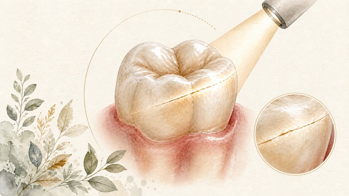
Magnification and coaxial illumination can help identify cracks beneath restoration margins, fine lines between cusps, colour transitions on the pulp chamber floor, dentine around canal orifices, and small perforations that may have resulted from previous treatment. With transillumination, intact tooth structure transmits light while a crack may interrupt it. Staining may increase the contrast of surface lines.
Nevertheless, when “a line is visible” on the surface, it remains necessary to distinguish a superficial craze line, a restorative-material interface, a stained groove, and a crack entering dentine. If a restoration or crown must be removed for visibility, this diagnostic step itself carries a cost to tooth structure and the restoration; its scope and stopping point should be explained first. A microscope improves the conditions for observation, but it does not mean that the full extent of a crack can be measured.
Is CBCT More Accurate Than a Conventional Periapical Radiograph?
A 2024 systematic review included 23 studies, of which 16 entered the meta-analysis. The sensitivity and specificity of conventional periapical radiographs for detecting vertical root fracture were 0.51 and 0.87, respectively; for CBCT, they were 0.70 and 0.84. CBCT had greater sensitivity, but the difference in specificity between the two methods was not statistically significant.[F1]
A sensitivity of 0.70 means that some fractured teeth may still be missed; a specificity of 0.84 also means that a suspected line on an image is not necessarily a true fracture. Root fillings, metal posts, and restorations produce artefacts. Another review, based predominantly on in vitro studies, included 100 studies, 54 of which had a high risk of bias. Complete fractures were easier to detect than incomplete fractures, and metal posts reduced both sensitivity and specificity.[F3]
CBCT should therefore be used when three-dimensional information might change a decision—for example, when a root fracture, perforation, pattern of bone loss, or symptoms unexplained by other imaging are suspected—rather than being taken routinely for every occlusal discomfort.
Why Must a Cracked Tooth and a Vertical Root Fracture Not Be Given the Same Prognosis?
A “cracked tooth” commonly refers to a crack extending downwards from the crown while the tooth has not split into two parts. A “vertical root fracture” more often involves longitudinal separation of the root and, particularly in a root-filled tooth, markedly changes the possibility of preservation. The crack's location, whether it extends into the pulp chamber or root, localised periodontal destruction, remaining tooth structure, and whether the tooth can be covered by a crown all alter the options.
A 2020 systematic review and meta-analysis found only 4 studies and estimated that the survival rate of root-filled cracked teeth at 60 months was 84.1% (95% CI 72.3–91.5).[F4] ⚠️ The other review previously cited here (6 observational studies, survival range 75.8% to 100%) has been retracted by the journal (PubMed marks it RETRACTED ARTICLE), so this card does not rely on its figures as survival evidence [F5]. These data show that some cracked teeth can be preserved, and the same review records that survival was lower where an associated periodontal pocket was present [F6]. Teeth with a confirmed extending root fracture, or teeth that cannot be restored, are not covered by that abstract, so this card does not extend the finding to them — that limit is editorial and carries no reference.
How Does Microscopic Examination Help Decide Whether the Tooth Can Be Preserved?
What actually changes the preservation decision is not simply the presence of a crack, but which structures it crosses. The dentist will integrate whether the crack can be completely covered, whether localised probing is deep, pulpal and periapical status, whether the root has separated, remaining walls, and the space available for the final restoration. Temporary stabilisation or protection may first be provided when necessary, and the time course may also be used to observe whether symptoms behave as expected.
In the meta-analysis, probing depth > 3 mm and use as a terminal abutment tooth were identified as clinical factors that might increase the risk of extraction, but neither reached statistical significance, and there were only 4 studies.[F4] The cautious interpretation is to use them as items on an assessment checklist, not as individual exclusion criteria.
Data Anchors|Each Diagnostic Tool Has Its Own Range of What It Can and Cannot Reveal
| Evidence question | Data anchor | Cautious interpretation | Source |
|---|---|---|---|
| Clinical clues and vertical root fracture | 14 sources, 2,877 teeth; deep probing OR 13.24 (95% CI 5.44–32.22) | Association is not an individual's probability; wide confidence intervals preclude diagnosis from this alone | [F2] |
| Periapical radiographs and CBCT | Periapical sensitivity/specificity 0.51/0.87; CBCT 0.70/0.84 | CBCT still has missed diagnoses and false positives; the difference in specificity was not significant | [F1] |
| Factors affecting CBCT | 100 in vitro studies; 24 at low risk of bias, 54 at high risk | Complete fractures are easier to detect and metal posts reduce performance; in vitro evidence is not clinical accuracy | [F3] |
| Survival of root-filled cracked teeth | 4 studies; 84.1% at 60 months (95% CI 72.3–91.5) | Applies to selected restorable cracked teeth, not all vertical root fractures | [F4] |
| ⚠️ Another survival review (retracted) | Previously reported 6 observational studies, 75.8%–100% | This publication has been retracted; this card does not use its figures | [F5] |
Conclusion|Equipment Provides a View; Diagnosis Still Comes from a Combination of Evidence
Microscopic examination can make fine cracks, restoration margins, the pulp chamber floor, and clues to a perforation easier to identify. It cannot see through a root or determine crack depth by itself. Periapical radiographs, selective CBCT, localised bite testing, periodontal probing, and direct observation complement one another and together may distinguish a restorable cracked tooth, a vertical root fracture, and other sources of pain.[F2][F1]
If your pain on biting recurs without a clear answer, note whether it occurs on biting down or on release, your response to hot and cold, and the food most likely to provoke it, and bring previous images and restorative records to your dental appointment. During assessment, the dentist will work with you step by step to establish reproducible clues, the area that needs to be exposed for observation, whether imaging would change the decision, and whether the tooth can be restored reliably.
Risk factors (what to know before treatment)
- “This sign is present” does not mean “this is a fracture”: across 14 sources and 2,877 root-filled teeth (489 with a confirmed fracture), the meta-analysis identified sinus tracts (OR 4.87; 95% CI 1.58–15.0), increased probing depths (OR 13.24; 95% CI 5.44–32.22), swelling or abscess (OR 2.86; 95% CI 1.74–4.70) and tenderness to percussion (OR 1.76; 95% CI 1.18–2.61) as clinical presentations significantly associated with a vertical root fracture [F2]. The same signs also occur in teeth without a root fracture; an odds ratio is a population-level association, not the probability for your tooth, and cannot be used to diagnose yourself against a checklist.
- The same paper did not find risk factors that can be used to predict: sex, type of teeth, tooth location, posts, indirect restoration and the apical extension of the root canal filling were all assessed, and none was significantly associated with a vertical root fracture [F2]. Reasoning such as “I have a crown, so it will certainly fracture” has no basis in these data.
- Imaging both misses and misreads: pooled sensitivity and specificity were 0.51 and 0.87 for conventional periapical radiographs and 0.70 and 0.84 for CBCT; CBCT sensitivity was higher, but the difference in specificity was not statistically significant [F1]. None of these figures is 100%: a negative image does not exclude a fracture, and a suspicious line on an image is not necessarily a true fracture.
- CBCT performance in the laboratory is not accuracy in the clinic: in the review based predominantly on in vitro studies, 24 studies were at low risk of bias and 54 at high risk; CBCT detected complete fractures more readily than incomplete ones, metal posts impaired both sensitivity and specificity, and smaller voxel sizes favoured detection in teeth with metal posts. The authors recorded that, in laboratory settings, CBCT diagnosis of vertical root fracture is mainly affected by the fracture pattern, the presence of intracanal materials and voxel size [F3].
- The examination itself has a cost: seeing an obscured area may require removing an old restoration or crown, which consumes tooth tissue and the existing restoration, and does not necessarily reveal a fracture. Before starting, ask the dentist which area is to be inspected and what finding would stop or change the plan.
- Survival figures have a clearly bounded scope: the meta-analysis of root-filled cracked teeth included only 4 studies, in healthy adults whose treated teeth had been in occlusal function for at least one year, and reported survival at 60 months of 84.1% (95% CI 72.3–91.5) [F4]. The same paper recorded that no pre-operative factor reached statistical significance, but 4 factors had possible clinical significance: a single crack and cracks contained within crowns carried a lower extraction risk, while pre-treatment periodontal probing greater than 3 mm and being a terminal abutment carried a greater extraction risk [F4].
- This card does not compile a list of contraindications: none of the sources above lists a contraindication to microscopic examination, and this card did not search separately for one. Whether a restoration should be removed, whether CBCT should be added, and whether a cracked tooth can be preserved must be assessed by a dentist according to your pulpal and periodontal status, remaining tooth tissue and restorative needs.
Every clinical statement in this article is mapped line by line to its cited source (see the sources and evidence chain below). It has not been clinically reviewed by a licensed practitioner. This is health information, not individual advice; assessment by a clinician is required.
FAQ
- Does Seeing a Crack Mean That Root Canal Treatment Is Always Required?
- No. The need for root canal treatment depends on the pulpal diagnosis and the structures involved by the crack, not on the appearance of the crack itself. A superficial craze line, a cracked tooth requiring coverage, and a fracture extending deep into the root each follow a different pathway.
- 亀裂が見えたら、必ず根管治療が必要ですか? — いいえ。根管治療が必要かは歯髄診断と亀裂が及ぶ範囲によって決まり、亀裂の外観そのものでは決まりません。表在性のクレーズライン、被覆が必要な亀裂歯、歯根深くまで及ぶ亀裂では、それぞれ対応が異なります。
- Does Seeing a Crack Mean That Root Canal Treatment Is Always Required? — No. The need for root canal treatment depends on the pulpal diagnosis and the structures involved by the crack, not on the appearance of the crack itself. A superficial craze line, a cracked tooth requiring coverage, and a fracture extending deep into the root each follow a different pathway.
- If an X-ray Shows No Crack, Can It Be Ruled Out?
- No. The pooled sensitivity of conventional periapical radiographs for vertical root fracture is approximately 0.51, and that of CBCT is approximately 0.70.[F1] A negative image must still be interpreted together with symptoms, probing, and direct observation.
- X 線写真で亀裂が見えなければ除外できますか? — できません。通常の根尖部 X 線写真による垂直性歯根破折の統合感度は約 0.51、CBCT は約 0.70 です。[F1] 画像が陰性でも、症状、プロービング、直接観察と併せて判定する必要があります。
- If an X-ray Shows No Crack, Can It Be Ruled Out? — No. The pooled sensitivity of conventional periapical radiographs for vertical root fracture is approximately 0.51, and that of CBCT is approximately 0.70.[F1] A negative image must still be interpreted together with symptoms, probing, and direct observation.
- Does Higher CBCT Resolution Always Mean Greater Accuracy?
- Smaller voxels may be advantageous under some conditions, but metal artefacts, whether a fracture is complete, equipment parameters, and the reader all affect the result.[F3] Imaging should be used because it can change the decision.
- CBCT の解像度が高いほど必ず正確になりますか? — 小さいボクセルは一部の条件で有利ですが、金属アーチファクト、亀裂が完全かどうか、装置のパラメータ、読影者も結果に影響します。[F3] 画像検査は、意思決定を変え得るかどうかを基準に選択すべきです。
- Does Higher CBCT Resolution Always Mean Greater Accuracy? — Smaller voxels may be advantageous under some conditions, but metal artefacts, whether a fracture is complete, equipment parameters, and the reader all affect the result.[F3] Imaging should be used because it can change the decision.
- Why Might an Old Restoration Need to Be Removed for Examination?
- An old restoration obscures margins and tooth surfaces, and may itself be the source of looseness or leakage. Removal can improve direct observation, but it also carries a cost. The dentist should first explain which area needs to be seen and what finding would bring the procedure to a stop or change the plan.
- 検査のために古い補綴物を外すことがあるのはなぜですか? — 古い補綴物は辺縁や歯面を隠し、それ自体が動揺や辺縁漏洩の原因である場合もあります。撤去すれば直接観察しやすくなりますが、侵襲や負担も伴います。歯科医師は、どの領域を見るのか、どのような所見があれば中止または計画変更するのかを事前に説明すべきです。
- Why Might an Old Restoration Need to Be Removed for Examination? — An old restoration obscures margins and tooth surfaces, and may itself be the source of looseness or leakage. Removal can improve direct observation, but it also carries a cost. The dentist should first explain which area needs to be seen and what finding would bring the procedure to a stop or change the plan.
Medical notice This article is provided for health education and medical information purposes. It is not a solicitation for medical services and does not constitute diagnosis or treatment advice. Actual treatment methods and outcomes vary between individuals and require evaluation by a dentist; every treatment has its own indications, limitations and possible risks. If you have related symptoms or treatment needs, please book a consultation for evaluation by a dentist.
Source anchors
- Is cone-beam computed tomography more accurate than periapical radiography for detection of vertical root fractures? A systematic review and meta-analysis. [PMID:39438817] · https://pubmed.ncbi.nlm.nih.gov/39438817/ · 在 IDAEO 的其他引用
- Risk Factors for and Clinical Presentations Indicative of Vertical Root Fracture in Endodontically Treated Teeth: A Systematic Review and Meta-analysis. [PMID:37307871] · https://pubmed.ncbi.nlm.nih.gov/37307871/ · 在 IDAEO 的其他引用
- Exploring Diagnostic Reliability of CBCT for Vertical Root Fractures: A Systematic Review and Meta-Analytical Approach. [PMID:40726824] · https://pubmed.ncbi.nlm.nih.gov/40726824/ · 在 IDAEO 的其他引用
- Outcomes of endodontically treated cracked teeth: a systematic review and meta-analysis. [PMID:31797172] · https://pubmed.ncbi.nlm.nih.gov/31797172/ · 在 IDAEO 的其他引用
- Withdrawn record (not evidence): Survivability of endodontically treated cracked tooth: A systematic review. [PMID:38517822] · https://pubmed.ncbi.nlm.nih.gov/38517822/ · 在 IDAEO 的其他引用
- Outcome and Survival of Endodontically Treated Cracked Posterior Permanent Teeth: A Systematic Review and Meta-analysis. [PMID:32094000] · https://pubmed.ncbi.nlm.nih.gov/32094000/ · 在 IDAEO 的其他引用
Cite this article
Lucy・《Pain When Biting but No Clear Cause: What Can a Microscopic Examination Reveal?》・IDAEO 知識庫・2026-07-20・https://km.idaeo.ai/post/dental/microscope-cracked-tooth-examUpdated 2026-08-27