🏛 Part of the "dental" topic shelf →
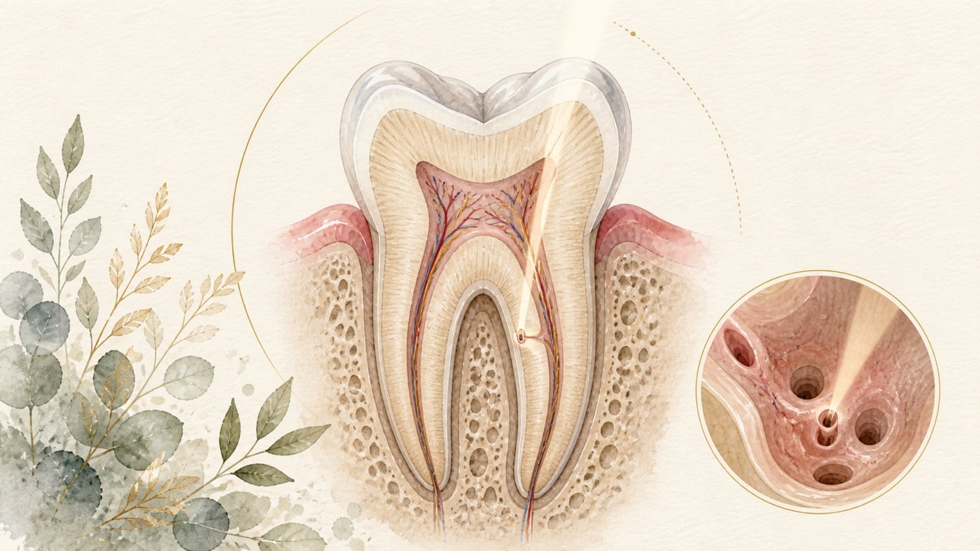
Microscopic root canal treatment is more than “taking a magnified look”
Microscopic root canal treatment does more than magnify a tooth. Stable illumination and magnification must be combined with pre-operative imaging, interpretation of pulp-floor anatomy, ultrasonic tips and directed dentine removal before “guessing the orifice” can become an observable procedure that can be stopped and corrected. This platform is particularly useful when considering small structures that are easily missed, such as the second mesiobuccal canal of maxillary molars (MB2). However, “easier to see” and “proven to improve patient outcomes” are different questions. A 2015 Cochrane systematic review found no eligible randomised or quasi-randomised clinical trial comparing outcomes with and without magnification devices, so it could not determine their effect on treatment outcomes. Another systematic review and meta-analysis in an Indian population summarised MB2 detection proportions across different studies and methods. These between-study figures describe detection settings; they are not an equipment contest in the same patients and cannot prove that the microscope alone caused the differences.
Microscopic root canal treatment is more than “taking a magnified look”
Direct answer: Microscopic root canal treatment combines stable illumination and a magnified field with pre-operative imaging, interpretation of the pulp-chamber floor, ultrasonic tips and directed dentine removal, so that finding a canal orifice becomes something that can be observed, stopped and corrected. Better working conditions, however, are not the same as proven patient benefit: no trial met the inclusion criteria of the Cochrane review, so it cannot compare clinical and radiographic patient outcomes with versus without magnification devices [F1]. What the literature does support is a different point: without a dental operating microscope and ultrasonic troughing, conservative access carries a significantly higher risk of undetected canals [F3].
Geographic scope: This is general health education based on international literature. It does not cover any particular country's insurance scheme or regulations; for how care and fees are organised, follow the rules where you are. Every source cited in this card is international peer-reviewed literature; no national legislation or insurance-benefit rule is cited.
TL;DR | The microscope provides a controlled field, but the evidence does not permit the instrument to become an outcome promise
Microscopic root canal treatment does more than magnify a tooth. Stable illumination and magnification must be combined with pre-operative imaging, interpretation of pulp-floor anatomy, ultrasonic tips and directed dentine removal before “guessing the orifice” can become an observable procedure that can be stopped and corrected. This platform is particularly useful when considering small structures that are easily missed, such as the second mesiobuccal canal of maxillary molars (MB2).
However, “easier to see” and “proven to improve patient outcomes” are different questions. A 2015 Cochrane systematic review found no eligible randomised or quasi-randomised clinical trial comparing outcomes with and without magnification devices, so it could not determine their effect on treatment outcomes. [F1] Another systematic review and meta-analysis in an Indian population summarised MB2 detection proportions across different studies and methods. These between-study figures describe detection settings; they are not an equipment contest in the same patients and cannot prove that the microscope alone caused the differences. [F2]
Importantly, an access cavity must not be forced smaller in the name of “conservatism”. A systematic review and meta-analysis of conservative access cavities included 33 studies, with an overall moderate risk of bias and low certainty of evidence. Without assistance from a dental operating microscope and ultrasonic troughing, conservative access carried a significantly higher risk of undetected canals. [F3] Refined microscopic endodontics is not about making the opening as small as possible. Every removal should have a reason, without sacrificing identification, debridement or instrument control.
Main discussion | Work backwards from where a canal may be hidden to the field and access required
Which step does the microscope actually change?
A canal orifice may be hidden in developmental grooves on the pulp chamber floor, beneath secondary dentine, or behind old restorative material and calcification. Light and the line of sight are less able to reach deep areas with the unaided eye. A microscope provides coaxial illumination and magnification, making colour transitions, fine grooves, reflections from instruments and tissue boundaries easier to distinguish. Ultrasonic tips can then remove overlying material layer by layer under direct vision, making the extent of removal more purposeful.
This does not mean that a microscope can see through intact dentine, or that increasing magnification will reveal every canal. The clinician must still build a hypothesis from tooth position, common anatomy, radiographs taken at different angles and traces of previous treatment, and consider limited-field CBCT only when necessary. Imaging provides three-dimensional clues, the microscope permits real-time observation inside the tooth, and ultrasonics provide precise removal. Each has a role, a margin of error and limits of use.
How should pooled MB2 figures be read safely?
A 2022 systematic review and meta-analysis focused on maxillary first molars in an Indian population. The researchers screened 534 records and included 16 studies using methods such as CBCT, radiographs, the unaided eye, a dental operating microscope, and a microscope combined with ultrasonic instruments. Pooled MB2 proportions were 64.76% with CBCT, 26.5% with the unaided eye, 60.4% with direct vision plus magnification, and 71.9% with direct vision, magnification and ultrasonics. [F2]
These four percentages do not come from one randomised head-to-head trial. Each method was drawn from different studies, samples and operating conditions, and anatomical presence on CBCT is not the same as successful clinical access, debridement and obturation. The population was also limited to India, so the figures cannot be transferred directly as fixed prevalence for patients in Taiwan. They best support two points: MB2 is not rare, and reported detection varies with study method and viewing conditions.
It would therefore be wrong to subtract 26.5% from 71.9% and claim that the microscope or ultrasonics “increased treatment effectiveness” by that amount. A cautious statement is that detection proportions were higher in studies incorporating magnification and ultrasonics, but this meta-analysis cannot isolate the contributions of equipment, operator, sample and study design.
Does a smaller access cavity always preserve more tooth tissue?
“Removing less” is a valuable direction, but it cannot be separated from the tasks of complete root canal treatment. A 2021 systematic review and meta-analysis included 33 articles and classified access cavities as traditional, conservative, ultraconservative or truss designs. It assessed fracture resistance, cleaning and disinfection, procedural errors, canal detection, treatment time, apically extruded debris and root filling. Overall risk of bias was rated moderate and certainty of evidence low. [F3]
The meta-analysis found greater fracture resistance for conservative or truss access when all marginal ridges were preserved. If one or two marginal ridges had already been lost, access type made no significant difference to fracture resistance. Conversely, a conservative access cavity without a dental operating microscope and ultrasonic troughing had a significantly higher risk of an undetected canal. The authors did not support routine use of truss access and recommended ultraconservative access only in limited settings. [F3]
The clinical question in conserving tooth tissue is therefore not “can another millimetre be saved?” but whether the current access allows the canals to be identified, instruments to enter and leave under control, and debridement and obturation to be completed. When the crown is already extensively damaged, forcing the access smaller may provide no mechanical advantage. If the field is sacrificed, the cost of a missed canal or procedural error must instead be included.
Why can systematic reviews still not answer the patient-outcome question?
The Cochrane review asked a direct question: compared with no magnification, does using a microscope, endoscope or magnifying loupes for root canal treatment change clinical and radiographic patient outcomes? The investigators searched several databases and trial registries and specified randomised or quasi-randomised trial designs. Up to 13 October 2015, the number of eligible trials was zero. [F1]
“Zero” does not mean that microscopes are ineffective or effective. It means there was then no eligible comparative trial capable of answering that patient-outcome question. MB2 detection proportions, in-vitro fracture tests of access designs and clinical treatment outcomes must therefore not be conflated. These three reviews also cannot answer whether post-operative pain, complete canal identification or long-term outcomes differ.
For which patient problems might a microscopic field genuinely change decisions?
When tooth anatomy is complex, MB2 is suspected in a maxillary molar, the pulp chamber is obscured by calcification or restorative material, or untreated space is suspected after previous treatment, the clinician can first explain which structure is being sought and the imaging and anatomical basis for that search. Only then should the access size, use of ultrasonics, timing of additional three-dimensional imaging, and stopping or referral threshold be determined.
If the access is too small to see the pulp chamber floor, enlarging it restores recognisability rather than abandoning tissue conservation. If the view and direction are no longer adequate, stopping exploration is itself part of preservation. True minimally invasive care aligns diagnosis, vision, instruments and restorative requirements rather than pursuing one fixed outline.
Data anchors | The three reviews each answer only one level of the question
| Evidence question | Data anchor | Cautious interpretation | Source |
|---|---|---|---|
| Do magnification devices improve patient outcomes? | Searches to 2015-10-13; zero eligible randomised or quasi-randomised trials | This is an evidence gap, not proof of effectiveness or ineffectiveness, and cannot be extended to pain | [F1] |
| MB2 detection by different methods | 16 Indian-population studies; CBCT 64.76%, unaided eye 26.5%, eye plus magnification 60.4%, with ultrasonics 71.9% | Pooled proportions across studies and methods, not a head-to-head trial; no causal or efficacy claim is justified | [F2] |
| Trade-off between conservative access and a missed canal | 33 articles; moderate risk of bias and low certainty; significantly higher risk of an undetected canal when conservative access lacked microscope and ultrasonic assistance | Smaller is not always better; the field must remain sufficient for identification, debridement and instrument control | [F3] |
Conclusion | Good magnification does not make the instrument the protagonist; it gives each step a basis
A microscopic field can help identify deep detail and ultrasonics can make local removal more controlled, but current reviews remind us that MB2 detection, access design and patient outcomes are three different levels. Percentages pooled across methods cannot establish causality, low-certainty access studies cannot become a fixed template, and the absence of an eligible Cochrane trial cannot be ignored. [F2][F3][F1]
If you face complex root canal anatomy, a possible missed canal or unanswered questions after previous treatment, bring radiographs from different times and your treatment records to your dental appointment. Ask the dentist to clarify the diagnostic problem first, then discuss the required field, imaging, access extent, ultrasonic assistance and referral threshold, so that “seeing more clearly” is translated into verifiable decisions rather than an equipment name.
Risk factors (what to know before treatment)
- The core risk in this topic is reading “easier to see” as “a better result”: the Cochrane review set inclusion criteria for “with versus without a microscope, endoscope or magnifying loupes”, and no article satisfied them; the authors recorded that, considering the high number of factors that may have a significant impact on the success of an endodontic surgical procedure, it is unknown if and how the type of magnification device affects the treatment outcome [F1]. This is an evidence gap. It can be read neither as “proven effective” nor as “proven ineffective”.
- Pooled MB2 proportions are not efficacy, and not the prevalence where you live: those figures come from 16 studies in an Indian population — CBCT 64.76%, the unaided eye 26.5%, the unaided eye plus magnification 60.4%, and with ultrasonic instrumentation added 71.9%; in its conclusion the same paper compares this with a global prevalence of 73.8% and calls for further well-designed studies to establish the prevalence in the Indian population [F2]. Proportions drawn across studies and methods cannot be subtracted into a treatment effect.
- Forcing the access cavity smaller can carry its own cost: the meta-analysis recorded a significantly higher risk of undetected canals with conservative access when it was not assisted by a dental operating microscope and ultrasonic troughing; the authors' conclusion also recorded that decreasing the access extent does not necessarily present mechanical and biological advantages, especially when one or more surfaces of the tooth structure are lost [F3].
- Certainty of evidence must be read together with the conclusion: the access-design review included 33 articles, with overall risk of bias rated moderate and level of evidence rated low; the authors recorded that the available evidence does not support the application of truss access, while ultraconservative access might be applied in limited occasions [F3].
- Questions these three sources cannot answer: post-operative pain and long-term survival are outside the outcomes these sources report [F1][F2][F3]. Asking the equipment itself to supply those answers goes beyond the evidence.
- This card does not compile a list of contraindications: none of the three sources lists a contraindication to using magnification devices, and this card did not search separately for one. Whether a microscopic field is needed, at what magnification, whether ultrasonics or additional three-dimensional imaging should be added, and when exploration should stop or be referred, must be assessed by a dentist according to your tooth position, anatomy, previous treatment and restorative needs.
Every clinical statement in this article is mapped line by line to its cited source (see the sources and evidence chain below). It has not been clinically reviewed by a licensed practitioner. This is health information, not individual advice; assessment by a clinician is required.
FAQ
- Does a microscope guarantee that every canal will be found?
- No such promise can be made. The MB2 meta-analysis found different detection proportions with different methods, but it was not a randomised comparison in the same patients and no method reached 100 per cent. [F2] Calcification, unusual anatomy, previous treatment and operator conditions still affect exploration.
- 顕微鏡を使えば全ての根管を見つけられますか? — 約束できません。MB2 のメタアナリシスでは方法別の検出割合が異なりましたが、同じ患者のランダム化比較ではなく、100パーセントに達した方法もありません。[F2] 石灰化、異常解剖、既往治療、術者条件が探索に影響します。
- Does a microscope guarantee that every canal will be found? — No such promise can be made. The MB2 meta-analysis found different detection proportions with different methods, but it was not a randomised comparison in the same patients and no method reached 100 per cent. [F2] Calcification, unusual anatomy, previous treatment and operator conditions still affect exploration.
- Does an MB2 proportion of 64.76% on CBCT mean that all remaining teeth lack MB2?
- No. This is a pooled proportion from several studies in an Indian population and is affected by the sample, equipment, interpretation criteria and image quality. [F2] It is not a diagnostic threshold for an individual tooth and does not replace clinical interpretation.
- CBCT の MB2 割合が 64.76%なら、残りの歯には MB2 がありませんか? — 違います。インド人集団の複数研究による統合割合で、標本、装置、判読基準、画像品質に左右されます。[F2] 一本の歯の診断閾値ではなく、臨床判断にも代わりません。
- Does an MB2 proportion of 64.76% on CBCT mean that all remaining teeth lack MB2? — No. This is a pooled proportion from several studies in an Indian population and is affected by the sample, equipment, interpretation criteria and image quality. [F2] It is not a diagnostic threshold for an individual tooth and does not replace clinical interpretation.
- Has microscopic root canal treatment been proved to improve patient outcomes?
- The 2015 Cochrane review found no eligible randomised or quasi-randomised trial, so it could not determine the effect of magnification devices on clinical and radiographic outcomes. [F1] A technical advantage in visibility cannot be rewritten directly as patient benefit.
- マイクロスコープ根管治療は患者転帰を改善すると証明されていますか? — 2015 年の Cochrane レビューは適格なランダム化・準ランダム化試験を見つけられず、拡大装置が臨床・画像転帰へ及ぼす影響を判断できませんでした。[F1] 技術的な視野の利点を患者への治療効果に直接書き換えられません。
- Has microscopic root canal treatment been proved to improve patient outcomes? — The 2015 Cochrane review found no eligible randomised or quasi-randomised trial, so it could not determine the effect of magnification devices on clinical and radiographic outcomes. [F1] A technical advantage in visibility cannot be rewritten directly as patient benefit.
- Is a tooth more durable when the access cavity is smaller?
- Not necessarily. The systematic review found no significant difference in fracture resistance between access types after loss of marginal ridges, while conservative access without microscope and ultrasonic assistance carried a higher risk of undetected canals. [F3] An access should be as small as practical, not so small that it obstructs treatment.
- アクセス窩が小さいほど歯は長持ちしますか? — 必ずしもそうではありません。辺縁隆線が既に失われている場合、アクセス窩間の破折抵抗に有意差はなく、顕微鏡・超音波を伴わない保存的アクセスでは未検出リスクが高まりました。[F3] アクセス窩は治療を妨げない範囲で小さくします。
- Is a tooth more durable when the access cavity is smaller? — Not necessarily. The systematic review found no significant difference in fracture resistance between access types after loss of marginal ridges, while conservative access without microscope and ultrasonic assistance carried a higher risk of undetected canals. [F3] An access should be as small as practical, not so small that it obstructs treatment.
- When should referral be considered?
- Early referral should be discussed when an additional canal is suspected, calcification is deep, previous treatment has altered pulp-chamber anatomy, or the current field no longer provides a safe direction. You can ask the clinician to explain the present hypothesis, imaging basis, intended extent of removal and stopping conditions.
- いつ紹介を検討しますか? — 追加根管が疑われる、石灰化が深い、既往治療で髄室解剖が変わった、現在の視野で安全な方向が得られない場合は、早めに紹介を相談します。現在の仮説、画像根拠、予定する除去範囲、中止条件を歯科医師に説明してもらえます。
- When should referral be considered? — Early referral should be discussed when an additional canal is suspected, calcification is deep, previous treatment has altered pulp-chamber anatomy, or the current field no longer provides a safe direction. You can ask the clinician to explain the present hypothesis, imaging basis, intended extent of removal and stopping conditions.
Medical notice This article is provided for health education and medical information purposes. It is not a solicitation for medical services and does not constitute diagnosis or treatment advice. Actual treatment methods and outcomes vary between individuals and require evaluation by a dentist; every treatment has its own indications, limitations and possible risks. If you have related symptoms or treatment needs, please book a consultation for evaluation by a dentist.
Source anchors
- Magnification devices for endodontic therapy. [PMID:26650099] · https://pubmed.ncbi.nlm.nih.gov/26650099/ · 在 IDAEO 的其他引用
- Detection of second mesiobuccal canals in maxillary first molars of the Indian population - a systematic review and meta-analysis. [PMID:35246612](⚠️ 該文有已發表勘誤 PMID:35750741;本站已取得勘誤全文:該勘誤更正的是該期多篇文章的**版權標示**(copyright line 與 copyright date 的排版錯誤),期刊明載為製作端錯誤,**未更動任何研究數據或結論**,因此不影響本卡引用之數值) · https://pubmed.ncbi.nlm.nih.gov/35246612/ · 在 IDAEO 的其他引用
- Current strategies for conservative endodontic access cavity preparation techniques-systematic review, meta-analysis, and decision-making protocol. [PMID:34623506] · https://pubmed.ncbi.nlm.nih.gov/34623506/ · 在 IDAEO 的其他引用
Cite this article
Lucy・《Microscopic root canal treatment is more than “taking a magnified look”》・IDAEO 知識庫・2026-07-20・https://km.idaeo.ai/post/dental/microscope-assisted-root-canalUpdated 2026-08-26