

🏛 Part of the "dental" topic shelf →
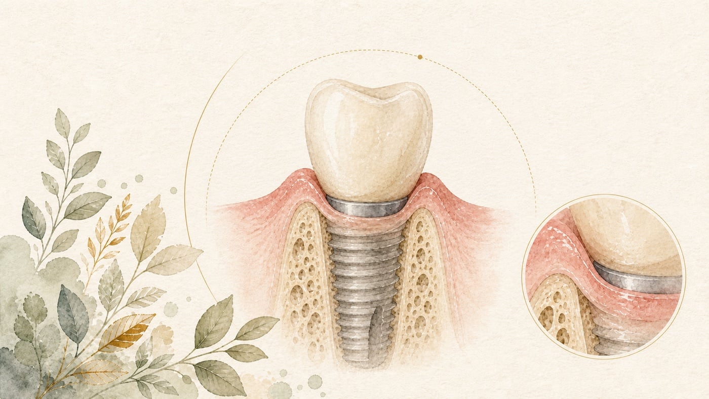
Gum recession and a visible metal edge after implant treatment? Three levels of assessment: appearance, cleaning, and hard and soft tissues
A dark or grey edge around an implant crown may reflect two overlapping issues: soft-tissue recession and colour showing through thin mucosa. Distinguishing them requires assessment of appearance, cleanability, and hard- and soft-tissue conditions. A prospective study that followed 58 posterior implant restorations for 1 year reported statistically significant correlations between buccal mucosal recession and the initial buccal mucosal width (r = −0.381), initial width-to-height ratio (r = −0.422), and buccal bone width (r = −0.290).
Gum recession and a visible metal edge after implant treatment? Three levels of assessment: appearance, cleaning, and hard and soft tissues
Direct answer: First separate whether the gingival margin has receded or whether colour is showing through thin mucosa — the two are managed differently. Where the problem is a buccal peri-implant soft tissue dehiscence, soft tissue augmentation appears to positively influence esthetic outcomes, but the review states that this is based on limited evidence, and complete coverage is not a given. [F3]
Geographic scope: global. Every figure here is taken from randomized controlled trials, systematic reviews and meta-analyses in international journals; none of it is written around the rules or funding arrangements of any single region. Which materials and techniques you can actually obtain still depends on your local care setting and your dentist's clinical judgement.
TL;DR|‘Seeing metal’ usually reflects two overlapping issues
You may look in the mirror and notice that the area where your implant crown meets the gum has darkened, that a grey edge is showing, or even that the surface feels uneven. In the literature, this is not a single problem; it reflects the overlap of soft-tissue recession and colour showing through. Determining which situation applies requires assessment at three levels.
First, recession is closely related to your original hard- and soft-tissue conditions. A prospective study that followed 58 posterior implant restorations for 1 year reported statistically significant correlations between buccal mucosal recession and the initial buccal mucosal width (r = −0.381), initial width-to-height ratio (r = −0.422), and buccal bone width (r = −0.290). The study concluded that sites with thin mucosa, thin bone, and a low width-to-height ratio are more prone to buccal mucosal recession and vertical bone loss [F1].
Second, the direction of effect favours one crown shape (concave rather than convex), but the meta-analysis did not reach significance. A systematic review and meta-analysis of 4 randomised controlled trials involving 144 implants with 12 months of follow-up reported a pooled mean difference of −0.31 mm (95% CI −0.63 to 0.02; p = 0.064)—the confidence interval crossed 0, so the result was not statistically significant [F2].
Third, can it be repaired? Improvement is possible, but complete coverage is not inevitable. A systematic review and meta-analysis of 5 studies involving 87 patients reported that, after soft-tissue augmentation, defect depth decreased by a mean of 2.2 mm (95% CI 1.76 to 2.69) and the complete coverage rate was 71% (95% CI 59% to 82%). The review explicitly stated that these findings were based on limited evidence [F3].
The three levels are unpacked below.
1. First distinguish between gum recession and colour showing through
Clinically, there are two reasons why ‘metal is visible’, and their management is entirely different.
Source A: soft-tissue recession. The gingival margin moves apically, exposing an abutment or implant neck that was previously covered. This is a positional problem.
Source B: colour showing through thin mucosa. There may be no obvious gingival recession, but the soft tissue is thin enough for the colour of the metal abutment underneath to show through, making the area look grey or dark. This is a problem of thickness and material.
Abutment material really does affect aesthetic ratings
A randomised controlled clinical trial placed single implant-supported restorations in the anterior maxilla (incisors, canines, and premolars) of 30 patients, who were randomly assigned to receive either zirconia or titanium abutments. Aesthetic (Implant Crown Aesthetic Index, ICAI), clinical, radiographic, and patient-reported outcomes were recorded at baseline (1 month after placement of the definitive restoration), 1 year, and 5 years. The trial was registered at ClinicalTrials.gov (NCT02315794), and 25 participants completed both the 1-year and 5-year follow-ups [F4]:
- ICAI scores showed statistically significantly better aesthetic outcomes with zirconia abutments than with titanium abutments [F4]
- Between 1 year and 5 years, the crown component of the aesthetic subanalysis worsened, whereas the mucosal component improved [F4]
- Bone levels did not change significantly; however, plaque index, bleeding on probing, and probing depth worsened in both groups [F4]
- The trial concluded that, at 5 years, standard zirconia abutments achieved better aesthetic outcomes, but clinical performance was similar [F4]
It is worth pausing over the phrase ‘better aesthetics but similar clinical performance’. It means that changing to a zirconia abutment improves the visible colour and overall aesthetic rating, not the health of the implant or its bone level [F4]. If your main concern is a grey colour, this is an evidence-supported avenue; if the problem is a receded gingival position, changing the abutment material does not correct that position.
A cautious reading: this was a randomised controlled trial with only 30 patients, of whom 25 completed follow-up [F4]. The sample was small, and follow-up extended only to 5 years. Another easily overlooked detail is that plaque index, bleeding on probing, and probing depth worsened in both groups over the 5 years [F4]. This reminds us that the choice of abutment material cannot replace daily cleaning and regular maintenance.
2. Conditions associated with recession: what your hard and soft tissues were like to begin with
Of the three levels, this one best explains ‘why me?’.
Thin sites are more prone to recession
A prospective study included 58 posterior implant restoration cases and assessed them when the restoration was fitted (T0) and 1 year later (T1). It measured buccal mucosal width (BMW), buccal bone width (BBW), implant buccal inclination, and emergence angle, then analysed which variables affected mucosal recession (MR) and vertical bone loss (VBL) [F1]:
- Baseline values were buccal mucosal width 2.93 ± 1.01 mm and buccal bone width 1.50 ± 0.82 mm [F1]
- At 1 year, the buccal mucosal width-to-height ratio was 1.23 ± 0.38, significantly lower than the baseline value of 1.42 ± 0.45 [F1]
- Buccal mucosal recession was −0.22 ± 0.47 mm, and vertical bone loss was 0.81 ± 0.80 mm [F1]
- Recession was statistically significantly correlated with three factors: initial mucosal width (r = −0.381), initial width-to-height ratio (r = −0.422), and buccal bone width (r = −0.290) [F1]
- Vertical bone loss was statistically significantly correlated with four factors: initial mucosal width (r = −0.421), initial width-to-height ratio (r = −0.305), implant buccal inclination (r = 0.507), and buccal bone width (r = −0.556) [F1]
- The study concluded that, within its scope, implant sites with thin buccal mucosa, thin buccal bone, and a low width-to-height ratio were more prone to buccal mucosal recession and vertical bone loss [F1]
The two most useful signals in this section are:
First, the correlation coefficient between ‘implant buccal inclination’ and vertical bone loss was 0.507 — the only positive correlation among the four statistically significant variables: the greater the inclination, the greater the bone loss [F1]. It was not, however, the largest in absolute terms: within the same set, buccal bone width at −0.556 had the larger absolute value [F1]. This means that the three-dimensional position of the implant itself is a factor independent of soft-tissue thickness.
Second, the correlation coefficient between buccal bone width and vertical bone loss was −0.556, the strongest negative correlation [F1]. The thinner the bone, the greater the vertical bone loss.
A cautious reading (the limits on extrapolating this section are important): this study involved the posterior region [F1], whereas concern about visible metal most often arises in the anterior aesthetic region. Data from the posterior region can indicate the direction of the mechanism (thin sites are more prone to recession), but cannot be used directly as a quantitative expectation for the anterior region.
Moreover, these are correlation coefficients. The strongest was 0.556, which corresponds to a coefficient of determination of approximately 31%; in other words, nearly seven-tenths of the variation came from other factors. Correlation is not causation, and a sample of 58 cases followed for 1 year cannot support stronger inferences. Finally, mean recession over 1 year was only 0.22 mm [F1]. This was an average, and individual variation may be substantial (the standard deviation of 0.47 mm exceeded the mean itself).
Width of keratinised mucosa: associated with cleaning and inflammation
Another frequently discussed soft-tissue measure is keratinised mucosal width—the width of the firmer, pink band of gingiva that does not move when pulled.
A systematic review and meta-analysis searched PubMed, Web of Science, CNKI, and VIP up to May 2024 and calculated mean differences and 95% confidence intervals for plaque index (PI), gingival index (GI), bleeding index (BI), probing depth (PD), clinical attachment loss (CAL), and bone loss (BL). A total of 30 articles were included [F5]:
- Implants with an adequate keratinised mucosal width (≥ 2 mm) had a significantly lower plaque index: mean difference −0.30 (95% CI −0.42 to −0.17) [F5]
- Gingival index: mean difference −0.26 (95% CI −0.38 to −0.13) [F5]
- Bleeding index: mean difference −0.20 (95% CI −0.33 to −0.07) [F5]
- Bone loss: mean difference −0.27 (95% CI −0.42 to −0.12) [F5]
- The analysis concluded that an adequate width of keratinised mucosa around implants (≥ 2 mm) was associated with less plaque accumulation, tissue inflammation, and bone loss [F5]
Note the role of this study: it concerns cleaning and inflammation, not aesthetic recession. All four significant findings point towards ‘easier to clean, less inflammation, and less bone loss’, which is a functional rationale [F5].
A cautious reading: the analysis explicitly reported significant heterogeneity among the included studies, although sensitivity analyses indicated that the results were robust [F5]. It also originally calculated six outcomes (PI, GI, BI, PD, CAL, and BL), but the results section of the abstract listed only four as significant—probing depth and clinical attachment loss were not listed among the significant findings. This should be remembered when interpreting the conclusion that ‘keratinised mucosa is important’. The four effect sizes were −0.30, −0.26, −0.20, and −0.27; none was large.
3. Crown emergence profile: concave appears preferable, but the meta-analysis did not reach significance
This section contains the data that require the most careful interpretation.
A systematic review and meta-analysis assessed the effect of a concave versus convex emergence profile on mid-buccal mucosal stability. It included only randomised controlled trials, was registered with PROSPERO (CRD420251139042), searched MEDLINE (PubMed) and Embase up to 7 May 2026, assessed risk of bias with RoB 2, and used a random-effects model to calculate the weighted mean difference [F2]:
- It included 4 randomised controlled trials, 144 implants, and 12 months of follow-up; 128 implants entered the quantitative synthesis (66 in the concave/modified group and 62 in the convex/non-concave group) [F2]
- The pooled mean difference was −0.31 mm (95% CI −0.63 to 0.02; p = 0.064), indicating a trend towards more mid-buccal mucosal recession with a convex emergence profile [F2]
- Between-study heterogeneity was low to moderate (I² = 28%), indicating that the results of the studies were fairly consistent [F2]
- Sensitivity analyses confirmed the direction of the effect, with pooled estimates ranging from −0.12 to −0.43 mm [F2]
- After one study was excluded, the result became statistically significant in favour of a concave profile: −0.43 mm (95% CI −0.70 to −0.16; p = 0.002) [F2]
- The review concluded that a convex emergence profile was associated with a trend towards increased mid-buccal mucosal recession; a concave profile was preferable, and emergence-profile design should be regarded as part of prosthetic and surgical planning [F2]
A cautious reading (each point here must be considered separately):
First, the primary analysis did not reach statistical significance. The 95% confidence interval was −0.63 to 0.02, its upper bound crossed 0, and p = 0.064 was also above the conventional threshold of 0.05 [F2]. This means that the possibility of no difference between the two profiles cannot be ruled out. The source paper used the wording ‘a trend toward’, not ‘significantly better’—that distinction matters.
Second, significance was reached only after one study was excluded. The value −0.43 mm (p = 0.002) was a sensitivity-analysis finding obtained after one study had been removed [F2]. In a meta-analysis of only 4 trials, removing one eliminates one-quarter of the data; such a finding is exploratory and carries less weight than the primary analysis.
Third, the effect size itself was small. Even the largest sensitivity-analysis estimate, −0.43 mm, represents a difference of less than half a millimetre [F2]. It may be statistically meaningful, but whether it produces a visible aesthetic difference is a separate question.
Fourth, the evidence base was thin. There were only 4 randomised controlled trials, 144 implants, and 12 months of follow-up [F2]. In the context of soft-tissue stability, 12 months is short-term follow-up.
The honest interpretation, therefore, is that a concave emergence profile appears favourable in direction, and low heterogeneity (I² = 28%) suggests that this direction was fairly consistent; however, current evidence is insufficient to establish a statistically significant advantage, and the effect size was also below 0.5 mm [F2]. Treating it as ‘a design option worth considering during planning’ is reasonable. Treating it as ‘changing to a concave profile will prevent recession’ goes beyond what the evidence supports.
4. If recession has occurred and metal is already visible, can it be repaired?
Improvement is possible, and the literature provides numerical estimates of that improvement; however, ‘complete coverage’ is not inevitable.
Real-world outcomes of coverage surgery
A systematic review and meta-analysis evaluated soft-tissue augmentation (STA) for buccal peri-implant soft-tissue dehiscence (PSTD). Its protocol followed the PRISMA checklist, and it included cases without coexisting peri-implantitis, randomised clinical trials, and prospective studies with at least 6 months of follow-up. A total of 8 publications covering 5 studies (2 randomised controlled trials and 3 prospective studies), published between 2013 and 2024 and involving 87 patients, were included. All studies used a coronally advanced flap (CAF) with a connective tissue graft (CTG) or a substitute material; one arm of one randomised controlled trial used a tunnel technique, and two studies also replaced prosthetic components. Of the studies, 3 were judged to have a low risk of bias, and 10 meta-analyses were performed [F3]:
- The final professionally assessed aesthetic score was 7.7 points (0-to-10 scale; 95% CI 6.63 to 8.83) [F3]
- The pooled patient-reported aesthetic value (0-to-100 visual analogue scale) was 60.8 (95% CI 46.56 to 75.01), with moderate to high heterogeneity; the source states only that the procedures showed improvements in terms of patient-reported esthetics and gives this pooled value, without specifying whether 60.8 is the post-treatment score or the size of the improvement, so this card does not choose one reading for it [F3]
- The estimated reduction in defect depth was 2.2 mm (95% CI 1.76 to 2.69) [F3]
- The estimated proportion achieving complete coverage was 71% (95% CI 59% to 82%) [F3]
- The review concluded that, based on limited evidence, soft-tissue augmentation appeared to have a positive effect on both professionally assessed and patient-reported aesthetic outcomes [F3]
How should ‘71% complete coverage’ be read? Its 95% confidence interval was 59% to 82% [F3], meaning that approximately 1 to 2 people out of every 5 to 6 would not achieve complete coverage. This is a fairly good result, but it is clearly not a guarantee of complete coverage. When discussing treatment at a follow-up visit, the interval is more important to remember than the single figure of 71%.
A cautious reading: the review itself used the phrase ‘based on limited evidence’ in its conclusion [F3]. The reasons are clear: there were only 5 studies and 87 patients; only 2 studies were randomised controlled trials, and 3 were judged to have a low risk of bias (meaning that the other two were not) [F3]. In addition, the pooled patient-reported aesthetic outcome had moderate to high heterogeneity [F3], and its confidence interval ran from 46.56 to 75.01—a span of nearly 30 points, indicating limited precision.
One further inclusion criterion must be noted: the review included only cases without coexisting peri-implantitis [F3]. If recession is accompanied by infection, these figures do not apply — and whether infection is present is not only a question of which literature applies; it changes the order of priorities: if there is persistent redness and swelling around the implant, pus on pressure or spontaneously, bleeding at times other than brushing, pain on biting, or a sense that the implant is loose, contact a dentist promptly for an examination rather than booking esthetic surgery first (this is an editorial triage note from this card, not from the sources listed above).
Which material should be used?
A PRISMA-guided systematic review assessed the medium- to long-term predictability (6 to 60 months) of peri-implant soft-tissue augmentation techniques. It included randomised controlled trials with at least 10 participants per group that used bilaminar or apically positioned flap techniques, and compared autogenous grafts with substitute materials (collagen matrices, acellular dermal matrices, and volume-stable collagen matrices). A total of 27 randomised controlled trials met the inclusion criteria [F6]:
- Implant survival exceeded 95% in every study [F6]
- Connective tissue grafts (CTG) consistently produced the greatest gains in keratinised mucosal width, mucosal thickness, soft-tissue margin stability, and aesthetic outcomes, particularly in the anterior and aesthetic regions [F6]
- Overall, autogenous grafts performed better in maintaining marginal bone levels; volume-stable collagen matrices showed acceptable long-term stability and caused less donor-site discomfort [F6]
- The review concluded that connective tissue grafting remained the most predictable method for long-term soft-tissue stability and aesthetics. Acellular dermal matrices, collagen matrices, and volume-stable collagen matrices were useful alternatives when reducing discomfort was the priority, but their outcomes were more variable [F6]
In practical terms, this means that if your main objective is soft-tissue thickness and marginal stability in the anterior aesthetic region, a connective tissue graft taken from your own mouth is currently the most predictable option [F6]. Substitute materials have the advantage of avoiding a second donor-site wound, at the cost of greater variability in outcomes [F6]. This is a classic trade-off, with no single correct answer.
5. A framework for assessment at your follow-up visit
These three levels can be translated into specific points to take to your dental appointment.
Three things you can describe proactively
1. Has the position changed, or has the colour changed? Look in the mirror: compared with the adjacent natural teeth, has the gingival margin moved upwards (or downwards)? Or is its position about the same, with a grey or dark band? These two situations point towards different management approaches—the former lies within the scope of soft-tissue augmentation [F3], whereas the latter may be related to abutment material and mucosal thickness [F4].
2. When did you notice it, and is it continuing to change? Prospective data indicate that mean buccal mucosal recession over 1 year was 0.22 ± 0.47 mm, with concurrent vertical bone loss of 0.81 ± 0.80 mm [F1]. Change is generally gradual, so knowing when you first noticed it is useful information.
3. Is there associated bleeding, swelling, or discomfort? Treat this first as a signal to be seen, not merely as a question of which evidence applies: persistent redness and swelling of the gum, pus on squeezing or spontaneously, bleeding at times other than brushing, pain around the implant or a sense that it is loose all mean you should arrange a dental examination promptly (this is an editorial triage note from this card, not from the sources listed above). At the level of the evidence, the soft-tissue augmentation meta-analysis included only cases without coexisting peri-implantitis [F3]. If infection is present, the order of management priorities is different.
Four questions you can ask
Question 1: Is my keratinised mucosa wide enough? The meta-analysis reported that a keratinised mucosal width ≥ 2 mm was associated with less plaque (mean difference −0.30), gingival inflammation (−0.26), bleeding (−0.20), and bone loss (−0.27) [F5]. This is a marker of cleanability and health, not a direct measure of aesthetics.
Question 2: How thick are my buccal bone and mucosa? Prospective data suggest that sites with thin mucosa, thin bone, and a low width-to-height ratio are more prone to recession, and that the strongest correlation was between buccal bone width and vertical bone loss (r = −0.556) [F1]. Imaging is usually required to assess this.
Question 3: If I have soft-tissue augmentation, how much improvement can I expect? The figures in the literature are: a mean reduction in defect depth of 2.2 mm (95% CI 1.76 to 2.69), a complete coverage rate of 71% (95% CI 59% to 82%), and a professional aesthetic score of 7.7 / 10 [F3]. Remember the range from 59% to 82%, rather than only the figure of 71%.
Question 4: Should tissue from my own mouth or a substitute material be used? Connective tissue grafting produced the greatest gains in keratinised mucosal width, mucosal thickness, marginal stability, and aesthetics, particularly in the anterior aesthetic region. Substitute materials caused less donor-site discomfort but had more variable outcomes [F6]. This is a trade-off worth discussing in light of your priorities.
All of these questions can be answered properly only after a dentist has performed a clinical examination and any necessary imaging, including assessment of hard- and soft-tissue thickness. The purpose of this article is to help you understand what to describe, what to ask, and what expectations to bring to the answers.
Conclusion|First distinguish between recession and show-through, then consider whether surgery is appropriate
The three levels can be distilled into one sentence: when metal is visible after implant treatment, first distinguish whether the gingival position has receded or colour is showing through thin mucosa—the answer to the former lies in soft-tissue augmentation, whereas the latter may relate to abutment material and mucosal thickness.
The three strands of evidence differ markedly in strength and should be remembered separately:
- Association between hard- and soft-tissue conditions and recession: sites with thin mucosa, thin bone, and a low width-to-height ratio were more prone to recession. The strongest correlation was between buccal bone width and vertical bone loss (r = −0.556), followed by implant buccal inclination (r = 0.507) [F1]. However, these were 1-year correlational data from the posterior region; they cannot be interpreted as causal or directly extrapolated to the anterior region.
- Crown emergence profile: the direction favoured a concave profile and heterogeneity was low (I² = 28%), but the primary analysis was not significant (−0.31 mm, 95% CI −0.63 to 0.02, p = 0.064), and the effect size was below 0.5 mm [F2]. At present, this evidence supports only the position that it is ‘worth considering during planning’.
- Actual outcomes of repair: mean defect depth decreased by 2.2 mm, the complete coverage rate was 71% (95% CI 59% to 82%), and the professional aesthetic score was 7.7 / 10 [F3]. In material selection, connective tissue grafting was the most predictable option in the aesthetic region, whereas substitute materials traded less donor-site discomfort for greater variability in outcomes [F6].
One further point should not be overlooked: the 5-year randomised controlled trial reported that, regardless of which abutment was used, plaque index, bleeding on probing, and probing depth worsened over the 5 years [F4]. This reminds us that even the best choices of material and shape cannot replace daily cleaning and regular maintenance.
The next step is straightforward: bring three pieces of information to your appointment—whether the position has receded or the colour has changed, when you first noticed it, and whether it is accompanied by bleeding or discomfort. You are welcome to book a comprehensive assessment with a dentist. First establish which level applies to your situation and assess the thickness of the hard and soft tissues; then discuss whether to adjust the restoration, undertake soft-tissue augmentation, or begin with enhanced maintenance and monitoring. Diagnosis should precede intervention, so that surgery does not leave the true cause unaddressed.
Risk factors (what to know before treatment)
- Indications and the boundary of applicability: the esthetic outcome data for soft tissue augmentation come from randomized clinical trials and prospective studies on implants without peri-implantitis, with a follow-up of at least 6 months [F3]. If the recession is accompanied by infection, these figures do not apply.
- Check first for signs of infection (editorial triage note from this card, not from the sources listed above): persistent redness and swelling around the implant, pus on pressure or spontaneously, bleeding at times other than brushing, pain on biting, persistent bad breath, or a sense that the implant is loose all mean you should contact a dentist promptly for an examination rather than booking esthetic surgery first; if there is fever, swelling that keeps spreading, or any effect on swallowing or breathing, seek care immediately.
- Who is more likely to be affected: the prospective study concluded that, within the scope of that study, implant sites in the posterior region presenting a thin buccal mucosa width, a thin buccal bone width and a small width/height ratio are more prone to exhibit buccal mucosa recession and vertical bone loss [F1]. That is posterior-region data and should not be extrapolated directly into quantitative expectations for the anterior esthetic zone.
- Adverse effects and complications: the review recorded that substitute materials (acellular dermal matrix, collagen matrix, volume-stable collagen matrix) are useful alternatives when reduced morbidity is prioritized, although their outcomes are more variable [F6]. The other side of “less donor-site morbidity” is that taking autogenous tissue adds a second surgical site.
- What cannot be expected: in the emergence-profile meta-analysis the primary analysis did not reach statistical significance, and the wording used was a “trend” rather than an established advantage [F2]. Changing a crown to a concave profile therefore cannot be treated as a way to avoid recession.
- Maintenance and long-term follow-up: in the randomized controlled trial with 5 years of follow-up, the plaque index, bleeding on probing and probing depths worsened in both groups [F4]; the meta-analysis also found that an adequate width of keratinised mucosa (≥ 2 mm) around dental implants is associated with reduced plaque accumulation, tissue inflammation and bone loss [F5]. The choice of material and contour does not replace daily cleaning and regular maintenance.
Every clinical statement in this article is mapped line by line to its cited source (see the sources and evidence chain below). It has not been clinically reviewed by a licensed practitioner. This is health information, not individual advice; assessment by a clinician is required.
FAQ
- Does a visible metal edge after implant treatment mean that the implant has failed?
- Not necessarily. In the literature, ‘visible metal’ corresponds to two different situations: **soft-tissue recession** (a positional change that exposes the abutment or implant neck) and **colour showing through** (thin mucosa allowing the colour of the underlying metal to show). The latter does not necessarily involve recession. Notably, a 5-year randomised controlled trial found a significant difference in aesthetic scores between zirconia and titanium abutments, but **clinical performance was similar and bone levels did not change significantly** [F4]. This shows that aesthetics and implant health can be separate issues. **The actual diagnosis must be made by a dentist on the basis of a clinical examination and any necessary imaging.**
- インプラント治療後に金属の縁が見えるのは、インプラントが失敗したということですか。 — 必ずしもそうではありません。文献上、「金属が見える」状態は 2つに分かれます。**軟組織の退縮**(位置が変化し、アバットメントまたはインプラント頸部が露出すること)と、**色の透過**(粘膜が薄く、その下にある金属の色が透けて見えること)です。後者は退縮を伴わない場合もあります。注目すべき点として、5年間のランダム化比較試験では、ジルコニア製とチタン製アバットメントの審美評価に有意差がありましたが、**臨床的な成績は同等で、骨の高さに有意な変化はありませんでした** [F4]。これは、審美上の問題とインプラントの健康状態を分けて考えられることを示しています。**実際の判断には、歯科医師による臨床検査と必要な画像検査が必要です。**
- Does a visible metal edge after implant treatment mean that the implant has failed? — Not necessarily. In the literature, ‘visible metal’ corresponds to two different situations: **soft-tissue recession** (a positional change that exposes the abutment or implant neck) and **colour showing through** (thin mucosa allowing the colour of the underlying metal to show). The latter does not necessarily involve recession. Notably, a 5-year randomised controlled trial found a significant difference in aesthetic scores between zirconia and titanium abutments, but **clinical performance was similar and bone levels did not change significantly** [F4]. This shows that aesthetics and implant health can be separate issues. **The actual diagnosis must be made by a dentist on the basis of a clinical examination and any necessary imaging.**
- Why did this happen to me when my friend did not have the same problem after implant treatment?
- It is closely related to the original hard- and soft-tissue conditions. A 1-year prospective study of 58 cases reported significant correlations between buccal mucosal recession and **initial buccal mucosal width (r = −0.381), initial width-to-height ratio (r = −0.422), and buccal bone width (r = −0.290)**. Vertical bone loss was most strongly correlated with **implant buccal inclination (r = 0.507)** and **buccal bone width (r = −0.556)** [F1]. The study concluded that **sites with thin mucosa, thin bone, and a low width-to-height ratio were more prone to recession** [F1]. **A cautious reading: these data came from the posterior region and can indicate the direction of the mechanism, but should not be directly extrapolated into quantitative expectations for the anterior region. Moreover, the strongest correlation coefficient, 0.556, explains only about three-tenths of the variation, leaving nearly seven-tenths attributable to other factors.**
- なぜ自分に起きたのでしょうか。友人のインプラントには同じ問題がありません。 — もともとの硬組織・軟組織の状態と強く関連します。58症例を 1年間追跡した前向き研究では、頬側粘膜の退縮と、**初期頬側粘膜幅(r = −0.381)、初期幅高比(r = −0.422)、頬側骨幅(r = −0.290)**との間に有意な相関が記録されました。また、垂直的骨喪失との相関が最も強かったのは、**インプラントの頬側傾斜角度(r = 0.507)**と**頬側骨幅(r = −0.556)**でした [F1]。この研究は、**粘膜が薄く、骨も薄く、幅高比が小さい部位ほど退縮しやすい**と結論づけています [F1]。**解釈上の注意点:これは臼歯部のデータであり、メカニズムの方向性は示せますが、前歯部の定量的な予測値として直接当てはめるべきではありません。また、最も強い相関係数 0.556でも、説明できる変動は約 3割にとどまり、7割近くは他の要因によるものです。**
- Why did this happen to me when my friend did not have the same problem after implant treatment? — It is closely related to the original hard- and soft-tissue conditions. A 1-year prospective study of 58 cases reported significant correlations between buccal mucosal recession and **initial buccal mucosal width (r = −0.381), initial width-to-height ratio (r = −0.422), and buccal bone width (r = −0.290)**. Vertical bone loss was most strongly correlated with **implant buccal inclination (r = 0.507)** and **buccal bone width (r = −0.556)** [F1]. The study concluded that **sites with thin mucosa, thin bone, and a low width-to-height ratio were more prone to recession** [F1]. **A cautious reading: these data came from the posterior region and can indicate the direction of the mechanism, but should not be directly extrapolated into quantitative expectations for the anterior region. Moreover, the strongest correlation coefficient, 0.556, explains only about three-tenths of the variation, leaving nearly seven-tenths attributable to other factors.**
- Would changing to an all-ceramic (zirconia) abutment improve the colour?
- There is supporting evidence for an improvement in aesthetic scores. A randomised controlled trial involving 30 patients in the anterior maxilla with 5 years of follow-up reported that **ICAI aesthetic scores were statistically significantly better with zirconia abutments than with titanium abutments**. At 5 years, **standard zirconia abutments achieved better aesthetic outcomes, but clinical performance was similar** [F4]. **However, the trial included only 30 patients, just 25 completed follow-up, the sample was small, and follow-up extended only to 5 years. It is also important that plaque index, bleeding on probing, and probing depth worsened in both groups over the 5 years**—abutment material cannot replace daily cleaning and regular maintenance [F4].
- オールセラミック(ジルコニア)製アバットメントに変えると、色は改善しますか。 — 審美評価については、裏付けとなるエビデンスがあります。上顎前歯部の患者 30人を対象に 5年間追跡したランダム化比較試験では、**ICAI審美指数において、ジルコニア製アバットメントはチタン製アバットメントより統計学的に有意に優れていました**。5年時点で、**標準的なジルコニア製アバットメントはより良好な審美的結果を達成しましたが、臨床的な成績は同等でした** [F4]。**ただし、この試験の患者は 30人のみで、追跡を完了したのは 25人です。サンプルサイズは小さく、追跡も 5年間に限られます。また、両群とも 5年間にプラーク指数、プロービング時出血、プロービングデプスが悪化したことにも注意が必要です**。アバットメントの材質は、日々の清掃や定期的なメインテナンスの代わりにはなりません [F4]。
- Would changing to an all-ceramic (zirconia) abutment improve the colour? — There is supporting evidence for an improvement in aesthetic scores. A randomised controlled trial involving 30 patients in the anterior maxilla with 5 years of follow-up reported that **ICAI aesthetic scores were statistically significantly better with zirconia abutments than with titanium abutments**. At 5 years, **standard zirconia abutments achieved better aesthetic outcomes, but clinical performance was similar** [F4]. **However, the trial included only 30 patients, just 25 completed follow-up, the sample was small, and follow-up extended only to 5 years. It is also important that plaque index, bleeding on probing, and probing depth worsened in both groups over the 5 years**—abutment material cannot replace daily cleaning and regular maintenance [F4].
- Will making the crown's emergence profile concave prevent gingival recession?
- The direction of effect appears favourable, but the evidence does not yet support that statement. A systematic review and meta-analysis of 4 randomised controlled trials, 144 implants, and 12 months of follow-up reported a pooled mean difference of **−0.31 mm (95% CI −0.63 to 0.02; p = 0.064)**—the **confidence interval crossed 0 and the result was not statistically significant**. The source paper described a ‘trend’, not a ‘significantly better’ result [F2]. The result became significant after one study was excluded (−0.43 mm, 95% CI −0.70 to −0.16, p = 0.002), but **this was a sensitivity analysis and, with only 4 trials, carries less weight than the primary analysis** [F2]. **Even the largest estimate was less than 0.5 mm.** It is reasonably characterised as ‘a design option worth considering during planning’.
- クラウンを凹面のエマージェンスプロファイルにすれば、歯肉退縮を防げますか。 — 効果の方向としては望ましいものの、現時点のエビデンスでは、そこまで断定できません。4件のランダム化比較試験、144本のインプラント、12か月の追跡を対象としたシステマティックレビューとメタアナリシスでは、統合平均差は**−0.31 mm(95% CI −0.63~0.02、p = 0.064)**でした。**信頼区間は 0をまたいでおり、統計学的有意差には達していません**。原著の表現は「傾向」であり、「有意に優れている」ではありません [F2]。1件の研究を除外すると有意差が得られました(−0.43 mm、95% CI −0.70~−0.16、p = 0.002)が、**これは感度分析の結果であり、試験が 4件しかない状況では主解析よりエビデンスの強度が低くなります** [F2]。**さらに、最大の推定値を採用しても、差は 0.5 mm未満です。** 妥当な位置づけは、「計画時に検討する価値のある設計上の選択肢」です。
- Will making the crown's emergence profile concave prevent gingival recession? — The direction of effect appears favourable, but the evidence does not yet support that statement. A systematic review and meta-analysis of 4 randomised controlled trials, 144 implants, and 12 months of follow-up reported a pooled mean difference of **−0.31 mm (95% CI −0.63 to 0.02; p = 0.064)**—the **confidence interval crossed 0 and the result was not statistically significant**. The source paper described a ‘trend’, not a ‘significantly better’ result [F2]. The result became significant after one study was excluded (−0.43 mm, 95% CI −0.70 to −0.16, p = 0.002), but **this was a sensitivity analysis and, with only 4 trials, carries less weight than the primary analysis** [F2]. **Even the largest estimate was less than 0.5 mm.** It is reasonably characterised as ‘a design option worth considering during planning’.
- How wide does the keratinised mucosa need to be?
- The commonly used threshold in the literature is 2 mm, but this relates mainly to cleaning and inflammation rather than aesthetics. A systematic review and meta-analysis of 30 articles reported that implants with a keratinised mucosal width ≥ 2 mm had significantly lower **plaque index (mean difference −0.30, 95% CI −0.42 to −0.17), gingival index (−0.26, −0.38 to −0.13), bleeding index (−0.20, −0.33 to −0.07), and bone loss (−0.27, −0.42 to −0.12)** [F5]. **A cautious reading: the analysis reported significant heterogeneity among the included studies, although sensitivity analysis suggested that the findings were robust; the four effect sizes were all small; and, of the six outcomes originally calculated, only four were listed as significant in the abstract's results section.**
- 角化粘膜はどの程度の幅があれば十分ですか。 — 文献で一般的に用いられる基準は 2 mmですが、これは主に清掃と炎症に関するものであり、審美性の基準ではありません。30報を対象としたシステマティックレビューとメタアナリシスでは、角化粘膜幅≥ 2 mmのインプラントは、**プラーク指数(平均差 −0.30、95% CI −0.42~−0.17)、歯肉指数(−0.26、−0.38~−0.13)、出血指数(−0.20、−0.33~−0.07)、骨喪失(−0.27、−0.42~−0.12)がいずれも有意に低い**という結果でした [F5]。**解釈上の注意点:この解析では、採用研究間に有意な異質性が記録されています(感度分析では結果の頑健性が示されました)。4つの効果量はいずれも大きくありません。また、当初算出した 6項目のうち、抄録の結果欄で有意とされたのは 4項目のみです。**
- How wide does the keratinised mucosa need to be? — The commonly used threshold in the literature is 2 mm, but this relates mainly to cleaning and inflammation rather than aesthetics. A systematic review and meta-analysis of 30 articles reported that implants with a keratinised mucosal width ≥ 2 mm had significantly lower **plaque index (mean difference −0.30, 95% CI −0.42 to −0.17), gingival index (−0.26, −0.38 to −0.13), bleeding index (−0.20, −0.33 to −0.07), and bone loss (−0.27, −0.42 to −0.12)** [F5]. **A cautious reading: the analysis reported significant heterogeneity among the included studies, although sensitivity analysis suggested that the findings were robust; the four effect sizes were all small; and, of the six outcomes originally calculated, only four were listed as significant in the abstract's results section.**
- If recession has already occurred and the metal is visible, can it still be repaired?
- Substantial improvement is possible in most cases, but complete coverage is not inevitable. A systematic review and meta-analysis of 5 studies involving 87 patients reported an **estimated reduction in defect depth of 2.2 mm (95% CI 1.76 to 2.69)**, an **estimated complete coverage rate of 71% (95% CI 59% to 82%)**, and a final professionally assessed aesthetic score of **7.7 points (0-to-10 scale; 95% CI 6.63 to 8.83)** [F3]. **Remember the range from 59% to 82%: approximately 1 to 2 people out of every 5 to 6 did not achieve complete coverage.** The review itself stated that its conclusions were **based on limited evidence** (only 2 of the 5 studies were randomised controlled trials, and 3 were judged to have a low risk of bias), and it included **only cases without coexisting peri-implantitis** [F3].
- すでに退縮して金属が見えていても、元に戻せますか。 — 多くの場合、明らかな改善は可能ですが、完全被覆が必ず得られるわけではありません。5件の研究、87人の患者を対象としたシステマティックレビューとメタアナリシスでは、**欠損深さの推定減少量は 2.2 mm(95% CI 1.76~2.69)**、**完全被覆の推定割合は 71%(95% CI 59%~82%)**、専門家が評価した最終審美スコアは**7.7点(0~10点尺度、95% CI 6.63~8.83)**でした [F3]。**59%~82%という範囲もあわせて覚えておいてください。およそ 5~6人に 1~2人は完全被覆に達しません。** このレビュー自身が**限られたエビデンスに基づく**と述べています(5件の研究のうち、ランダム化比較試験は 2件のみで、バイアスリスク低と評価されたのは 3件です)。また、対象は**インプラント周囲炎を併発していない症例のみ**でした [F3]。
- If recession has already occurred and the metal is visible, can it still be repaired? — Substantial improvement is possible in most cases, but complete coverage is not inevitable. A systematic review and meta-analysis of 5 studies involving 87 patients reported an **estimated reduction in defect depth of 2.2 mm (95% CI 1.76 to 2.69)**, an **estimated complete coverage rate of 71% (95% CI 59% to 82%)**, and a final professionally assessed aesthetic score of **7.7 points (0-to-10 scale; 95% CI 6.63 to 8.83)** [F3]. **Remember the range from 59% to 82%: approximately 1 to 2 people out of every 5 to 6 did not achieve complete coverage.** The review itself stated that its conclusions were **based on limited evidence** (only 2 of the 5 studies were randomised controlled trials, and 3 were judged to have a low risk of bias), and it included **only cases without coexisting peri-implantitis** [F3].
- Should surgery use my own tissue or an artificial material?
- The two involve a trade-off rather than a simple distinction between better and worse. A systematic review of 27 randomised controlled trials with 6 to 60 months of follow-up reported that **connective tissue grafts (CTG) consistently produced the greatest gains in keratinised mucosal width, mucosal thickness, soft-tissue margin stability, and aesthetic outcomes, particularly in the anterior and aesthetic regions**. **Overall, autogenous grafts performed better in maintaining marginal bone levels** [F6]. Substitute materials (acellular dermal matrices, collagen matrices, and volume-stable collagen matrices) caused **less donor-site discomfort**; volume-stable collagen matrices showed acceptable long-term stability, but the review explicitly noted that outcomes with substitute materials were **more variable** [F6]. Implant survival exceeded 95% in every study [F6].
- 手術では、自分の組織と人工材料のどちらを使うのでしょうか。 — 両者の違いは単純な優劣ではなく、トレードオフです。27件のランダム化比較試験、6~60か月の追跡を対象としたシステマティックレビューでは、**結合組織移植(CTG)は、角化粘膜幅、粘膜厚、軟組織辺縁の安定性、審美的結果について一貫して最大の増加を示し、前歯部と審美領域で特に明瞭でした**。また、**自家移植は全体として辺縁骨レベルの維持に優れていました** [F6]。代替材料(無細胞真皮マトリックス、コラーゲンマトリックス、体積安定型コラーゲンマトリックス)では**採取部位の不快感が少なく**、体積安定型コラーゲンマトリックスは許容できる長期安定性を示しましたが、レビューは代替材料の**結果のばらつきがより大きい**と明記しています [F6]。すべての研究で、インプラント生存率は 95%を超えていました [F6]。
- Should surgery use my own tissue or an artificial material? — The two involve a trade-off rather than a simple distinction between better and worse. A systematic review of 27 randomised controlled trials with 6 to 60 months of follow-up reported that **connective tissue grafts (CTG) consistently produced the greatest gains in keratinised mucosal width, mucosal thickness, soft-tissue margin stability, and aesthetic outcomes, particularly in the anterior and aesthetic regions**. **Overall, autogenous grafts performed better in maintaining marginal bone levels** [F6]. Substitute materials (acellular dermal matrices, collagen matrices, and volume-stable collagen matrices) caused **less donor-site discomfort**; volume-stable collagen matrices showed acceptable long-term stability, but the review explicitly noted that outcomes with substitute materials were **more variable** [F6]. Implant survival exceeded 95% in every study [F6].
- How much will the gums recede each year?
- The amount was small in the available prospective data, but individual variation may be substantial. A study of 58 posterior implant cases followed for 1 year reported **buccal mucosal recession of −0.22 ± 0.47 mm and vertical bone loss of 0.81 ± 0.80 mm**. Over the same period, the buccal mucosal width-to-height ratio fell significantly from 1.42 ± 0.45 to 1.23 ± 0.38 [F1]. **A cautious reading: the standard deviation of recession (0.47 mm) exceeded the mean itself (0.22 mm), indicating substantial individual variation; these data also came from the posterior region with only 1 year of follow-up and should not be applied directly to the anterior aesthetic region or a longer timescale.**
- 歯肉は年間どの程度退縮しますか。 — 現在得られている前向きデータでは、その程度は小さいものの、個人差は大きい可能性があります。臼歯部のインプラント 58症例を 1年間追跡した研究では、**頬側粘膜退縮は −0.22 ± 0.47 mm、垂直的骨喪失は 0.81 ± 0.80 mm**でした。同じ期間に、頬側粘膜幅高比は 1.42 ± 0.45から 1.23 ± 0.38へ有意に低下しました [F1]。**解釈上の注意点:退縮量の標準偏差(0.47 mm)は平均値そのもの(0.22 mm)を上回っており、個人差がかなり大きいことを示しています。また、これは臼歯部を 1年間のみ追跡したデータであり、前歯部の審美領域や、より長い期間に直接当てはめるべきではありません。**
- How much will the gums recede each year? — The amount was small in the available prospective data, but individual variation may be substantial. A study of 58 posterior implant cases followed for 1 year reported **buccal mucosal recession of −0.22 ± 0.47 mm and vertical bone loss of 0.81 ± 0.80 mm**. Over the same period, the buccal mucosal width-to-height ratio fell significantly from 1.42 ± 0.45 to 1.23 ± 0.38 [F1]. **A cautious reading: the standard deviation of recession (0.47 mm) exceeded the mean itself (0.22 mm), indicating substantial individual variation; these data also came from the posterior region with only 1 year of follow-up and should not be applied directly to the anterior aesthetic region or a longer timescale.**
- Will this affect the health of the implant, or only how it looks?
- Both are possible, but they must be assessed separately. **Aesthetics**: a 5-year randomised controlled trial reported that abutment material affected aesthetic scores, but **clinical performance was similar and bone levels did not change significantly** [F4]. **Health**: a keratinised mucosal width ≥ 2 mm was associated with less plaque, inflammation, and bone loss [F5], while sites with thin mucosa and thin bone were also more prone to both **recession and vertical bone loss** [F1]. **The reasonable approach is therefore to assess ‘how it looks’ and ‘how healthy it is’ as separate lines of enquiry, rather than inferring one from the other.**
- これはインプラントの健康に影響しますか。それとも見た目だけの問題ですか。 — どちらにも影響する可能性がありますが、分けて評価する必要があります。**審美面**では、5年間のランダム化比較試験で、アバットメントの材質が審美評価に影響しましたが、**臨床的な成績は同等で、骨の高さに有意な変化はありませんでした** [F4]。**健康面**では、角化粘膜幅≥ 2 mmは、プラーク、炎症、骨喪失が少ないことと関連し [F5]、粘膜と骨が薄い部位では、**退縮と垂直的骨喪失**の双方が生じやすい傾向がありました [F1]。**したがって、「見た目がどうか」と「健康かどうか」を別々の観点から確認し、一方から他方を推測しないことが妥当です。**
- Will this affect the health of the implant, or only how it looks? — Both are possible, but they must be assessed separately. **Aesthetics**: a 5-year randomised controlled trial reported that abutment material affected aesthetic scores, but **clinical performance was similar and bone levels did not change significantly** [F4]. **Health**: a keratinised mucosal width ≥ 2 mm was associated with less plaque, inflammation, and bone loss [F5], while sites with thin mucosa and thin bone were also more prone to both **recession and vertical bone loss** [F1]. **The reasonable approach is therefore to assess ‘how it looks’ and ‘how healthy it is’ as separate lines of enquiry, rather than inferring one from the other.**
- How often should I have follow-up checks?
- The literature does not provide a universal interval; this must be decided by a dentist according to your circumstances. Three pieces of evidence can inform the discussion: soft-tissue and bone changes are gradual (mean recession of 0.22 mm and bone loss of 0.81 mm over 1 year) [F1]; the 5-year randomised controlled trial reported that **plaque index, bleeding on probing, and probing depth worsened in both groups during follow-up** [F4]; and the association between keratinised mucosal width and plaque, inflammation, and bone loss has been documented in a meta-analysis [F5]. If your hard and soft tissues are thin, follow-up frequency is worth discussing proactively.
- どのくらいの間隔で経過観察を受けるべきですか。 — 文献には一律の間隔は示されておらず、歯科医師が個々の状態に応じて判断する必要があります。ただし、相談の参考になる情報が 3つあります。軟組織と骨の変化は徐々に進みます(1年間の平均退縮量 0.22 mm、骨喪失量 0.81 mm)[F1]。5年間のランダム化比較試験では、**両群とも追跡期間中にプラーク指数、プロービング時出血、プロービングデプスが悪化しました** [F4]。さらに、角化粘膜幅とプラーク、炎症、骨喪失との関連は、メタアナリシスで記録されています [F5]。硬組織・軟組織が薄い場合には、経過観察の頻度について積極的に相談する価値があります。
- How often should I have follow-up checks? — The literature does not provide a universal interval; this must be decided by a dentist according to your circumstances. Three pieces of evidence can inform the discussion: soft-tissue and bone changes are gradual (mean recession of 0.22 mm and bone loss of 0.81 mm over 1 year) [F1]; the 5-year randomised controlled trial reported that **plaque index, bleeding on probing, and probing depth worsened in both groups during follow-up** [F4]; and the association between keratinised mucosal width and plaque, inflammation, and bone loss has been documented in a meta-analysis [F5]. If your hard and soft tissues are thin, follow-up frequency is worth discussing proactively.
Medical notice This article is provided for health education and medical information purposes. It is not a solicitation for medical services and does not constitute diagnosis or treatment advice. Actual treatment methods and outcomes vary between individuals and require evaluation by a dentist; every treatment has its own indications, limitations and possible risks. If you have related symptoms or treatment needs, please book a consultation for evaluation by a dentist.
Source anchors
- Influence of buccal mucosa width/height ratio, emergence profile and buccal bone width on peri-implant tissues: a prospective one-year study. [PMID:39799344] · https://pubmed.ncbi.nlm.nih.gov/39799344/ · 在 IDAEO 的其他引用
- Influence of Concave Versus Convex Emergence Profiles on Midfacial Mucosal Stability-A Systematic Review With Meta-Analysis. [PMID:42322345] · https://pubmed.ncbi.nlm.nih.gov/42322345/ · 在 IDAEO 的其他引用
- Impact of soft tissue augmentation procedures on esthetics and patient satisfaction in the treatment of peri-implant buccal soft tissue dehiscences: A systematic review and meta-analysis. [PMID:40673678] · https://pubmed.ncbi.nlm.nih.gov/40673678/ · 在 IDAEO 的其他引用
- Five-year outcomes of a randomized controlled clinical trial comparing single-tooth implant-supported restoration with either zirconia or titanium abutments. [PMID:36748305] · https://pubmed.ncbi.nlm.nih.gov/36748305/ · 在 IDAEO 的其他引用
- The relationship between adequate keratinized mucosa and peri-implant disease: a systematic review and meta-analysis. [PMID:40050830] · https://pubmed.ncbi.nlm.nih.gov/40050830/ · 在 IDAEO 的其他引用
- Soft Tissue Augmentation Around Dental Implants - a Systematic Review. [PMID:42416748] · https://pubmed.ncbi.nlm.nih.gov/42416748/ · 在 IDAEO 的其他引用
Cite this article
Lucy・《Gum recession and a visible metal edge after implant treatment? Three levels of assessment: appearance, cleaning, and hard and soft tissues》・IDAEO 知識庫・2026-07-20・https://km.idaeo.ai/post/dental/implant-gingival-recession-metal-showUpdated 2026-08-26