

🏛 Part of the "dental" topic shelf →
What Has to Be Assessed Before an Implant: From Bone Volume and Gingival Thickness to Angle and Depth
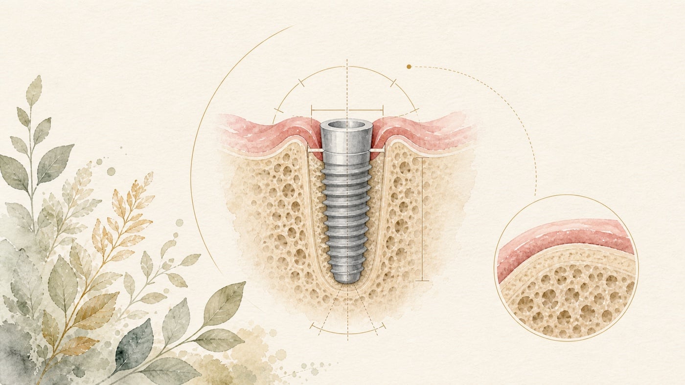
Assessment before an implant has to answer at least several different questions: whether the bone at the edentulous site can accommodate the planned implant, whether the soft tissue favours cleaning and stability, whether the angle and depth of the implant can be matched to the future crown, and how much deviation there may be between the planned position and the actual surgery. These questions influence one another, but they cannot substitute for one another. Gingiva that looks thick does not mean the bone volume is necessarily sufficient, and a mean imaging error derived from a population is not your personal safety margin. The dentist has to bring the intraoral examination, the three-dimensional imaging, the planned prosthetic position and the surgical approach into a single plan. For you, a more useful question than "how many millimetres of bone are enough" is this: which direction has been measured, how does that measurement relate to the planned implant and crown, which part lies closest to an important anatomical structure, and how is the dentist leaving room for measurement and surgical deviation?
What Has to Be Assessed Before an Implant: From Bone Volume and Gingival Thickness to Angle and Depth
Direct answer: Assessment before an implant brings bone, soft tissue, imaging and the position of the future crown into a single plan; there is no pass mark that applies to every site. The review records that implant stability was correlated with bone density, alveolar ridge width and implant size, with P values below 0.01, and that regions with thicker cortical bone demonstrated superior primary stability [F1]. Gingival thickness is only a clue: the pooled mean difference in alveolar bone thickness between thick and thin gingival phenotypes was 0.33 mm, with P below 0.01, and the association was more evident at the crest and decreased towards the apex [F2].
Geographic scope: This is general health education based on international literature. It does not cover any particular country's insurance scheme or regulations; for how care and fees are organised, follow the rules where you are. Every source cited in this card is international peer-reviewed literature; no national legislation or insurance-benefit rule is cited.
TL;DR — Implant planning is not about finding a pass mark; it is about aligning conditions in three dimensions
Assessment before an implant has to answer at least several different questions: whether the bone at the edentulous site can accommodate the planned implant, whether the soft tissue favours cleaning and stability, whether the angle and depth of the implant can be matched to the future crown, and how much deviation there may be between the planned position and the actual surgery.
These questions influence one another, but they cannot substitute for one another. Gingiva that looks thick does not mean the bone volume is necessarily sufficient, and a mean imaging error derived from a population is not your personal safety margin. The dentist has to bring the intraoral examination, the three-dimensional imaging, the planned prosthetic position and the surgical approach into a single plan.
For you, a more useful question than "how many millimetres of bone are enough" is this: which direction has been measured, how does that measurement relate to the planned implant and crown, which part lies closest to an important anatomical structure, and how is the dentist leaving room for measurement and surgical deviation?
Assessing bone volume: not just width or height
An implant has to be enclosed and supported by bone in three dimensions. Dentists are usually concerned with the width and height of the alveolar ridge, the outer bone plate, bone density, the morphology of the defect, and how these conditions relate to the size and position of the planned implant. Asking only whether "there is enough bone" makes it easy to lose sight of direction and position.
A systematic review that included six studies, two hundred and nine patients and four hundred and eighteen implants found that thicker cortical bone was associated with better primary stability; bone density, alveolar ridge width and implant size were also associated with stability, with P values below 0.01 [F1]. This shows that bone conditions are worth assessing before surgery, but the review does not provide a minimum bone width or minimum bone height applicable to every site.
The same review also observed that implants in native bone had higher ISQ values at baseline, at eight weeks and at twelve weeks, with P values of 0.011, 0.013 and below 0.001 respectively [F1]. These are population-level associations and follow-up results; they cannot be worked backwards into your personal stability, nor can implant size or the timing of loading be decided on thicker cortical bone alone.
Gingival thickness: a clue, not a stand-in for bone volume
Gingival phenotype can give a preliminary clue from the intraoral examination. A systematic review and meta-analysis showed a pooled mean difference in alveolar bone thickness of 0.33 mm between thick and thin gingival phenotypes, with P below 0.01; across the studies, the correlation between gingival thickness and alveolar bone thickness ranged from 0.11 to 0.49 [F2].
The authors also point out that the evidence remains contradictory, and that the association is more evident at the crest and weakens towards the apex [F2]. Thick gingiva may therefore suggest a thicker alveolar bone plate, but it cannot replace an actual three-dimensional assessment of bone volume; nor does thin gingiva mean that you certainly cannot have an implant.
Another review compared soft-tissue thickness directly before implant placement, including six studies and three hundred and fifty-four implants followed for ten to fourteen months; of these, one hundred and ninety-four had a soft-tissue thickness of at least 2 mm and one hundred and sixty were below 2 mm [F3]. The meta-analysis reported a between-group difference of 0.54 with a P value of 0.027, but the abstract does not clearly state the direction or the unit of that figure, and heterogeneity was above 50% [F3].
That abstract cannot therefore be rewritten as "a gingival thickness of 2 mm is safe", nor can it be claimed that the thick group certainly loses 0.54 mm less [F3]. It is better suited to supporting the measurement of soft tissue before surgery, and to reminding the dentist to build short-term marginal bone change into the plan.
Imaging and measurement: instruments have error, and error has a context
CBCT is often used to look at hard tissue and important anatomical structures, but boundary identification in the image, voxel size, metal interference and study settings all affect the measurement. [F7] A systematic review included twenty-two studies, of which nine were animal, nine in-vitro and four human studies; the in-vitro meta-analysis showed that CBCT underestimated buccal bone thickness by a mean of 0.15 mm, with a confidence interval of 0.03 to 0.26 mm of underestimation [F4].
All the studies included in that review were rated at high risk of bias, and what the authors discuss is the visualisation and monitoring of the peri-implant buccal bone plate — not a claim that pre-operative CBCT has no use [F4]. Still less can a mean underestimation of 0.15 mm be treated as a fixed correction factor for every individual [F4].
Instruments that do not use ionising radiation have also been studied. A meta-analysis of twelve studies showed that, relative to CBCT, MRI gave an implant-tip deviation of 0.30 mm with a confidence interval of -0.08 to 0.68; an entry deviation of 0.38 mm with a confidence interval of 0.04 to 0.71; and an angulation mean difference of 0.81° with a confidence interval of -0.50 to 2.12 [F5]. Ultrasound gave a deviation of 0.04 mm in soft-tissue measurement, with a confidence interval of -0.04 to 0.13 [F5].
These mean values can be used to understand how the instruments perform; they cannot be used to declare that your bone or soft tissue only needs the same distance left in reserve. Equipment, scanning protocol, anatomical site and the clinician's interpretation still change the individual situation [F5].
Angle and depth: working back to the implant position from the future crown
Ideal planning does not mean first finding somewhere in the bone the implant will fit and then forcing a crown onto it. The implant platform, long axis and apex all have to be considered together with the future crown, the occlusion, the space needed for cleaning and the neighbouring anatomical structures. This is also why the dentist may use digital planning, guided surgery or dynamic navigation.
A systematic review and meta-analysis comparing freehand with computer-aided implant surgery reported mean angular, entry and apex deviations for freehand of 7.46°, 1.56 mm and 2.22 mm, and for fully guided static CAIS of 2.57°, 0.72 mm and 0.88 mm [F6]. Pilot-drill static CAIS gave 5.94°, 1.13 mm and 1.43 mm, and dynamic CAIS gave 3.67°, 1.01 mm and 1.36 mm [F6].
The authors point out that even with CAIS the apex deviation may still fall between 1 and 2 mm, and they therefore propose a 2 mm safety margin [F6]. That is a mean across studies and a surgical planning recommendation from the authors, not a personal safety line for every site; the actual distance to the sinus, the nerve, the roots of adjacent teeth and the bone walls still has to be judged from your own images and treatment design.
What a complete assessment usually has to join up
The dentist will first establish where the teeth are missing and where the future crown needs to appear, then work back from the prosthetic direction to the long axis, platform and depth of the implant. Next comes a check on three-dimensional bone coverage, soft-tissue thickness and access for cleaning, along with confirmation of the roots of adjacent teeth, the sinus, the nerve and similar structures.
If the existing bone or soft-tissue conditions do not match the ideal position, the discussion shifts to a different set of questions: can the implant design or position be adjusted, is bone grafting or soft-tissue treatment needed, should another method of restoring the missing tooth be chosen, and what does each of those cost. This is not simply a matter of topping a failing number up to a pass; it is a fresh check on whether the whole treatment plan is reasonable.
You can ask the dentist to point out the planned implant, the future crown and the important structures on the images, and to explain how the measurements affect the plan. Once you can see why this position was chosen, the steps and the limits of the treatment become easier to understand.
Data anchors — a mean is not your personal threshold
| Aspect assessed | Data anchor | How to read it safely | Source |
|---|---|---|---|
| Cortical bone and primary stability | Six studies, two hundred and nine people, four hundred and eighteen implants; bone conditions associated with stability, P < 0.01 [F1] | Supports assessing bone conditions; offers no universal minimum bone volume | [F1] |
| Gingival phenotype and bone plate | Pooled mean difference 0.33 mm, P < 0.01; correlation range 0.11 to 0.49 [F2] | A population-level association; gingival thickness cannot replace bone imaging | [F2] |
| Pre-operative soft-tissue thickness | Between-group difference of 0.54 for at least 2 mm versus below 2 mm, P = 0.027; heterogeneity above 50% [F3] | The abstract lacks direction and unit; it cannot be written up as a personal threshold or an amount of loss | [F3] |
| CBCT measurement of buccal bone | In-vitro pooled MD = -0.15 mm; 95% CI -0.26 to -0.03 [F4] | Mainly a laboratory mean, and the studies were at high risk of bias; not to be added to or subtracted from an individual measurement | [F4] |
| MRI and ultrasound | MRI tip 0.30 mm, entry 0.38 mm, angle 0.81°; ultrasound soft tissue 0.04 mm [F5] | Instrument accuracy relative to CBCT, not an outcome or a safety threshold | [F5] |
| Planned versus actual implant position | Freehand 7.46° / 1.56 mm / 2.22 mm; fully guided 2.57° / 0.72 mm / 0.88 mm [F6] | A technique mean cannot replace individual anatomy and safety margins | [F6] |
Conclusion — let the images, the hard and soft tissues and the future crown all say the same thing
The point of assessment before an implant is not to find you a pass mark from the internet, but to confirm whether the planned crown, the implant position, the three-dimensional bone conditions, the soft tissue and the important anatomical structures can be reconciled. A population mean error can remind the clinical team to leave room in the plan, but it cannot become your personal threshold.
If you are planning an implant, you can bring your existing images to your appointment and ask the dentist to point out the planned crown and implant position, the limits set by bone and soft tissue, and the steps each option would require you to take on. Understanding the plan first, and only then deciding whether to proceed with treatment, is a restrained and practical next step.
Risk factors (what to know before treatment)
- The measuring instruments themselves have error: the in-vitro meta-analysis of CBCT for buccal bone thickness showed a mean underestimation of 0.15 mm, with a confidence interval of 0.03 to 0.26 mm of underestimation, and every included study was rated at high risk of bias; the review concludes that CBCT cannot yet be recommended as a standard diagnostic tool for follow-up of the buccal bone plate at oral implants [F4].
- Guided surgery reduces deviation but does not remove it: mean angular, entry and apex deviations were 7.46°, 1.56 mm and 2.22 mm for freehand and 2.57°, 0.72 mm and 0.88 mm for fully guided static CAIS [F6]; because apex deviations of 1 to 2 mm have still been observed with CAIS, the authors recommend implementing a 2 mm safety margin [F6].
- The evidence on soft-tissue thickness covers only a short period: the review comparing soft-tissue thickness before placement included only six studies with a follow-up of ten to fourteen months, and the included studies had a high level of heterogeneity (I² above 50%); the authors conclude that initial soft-tissue thickness seems to influence marginal bone loss over a short follow-up period and state plainly that further studies are needed [F3].
- Gingival thickness is not a stand-in for bone volume: although the difference in alveolar bone thickness between thick and thin phenotypes was significant (0.33 mm, P below 0.01), the authors also point out that the evidence remains contradictory and that the association is more evident at the crest and decreases towards the apex [F2]. Thin gingiva does not mean an implant is impossible, and thick gingiva cannot replace three-dimensional imaging.
- The non-ionising instruments are still being standardised: relative to CBCT, MRI gave an implant-tip deviation of 0.30 mm (confidence interval -0.08 to 0.68), an entry deviation of 0.38 mm (confidence interval 0.04 to 0.71) and an angulation mean difference of 0.81° (confidence interval -0.50 to 2.12), while ultrasound gave a deviation of 0.04 mm in soft-tissue measurement (confidence interval -0.04 to 0.13) [F5]; the authors regard both as reliable non-ionising alternatives, but also state that further standardisation of protocols is needed to address variability in clinical workflows [F5].
- This card does not compile lists of indications or contraindications: no separate literature search on the indications, contraindications or systemic conditions relevant to implant treatment was carried out for this card. Whether your bone and soft-tissue conditions can support the planned implant position, whether bone or soft-tissue augmentation is needed, and what alternative ways of replacing a missing tooth exist, all have to be assessed by a dentist from your images and intraoral examination.
Every clinical statement in this article is mapped line by line to its cited source (see the sources and evidence chain below). It has not been clinically reviewed by a licensed practitioner. This is health information, not individual advice; assessment by a clinician is required.
FAQ
- How much bone does an implant need?
- There is no single figure supported by these reviews that applies to every site. The dentist has to assess bone width, height, the bone plate, density, the size of the planned implant, the crown position and the important anatomical structures together [F1].
- インプラントにはどれくらいの骨が必要ですか — これらのレビューによって裏づけられる、すべての部位に当てはまる単一の数値はありません。歯科医師は骨幅、高さ、骨壁、密度、予定するインプラント体のサイズ、クラウンの位置、重要な解剖学的構造をあわせて評価する必要があります [F1]。
- How much bone does an implant need? — There is no single figure supported by these reviews that applies to every site. The dentist has to assess bone width, height, the bone plate, density, the size of the planned implant, the crown position and the important anatomical structures together [F1].
- If my gingiva is thick, does that mean my bone is thick too?
- Not necessarily. The meta-analysis found that a thick phenotype was on average accompanied by a thicker bone plate, but the studies remain contradictory and the association weakens towards the apex [F2]. Gingival phenotype is a clue, not a stand-in for bone volume.
- 歯肉が厚ければ、骨も必ず厚いのですか — 必ずしもそうではありません。メタアナリシスでは厚い歯肉フェノタイプが平均してより厚い骨壁を伴っていましたが、研究にはなお矛盾があり、関連は根尖側に向かって弱まります [F2]。歯肉フェノタイプは手がかりであって、骨量の代役ではありません。
- If my gingiva is thick, does that mean my bone is thick too? — Not necessarily. The meta-analysis found that a thick phenotype was on average accompanied by a thicker bone plate, but the studies remain contradictory and the association weakens towards the apex [F2]. Gingival phenotype is a clue, not a stand-in for bone volume.
- Does the soft tissue have to be at least two millimetres before an implant is possible?
- A personal pass mark cannot be set from this abstract. The relevant review compared at least 2 mm with below 2 mm, but heterogeneity was high, and the between-group difference in the abstract does not clearly state its direction or unit [F3].
- 軟組織は少なくとも二ミリなければインプラントはできないのですか — この抄録から個人の合格ラインを決めることはできません。関連するレビューは 2 mm 以上と 2 mm 未満を比較していますが、異質性が高く、抄録中の群間差にも方向と単位が明示されていません [F3]。
- Does the soft tissue have to be at least two millimetres before an implant is possible? — A personal pass mark cannot be set from this abstract. The relevant review compared at least 2 mm with below 2 mm, but heterogeneity was high, and the between-group difference in the abstract does not clearly state its direction or unit [F3].
- If CBCT underestimates by 0.15 mm, can I just add it back when planning?
- No. This is a pooled mean drawn mainly from in-vitro studies, and every included study was at high risk of bias; it cannot be treated as a fixed correction factor for each patient or each machine [F4].
- CBCT が 0.15 mm 過小評価するなら、計画のときにそのまま足し戻せばよいのですか — それはできません。これは in-vitro の研究を中心とした統合平均値であり、組み入れられた研究はいずれもバイアスリスクが高いものでした。患者ごと、機器ごとの固定した補正値として扱うことはできません [F4]。
- If CBCT underestimates by 0.15 mm, can I just add it back when planning? — No. This is a pooled mean drawn mainly from in-vitro studies, and every included study was at high risk of bias; it cannot be treated as a fixed correction factor for each patient or each machine [F4].
- With digital guidance, is there no angular or depth deviation?
- There is still deviation. In the meta-analysis, the mean deviation with fully guided surgery was lower than with freehand, but that does not mean deviation disappears; individual anatomy, guide support and the surgical protocol still have to be built into the plan [F6].
- デジタルガイドを使えば、角度や深さのずれは生じないのですか — ずれは生じます。メタアナリシスでは fully guided の平均 deviation が freehand より小さいという結果でしたが、ずれが消えるという意味ではありません。個々の解剖、ガイドの支持、手術の手順は、なお計画に組み込む必要があります [F6]。
- With digital guidance, is there no angular or depth deviation? — There is still deviation. In the meta-analysis, the mean deviation with fully guided surgery was lower than with freehand, but that does not mean deviation disappears; individual anatomy, guide support and the surgical protocol still have to be built into the plan [F6].
- Can MRI or ultrasound replace CBCT completely?
- It is not appropriate to say so at present. The relevant meta-analysis considers that both have potential — MRI can approach CBCT for hard-tissue planning and ultrasound is suited to soft-tissue assessment — but clinical protocols still need standardisation [F5].
- MRI や超音波は CBCT を完全に置き換えられますか — 現時点でそう言い切るのは適切ではありません。関連するメタアナリシスは、両者に可能性があり、MRI の硬組織計画における成績は CBCT に近いこと、超音波は軟組織の評価に適していることを認めていますが、臨床の手順にはなお標準化が必要だとしています [F5]。
- Can MRI or ultrasound replace CBCT completely? — It is not appropriate to say so at present. The relevant meta-analysis considers that both have potential — MRI can approach CBCT for hard-tissue planning and ultrasound is suited to soft-tissue assessment — but clinical protocols still need standardisation [F5].
Medical notice This article is provided for health education and medical information purposes. It is not a solicitation for medical services and does not constitute diagnosis or treatment advice. Actual treatment methods and outcomes vary between individuals and require evaluation by a dentist; every treatment has its own indications, limitations and possible risks. If you have related symptoms or treatment needs, please book a consultation for evaluation by a dentist.
Source anchors
- The Interrelation between Cortical Bone Thickness and Primary and Secondary Dental Implant Stability: a Systematic Review. [PMID:40017687] · https://pubmed.ncbi.nlm.nih.gov/40017687/ · 在 IDAEO 的其他引用
- Evaluation of the association between gingival phenotype and alveolar bone thickness: A systematic review and meta-analysis. [PMID:34768057] · https://pubmed.ncbi.nlm.nih.gov/34768057/ · 在 IDAEO 的其他引用
- Influence of soft tissue thickness on marginal bone level around dental implants: A systematic review with meta-analysis and trial-sequential analysis. [PMID:36737243] · https://pubmed.ncbi.nlm.nih.gov/36737243/ · 在 IDAEO 的其他引用
- The accuracy of probing, ultrasound and cone-beam CT scans for determining the buccal bone plate dimensions around oral implants - A systematic review. [PMID:35612409] · https://pubmed.ncbi.nlm.nih.gov/35612409/ · 在 IDAEO 的其他引用
- Are dental magnetic resonance imaging and ultrasonography techniques reliable alternatives for treatment planning dental implants? A systematic review and meta-analysis. [PMID:40788371] · https://pubmed.ncbi.nlm.nih.gov/40788371/ · 在 IDAEO 的其他引用
- Freehand vs. computer-aided implant surgery: a systematic review and meta-analysis-part 1: accuracy of planned and placed implant position. [PMID:40314873] · https://pubmed.ncbi.nlm.nih.gov/40314873/ · 在 IDAEO 的其他引用
- Accuracy of linear measurements on CBCT images related to presurgical implant treatment planning: A systematic review. [PMID:30328204] · https://pubmed.ncbi.nlm.nih.gov/30328204/ · 在 IDAEO 的其他引用
Cite this article
Lucy・《What Has to Be Assessed Before an Implant: From Bone Volume and Gingival Thickness to Angle and Depth》・IDAEO 知識庫・2026-07-20・https://km.idaeo.ai/post/dental/implant-assessmentUpdated 2026-08-26