🏛 Part of the "dental" topic shelf →
Gingival Recession and Sensitivity: Observation, a Restoration or Root Coverage?
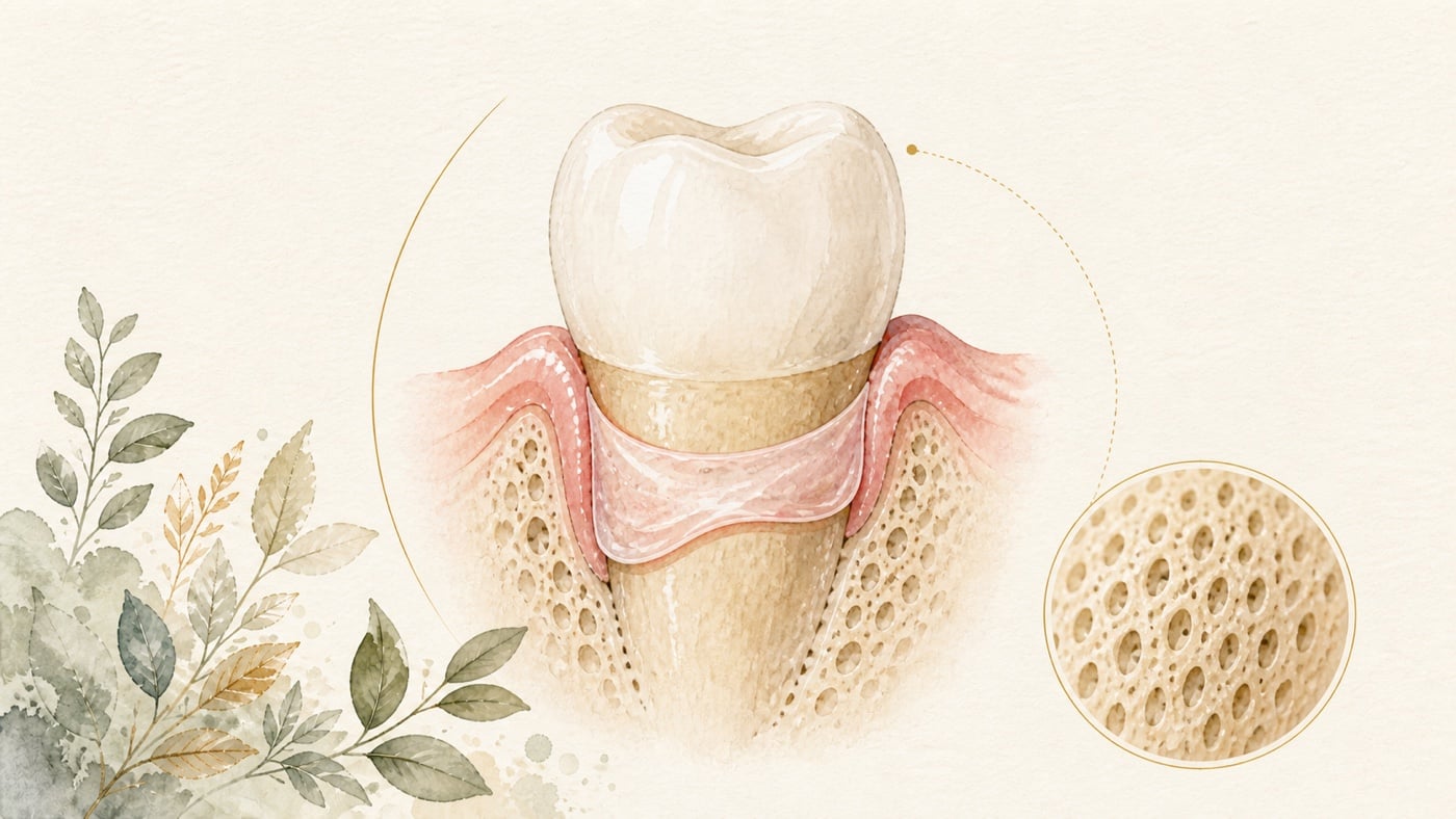
Root exposure after gingival recession may bring sensitivity to cold, discomfort during brushing, cervical tooth wear and aesthetic concerns at the same time, but these problems are not necessarily resolved by the same treatment. Observation and non-surgical treatment focus on stopping continuing irritation, improving cleaning methods and controlling sensitivity. A restoration treats the cervical defect and surface contour. Root-coverage surgery moves or adds soft tissue. Before choosing, the recession type, interdental attachment, whether a non-carious cervical lesion is already present, soft-tissue thickness and the symptom you most want to improve should first be established. For sensitivity associated with gingival recession, a systematic review and meta-analysis included 13 randomised trials, 701 patients and 1,086 recession sites. After root-coverage surgery, the proportion with suppression of sensitivity was 70.8%, with a 95% confidence interval of 64.4%–76.6%. This is an average across techniques and studies; it does not mean that every site will achieve the same improvement, and it did not directly compare surgery with simple observation or every non-surgical treatment for sensitivity.
Gingival Recession and Sensitivity: Observation, a Restoration or Root Coverage?
Direct answer: The three routes treat different layers, so first establish whether you want to improve the symptom, the cervical defect or the position of the gingival margin. A meta-analysis records suppression of dentine hypersensitivity in 70.8% (95% CI 64.4%–76.6%) after root-coverage surgery, with both recession reduction and the percentage of root coverage significantly associated with that suppression [F1]. In teeth that also have a non-carious cervical lesion, restoring the lesion does not affect the percentage of root coverage, but it significantly decreases dentine hypersensitivity [F2].
Geographic scope: This is general health education based on international literature. It does not cover any particular country's insurance scheme or regulations; for how care and fees are organised, follow the rules where you are. Every source cited in this card is international peer-reviewed literature; no national legislation or insurance-benefit rule is cited.
TL;DR | First Distinguish Sensitivity, the Defect and Gingival Position to Know Which Layer Needs Treatment
Root exposure after gingival recession may bring sensitivity to cold, discomfort during brushing, cervical tooth wear and aesthetic concerns at the same time, but these problems are not necessarily resolved by the same treatment. Observation and non-surgical treatment focus on stopping continuing irritation, improving cleaning methods and controlling sensitivity. A restoration treats the cervical defect and surface contour. Root-coverage surgery moves or adds soft tissue. Before choosing, the recession type, interdental attachment, whether a non-carious cervical lesion is already present, soft-tissue thickness and the symptom you most want to improve should first be established.
For sensitivity associated with gingival recession, a systematic review and meta-analysis included 13 randomised trials, 701 patients and 1,086 recession sites. After root-coverage surgery, the proportion with suppression of sensitivity was 70.8%, with a 95% confidence interval of 64.4%–76.6%.[F1] This is an average across techniques and studies; it does not mean that every site will achieve the same improvement, and it did not directly compare surgery with simple observation or every non-surgical treatment for sensitivity.
Where a non-carious cervical lesion is also present, restoration and surgery need not be mutually exclusive. The abstract of another review found that restoring the defect did not alter the percentage of root coverage after surgery, but could reduce dentine hypersensitivity and improve patients’ perceptions of appearance and satisfaction. The abstract provided no effect size, so only the direction can be interpreted; the magnitude of improvement cannot be claimed. The “satisfaction” half, however, is not supported by the review’s own text: the only study in it that recorded patient satisfaction reported no significant difference between the two groups, and that comparison was not “restored versus unrestored”. This card therefore uses only the appearance half.[F2]
Main Text | Three Routes, Each Answering a Different Question
When Can Observation and Non-surgical Treatment Be Tried First?
If the position of recession is stable, the tooth structure has no defect requiring restoration and the principal problem is occasional sensitivity, the dentist will generally first look for factors that may continue to irritate the root surface: brushing force and how the bristles contact it, dental plaque and gingival inflammation, acid exposure, loading, and existing restorative margins. The value of the non-surgical route lies in first addressing modifiable factors and then monitoring with consistent photographs, periodontal records and descriptions of symptoms.
‘Observation’ does not mean leaving it unattended. It should include a baseline record, situations that trigger sensitivity, cleanability and conditions for review. If the extent of root exposure continues to change, sensitivity interferes with cleaning, or aesthetic and functional concerns remain pronounced, there is then a reason to bring a restoration or soft-tissue surgery into the next discussion.
When Does a Restoration Treat the Defect rather than Move the Gingiva Back?
A non-carious cervical lesion changes the contour of the tooth surface and may obscure the cemento-enamel junction that should normally be identified. A restoration can rebuild part of the contour and treat exposed dentine or sensitivity, but does not increase gingival height by itself. If root coverage is also planned, the restorative margin, surface contour and position intended for surgical coverage need to be designed together.
For combined treatment, a systematic review initially identified 222 potentially relevant titles and abstracts, analysed 23 papers further, and finally used 13 for validity assessment and data extraction. Look closely at the evidence base: the review’s own text states that, of those 13, only 3 randomised trials directly compared restored with unrestored lesions. In the abstract the authors concluded that, in teeth with gingival recession and non-carious cervical lesions, restoring the defect first did not affect the percentage of root coverage, while restoration significantly reduced dentine hypersensitivity and improved patients’ perceptions of appearance and satisfaction. The text itself, however, supports only the appearance half: just one study recorded patient satisfaction, it found no significant difference between the two groups, and that comparison was not “restored versus unrestored”.[F2]
The abstract did not provide a numerical reduction in sensitivity, confidence intervals or details of follow-up in each group, so ‘significant’ cannot be rewritten as an expected magnitude of improvement. It is better suited to support a decision concept: restorations and root coverage treat different layers, and whether to combine them should depend on defect morphology, sensitivity and the surgical margin, rather than restoring every exposed root directly.
How Much Effect on Sensitivity Can Be Inferred for Root-coverage Surgery?
The meta-analysis of 13 randomised trials estimated a postsurgical proportion of sensitivity suppression of 70.8% (95% CI 64.4%–76.6%), with between-study heterogeneity of I² = 39.2%. The amount of recession reduction and the percentage of root coverage were significantly associated with suppression of sensitivity, with model estimates of 0.66 (0.10–1.23, p = 0.022) and 0.04 (0.01–0.08, p = 0.012), respectively.[F1]
In the same analysis, the proportion with suppression of sensitivity was 73.3% (65.6%–79.8%) for a coronally advanced flap combined with an autogenous connective-tissue graft and 61.4% (51.7%–70.3%) when combined with a xenogeneic collagen matrix, with between-group p = 0.048.[F1] This supports the possibility that material differences affect average results, but the surgical method cannot be selected from sensitivity alone. Donor-site burden, the number of recession sites, soft-tissue thickness, interdental attachment and patient preferences still need to be considered together.
How Should an Autogenous Connective-tissue Graft and Xenogeneic Substitute Be Weighed?
A newer systematic review and meta-analysis included 16 randomised trials, 632 patients and 1,878 sites of multiple gingival recessions. In the coding direction used by that analysis, the mean differences in root coverage for an autogenous connective-tissue graft compared with a xenogeneic collagen matrix were −13.4 and −11.05 percentage points at 6 and 12 months, respectively. The difference from a porcine acellular dermal matrix at 12 months was −11.63 percentage points; the authors interpreted all of these as favouring autogenous connective tissue. The autogenous group also had better complete root-coverage results at 6 months than the xenogeneic collagen matrix, with an RR of 0.71 (95% CI 0.62–0.82, coded as the xenogeneic material relative to the autogenous material).[F3]
These results focus on ‘multiple’ gingival recessions and cannot be applied directly to a single tooth. Follow-up was mainly at 6 and 12 months and is also insufficient to answer questions about longer-term stability. The review considered that xenogeneic materials may remain an alternative where comfort and satisfaction are prioritised, but the abstract did not give pooled figures for these patient-reported outcomes. The safe interpretation is that autogenous material had better clinical coverage results, while substitute material needs to be weighed against donor-site requirements and patient priorities; the choice cannot be made by comparing a single percentage.
Which Is Better, a Coronally Advanced Flap or the Tunnel Technique?
A systematic review comparing coronally advanced flaps with the tunnel technique included 26 studies, of which 14 entered the meta-analysis. For the tunnel technique with a connective-tissue graft compared with a coronally advanced flap with a connective-tissue graft, the effect difference in mean root coverage was −8.68, with a 95% CI from −17.19 to −0.17 and p = 0.0457. The authors interpreted this as favouring the tunnel technique according to their coding direction.[F4]
Other comparisons were not equally clear. The mean root-coverage difference between a tunnel technique plus other biomaterials and a tunnel technique using connective tissue alone was 4.17, with a 95% CI from −17.91 to 26.26 and p = 0.7110. Irrespective of adjunctive material, the direct comparison of the tunnel technique with a coronally advanced flap had an effect difference of 5.73, with a 95% CI from −8.90 to 13.55 and p = 0.685.[F4] Both confidence intervals cross 0 and are wide. The safe interpretation is that ‘no statistical difference was found and the estimates were imprecise’. This cannot be rewritten to mean that the techniques are equivalent, nor can one be claimed to be better.
The choice of technique must still return to the number of teeth, vestibular depth, tissue thickness, papillae and interdental attachment, defect morphology, donor-tissue choice and operator experience. Pooled averages provide direction, but do not replace an assessment of feasibility at the individual site.
Data Anchor Table | Each Figure Corresponds to a Treatment Aim
| Decision question | Data anchor | Safe interpretation | Source |
|---|---|---|---|
| Surgery and sensitivity | 13 RCTs, 701 people and 1,086 recession sites; sensitivity suppression 70.8% (95% CI 64.4%–76.6%) | An average across techniques that did not directly compare every non-surgical option, and not a prediction of individual improvement | [F1] |
| Combining restoration of a cervical lesion | Initial screening of 222 records, further analysis of 23 papers and extraction from 13 (of which only 3 randomised trials directly compared restored with unrestored lesions); restoration did not affect coverage percentage but improved sensitivity and esthetic scores | The abstract has no effect size for sensitivity, so only the direction can be cited; the only study recording satisfaction found no significant difference; restorations and soft-tissue surgery have different aims | [F2] |
| Tunnel technique and coronally advanced flap | 26 studies, with 14 pooled; in some comparisons the 95% CI crossed 0: −17.91 to 26.26 and −8.90 to 13.55 | Crossing 0 with a wide interval means that no difference was found and the estimate was imprecise, not that the treatments were equivalent | [F4] |
| Autogenous and xenogeneic materials | 16 RCTs, 632 people and 1,878 recession sites; autogenous material had better results for several coverage outcomes | Mainly multiple recessions and outcomes at 6 and 12 months; donor-site needs and patient priorities still need to be weighed | [F3] |
Conclusion | First Clarify Whether You Want to Improve the Symptom, Defect or Gingival Position
When sensitivity accompanies gingival recession, no single tool answers every problem at once. Observation and non-surgical treatment can first control continuing irritation and establish a record of change. A restoration rebuilds a cervical defect; root coverage treats soft-tissue position and thickness. Sensitivity can improve on average after surgery, but different materials and techniques each have evidence boundaries. In particular, when a confidence interval crosses 0, failure to find a difference cannot be described as the two treatments being equivalent.[F1][F4]
If you are concerned about both sensitivity during brushing and the appearance of exposed roots, you can ask your dentist to record, in sequence, the recession type, interdental attachment, cervical defect, soft-tissue thickness, current sensitivity triggers and the treatment aim that matters most to you. Then compare ‘observation and non-surgical control first’, ‘restoration of the tooth surface is required’ and ‘assessment for soft-tissue grafting’ in 3 columns. This comes closer to the choice that genuinely suits you than asking directly which operation is best.
Risk factors (what to know before treatment)
- A population average is not an individual expectation: the 70.8% (95% CI 64.4%–76.6%) proportion of sensitivity suppression after surgery is a pooled estimate across 13 randomised trials, 701 patients and 1,086 recessions, with between-study heterogeneity of I² = 39.2% [F1]. It describes an average at population level; it does not mean that every recession site improves to the same degree.
- The difference between materials answers only the sensitivity question: in the same analysis, sensitivity suppression was 73.3% (65.6%–79.8%) for a coronally advanced flap with an autogenous connective-tissue graft and 61.4% (51.7%–70.3%) with a xenogeneic collagen matrix, with between-group p = 0.048 [F1]. Sensitivity is only one outcome measure; donor-site burden, the number of recession sites and local conditions still have to be weighed by the dentist.
- Choosing between the two soft-tissue materials is not only about a coverage percentage: the review of 16 randomised trials, 632 patients and 1,878 recessions concluded that autogenous connective tissue continues to have superior clinical outcomes in multiple gingival recessions; the same paper describes xenogeneic materials as a promising alternative, particularly where patient comfort and satisfaction are prioritised [F3].
- Finding no difference is not the same as the two techniques being equal: the review comparing the tunnel technique with the coronally advanced flap included 26 studies, of which 14 entered the meta-analysis; among the mean root-coverage comparisons reported in the abstract, only the comparison of the tunnel technique with a connective-tissue graft against a coronally advanced flap with a connective-tissue graft reached statistical significance (−8.68, 95% CI −17.19 to −0.17, p = 0.0457) [F4]. The authors conclude that the tunnel technique with a connective-tissue graft might offer better root coverage than a coronally advanced flap with a connective-tissue graft, but that the selection of biomaterial requires consideration [F4].
- Evidence can only be used for what it can answer: the data in F3 focus on 'multiple' gingival recessions, mainly at 6 and 12 months [F3]; the abstract of F2 gives no effect size for the decrease in sensitivity, so only the direction can be read, not the magnitude; and of the 13 studies it included, only 3 randomised trials directly compared restored with unrestored lesions, while the only study recording patient satisfaction found no significant difference between the two groups [F2]. Longer-term stability, and feasibility at a single site, lie outside what these abstracts can answer.
- This card does not compile a list of contraindications: no separate literature search on surgical contraindications was carried out for this card. Whether root coverage is suitable, which technique and material to use, and whether a cervical defect should be restored first, have to be assessed by a dentist according to your recession type, interdental attachment, soft-tissue thickness and general health.
Every clinical statement in this article is mapped line by line to its cited source (see the sources and evidence chain below). It has not been clinically reviewed by a licensed practitioner. This is health information, not individual advice; assessment by a clinician is required.
FAQ
- Can I Start without Surgery for Sensitivity at an Exposed Root?
- The dentist can first establish whether the recession is stable, whether there is a cervical defect, and whether factors such as brushing, inflammation or acid exposure are present. If non-surgical treatment makes the symptoms acceptable and the site cleanable, it can be monitored from the baseline record. If the problem persists or conditions change, a restoration and root coverage can then be discussed.
- 露出根面がしみる場合、手術をせずに始められますか? — 歯科医師が退縮は安定しているか、歯頸部に欠損があるか、歯磨き、炎症、酸への曝露などの要因があるかを確認したうえで、非外科的処置から始められます。非外科的処置で症状を許容範囲にでき、部位を清掃できるなら、ベースライン記録を使って追跡できます。悩みが続く、または条件が変わる場合に、修復と根面被覆を話し合います。
- Can I Start without Surgery for Sensitivity at an Exposed Root? — The dentist can first establish whether the recession is stable, whether there is a cervical defect, and whether factors such as brushing, inflammation or acid exposure are present. If non-surgical treatment makes the symptoms acceptable and the site cleanable, it can be monitored from the baseline record. If the problem persists or conditions change, a restoration and root coverage can then be discussed.
- Will the Gingiva Return to Its Original Position after a Restoration?
- A restoration rebuilds the tooth surface and does not move the gingiva by itself. A review found that combining a restoration did not affect the percentage of root coverage after surgery, but may improve sensitivity and esthetic scores; the only study in that review which recorded patient satisfaction found no significant difference between the two groups.[F2] Whether a restoration is required depends on whether a cervical lesion genuinely requiring restoration is present.
- 修復後、歯肉は元の位置まで戻りますか? — 修復で再建するのは歯面であり、それだけで歯肉が移動することはありません。レビューでは、修復を併用しても手術後の根面被覆率は変わりませんでしたが、知覚過敏と外観評価を改善する可能性が示されました。ただし、このレビューの本文で患者満足度を記録した唯一の研究は 2 群間に有意差なしでした。[F2] 修復が必要かは、修復すべき歯頸部欠損が実際にあるかによって決まります。
- Will the Gingiva Return to Its Original Position after a Restoration? — A restoration rebuilds the tooth surface and does not move the gingiva by itself. A review found that combining a restoration did not affect the percentage of root coverage after surgery, but may improve sensitivity and esthetic scores; the only study in that review which recorded patient satisfaction found no significant difference between the two groups.[F2] Whether a restoration is required depends on whether a cervical lesion genuinely requiring restoration is present.
- Does Sensitivity Always Disappear after Root Coverage?
- The mean proportion with suppression of sensitivity across 13 randomised trials was 70.8%, so not every site improved.[F1] Symptoms may also be affected by defect morphology and other irritants, making it important to clarify the source of sensitivity and the primary aim before surgery.
- 根面被覆後、知覚過敏はすべてなくなりますか? — 13 件のランダム化試験を通じた平均知覚過敏抑制率は 70.8% であり、すべての部位で改善するという意味ではありません。[F1] 症状は欠損形態とほかの刺激要因にも影響される可能性があるため、術前に知覚過敏の原因と主な目標を明確にする必要があります。
- Does Sensitivity Always Disappear after Root Coverage? — The mean proportion with suppression of sensitivity across 13 randomised trials was 70.8%, so not every site improved.[F1] Symptoms may also be affected by defect morphology and other irritants, making it important to clarify the source of sensitivity and the primary aim before surgery.
- If a Xenogeneic Material Is More Comfortable, Is It Necessarily More Suitable for Me?
- Not necessarily. A meta-analysis of multiple recessions found better results for autogenous connective tissue in several root-coverage outcomes, while xenogeneic material may be an alternative when avoiding autogenous tissue harvest is important.[F3] The choice should balance the coverage aim, harvesting, site conditions and your preferences.
- 異種材料の方が快適なら、必ず私に適していますか? — 必ずしもそうではありません。複数歯退縮のメタアナリシスでは、自家結合組織が複数の根面被覆結果で優れていました。異種材料は、自家組織採取を避けることを重視する場合の代替選択肢になり得ます。[F3] 選択では、被覆目標、採取の負担、部位条件、あなたの希望を総合して判断する必要があります。
- If a Xenogeneic Material Is More Comfortable, Is It Necessarily More Suitable for Me? — Not necessarily. A meta-analysis of multiple recessions found better results for autogenous connective tissue in several root-coverage outcomes, while xenogeneic material may be an alternative when avoiding autogenous tissue harvest is important.[F3] The choice should balance the coverage aim, harvesting, site conditions and your preferences.
- Is the Tunnel Technique Necessarily Better than a Coronally Advanced Flap?
- That conclusion cannot be drawn. Although one comparison favoured the tunnel technique, confidence intervals in other analyses crossed 0 and were wide, so a difference was not demonstrated.[F4] The technique still needs to be selected according to local anatomy and the operator’s assessment.
- トンネル法は必ず歯冠側移動フラップより優れていますか? — そのようには結論できません。トンネル法に有利な比較が一つありましたが、ほかの分析では信頼区間が 0 をまたぎ、幅も広く、差は証明されませんでした。[F4] 術式は局所解剖と術者の評価に基づいて選ぶ必要があります。
- Is the Tunnel Technique Necessarily Better than a Coronally Advanced Flap? — That conclusion cannot be drawn. Although one comparison favoured the tunnel technique, confidence intervals in other analyses crossed 0 and were wide, so a difference was not demonstrated.[F4] The technique still needs to be selected according to local anatomy and the operator’s assessment.
Medical notice This article is provided for health education and medical information purposes. It is not a solicitation for medical services and does not constitute diagnosis or treatment advice. Actual treatment methods and outcomes vary between individuals and require evaluation by a dentist; every treatment has its own indications, limitations and possible risks. If you have related symptoms or treatment needs, please book a consultation for evaluation by a dentist.
Source anchors
- Effectiveness of surgical root coverage on dentin hypersensitivity: A systematic review and meta-analysis. [PMID:35634650] · https://pubmed.ncbi.nlm.nih.gov/35634650/ · 在 IDAEO 的其他引用
- Root coverage with the restoration of non-carious cervical lesions: A systematic review and meta-analysis. [PMID:38445442] · https://pubmed.ncbi.nlm.nih.gov/38445442/ · 在 IDAEO 的其他引用
- Comparative Effectiveness of Autogenous Connective Tissue Grafts and Xenogeneic Soft Tissue Substitutes for Multiple Gingival Recessions: A Systematic Review and Meta-Analysis. [PMID:41752765] · https://pubmed.ncbi.nlm.nih.gov/41752765/ · 在 IDAEO 的其他引用
- Root Coverage Techniques: Coronally Advancement Flap vs. Tunnel Technique: A Systematic Review and Meta-Analysis. [PMID:39590391] · https://pubmed.ncbi.nlm.nih.gov/39590391/ · 在 IDAEO 的其他引用
Cite this article
Lucy・《Gingival Recession and Sensitivity: Observation, a Restoration or Root Coverage?》・IDAEO 知識庫・2026-07-20・https://km.idaeo.ai/post/dental/gingival-recession-root-coverageUpdated 2026-08-26