🏛 Part of the "dental" topic shelf →
A calcified root canal: can the tooth still be retained?
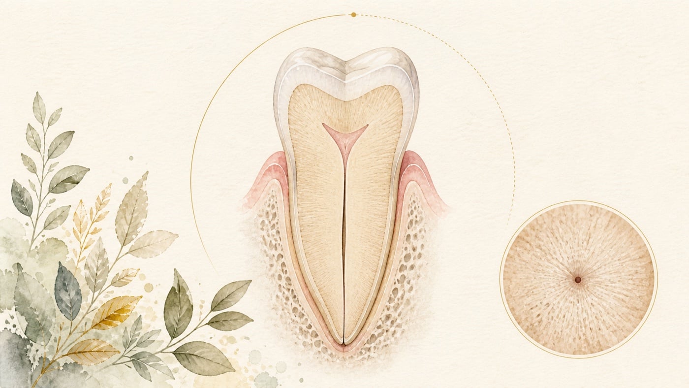
Canal calcification or pulp-canal obliteration (PCO) is progressive hard-tissue deposition that narrows the chamber and canals. Trauma, age and chronic irritation may contribute. A canal apparently “absent” on two-dimensional radiographs may retain a fine deep path; the challenge is deciding whether intervention is needed and, if so, establishing a controlled direction while conserving tooth tissue. The first decision is not microscope, CBCT or guide selection, but the reason to treat. A review of post-traumatic PCO found watchful waiting most common in asymptomatic cases and did not recommend preventive root canal treatment. A meta-analysis of 34 studies estimated post-traumatic PCO prevalence at 27.6% in permanent and 21.9% in primary teeth; prevalence is not the proportion requiring treatment. If symptoms, examination and imaging support intervention, microscopy, limited-field CBCT, static guides or dynamic navigation may assist. Current guided-endodontic reviews mainly contain case reports, series and in-vitro studies with low, heterogeneous evidence. A recent review included only 34 case reports and 41 patients, preventing general outcome estimates or tool comparisons. Technology follows diagnosis and stopping conditions; it is not a promise.
A calcified root canal: can the tooth still be retained?
Direct answer: Calcification does not mean root canal treatment is needed straight away. In the systematic review, which included only anterior permanent teeth with pulp canal obliteration due to dental trauma, watchful waiting was the most implemented clinical strategy; discoloured non-symptomatic calcified teeth were mostly managed with external bleaching, root canal treatment should not be implemented as a preventive intervention strategy, and symptomatic teeth should follow regular endodontic treatment pathways. [F1] Watchful waiting is not the same as leaving it alone: if spontaneous or night-time pain appears, if pain lingers after a hot or cold stimulus is removed, if pain on biting is increasing, if the gum swells or a small opening that discharges fluid appears on the gum, if discolouration keeps deepening, or if the tooth becomes mobile, waiting is no longer appropriate and the tooth should be reassessed by a dentist (this sentence is a general safety reminder compiled by this site, not a conclusion of the review above).
Geographic scope: global. All the evidence cited here comes from systematic reviews and meta-analyses in international journals; it does not describe the rules or funding arrangements of any particular country. Which examinations and procedures are actually available still depends on the care setting where you live and on your dentist's assessment.
TL;DR | Calcification requires reinterpretation; it is not an automatic instruction for root canal treatment
Canal calcification or pulp-canal obliteration (PCO) is progressive hard-tissue deposition that narrows the chamber and canals. Trauma, age and chronic irritation may contribute. A canal apparently “absent” on two-dimensional radiographs may retain a fine deep path; the challenge is deciding whether intervention is needed and, if so, establishing a controlled direction while conserving tooth tissue.
The first decision is not microscope, CBCT or guide selection, but the reason to treat. A review of post-traumatic PCO found watchful waiting most common in asymptomatic cases and did not recommend preventive root canal treatment. [F1] A meta-analysis of 34 studies estimated post-traumatic PCO prevalence at 27.6% in permanent and 21.9% in primary teeth; prevalence is not the proportion requiring treatment. [F2]
If symptoms, examination and imaging support intervention, microscopy, limited-field CBCT, static guides or dynamic navigation may assist. Current guided-endodontic reviews mainly contain case reports, series and in-vitro studies with low, heterogeneous evidence. [F3][F4][F5] A recent review included only 34 case reports and 41 patients, preventing general outcome estimates or tool comparisons. [F6] Technology follows diagnosis and stopping conditions; it is not a promise.
Main discussion | Distinguish “follow” from “enter” before deciding how to enter
How common is calcification after trauma?
A 2024 meta-analysis included 34 moderate-to-high-quality observational studies. Pooled prevalence was 27.6% (95% confidence interval 18.7% to 37.7%) in permanent teeth and 21.9% (95% confidence interval 16.0% to 28.4%) in primary teeth. By trauma type it was 78.6% after permanent-root fracture and 29.4% after lateral luxation of primary teeth. [F2]
These values answer frequency of radiographic calcification, not treatment need. Trauma type, age, follow-up and imaging criteria affect the estimates, and the confidence intervals also show the uncertainty between studies. Spontaneous pain, biting discomfort, periapical change, pulp status and serial imaging inform the individual decision.
With no symptoms but discolouration, should root canal treatment be done first?
A PRISMA systematic review registered with PROSPERO, focused on the clinical management of post-traumatic pulp canal obliteration in anterior teeth, found only 20 case reports, 27 patients and 33 teeth. Watchful waiting was most common; asymptomatic discoloured teeth were usually externally bleached, and preventive root canal treatment was not recommended. [F1]
Pulp necrosis was 36.4%, but this came from published case reports, not all calcified teeth after trauma. Cases that are unusual, complex or have been treated are more likely to be published, so this figure cannot be used to predict whether an individual tooth will become necrotic in the future. Establish symptom and imaging baselines and intervene only when a treatment diagnosis emerges.
While waiting and watching, what means you can no longer wait?
Watchful waiting only works if someone is actually watching and knows when to stop and reassess. The reviews cited here do not set out stopping conditions that a patient can check against, so the list below does not come from those papers. It is a general safety reminder compiled by this site, intended to show when to go back for a dental reassessment; it is not a way for you to reach a diagnosis yourself. Contact your dentist and arrange a reassessment if any of the following appears:
- Pain that starts on its own without any external stimulus, or pain that wakes you from sleep
- Pain that lingers after a hot or cold stimulus has been removed
- Increasing discomfort when biting or when the tooth is touched
- Gum swelling or tenderness, or a small opening on the gum that discharges fluid (a sinus tract)
- Discolouration that keeps deepening rather than staying stable
- Mobility of that tooth that was not there before
The test is whether something is changing and continuing to change, not how much time has passed. This card deliberately gives no observation period: if any item above is happening or getting worse, that in itself is the reason to be seen, and there is no interval to complete first. If facial swelling is expanding rapidly, or there is difficulty swallowing or breathing, severely limited mouth opening or fever, that is no longer within the scope of observation and emergency dental or emergency department care should be sought immediately.
Once treatment is needed, what do microscopy and CBCT each answer?
Microscopy provides deep illumination and magnification for chamber colour, grooves and calcification boundaries; ultrasonic tips remove obstruction layer by layer. Limited-field CBCT shows the three-dimensional relation of root, possible canal and surrounding structures. One is live vision, the other planning information; neither replaces the other.
CBCT is not routine for every calcified tooth because resolution, metal artefact, radiation and interpretation errors matter. Use it when three-dimensional information may change direction, depth, referral or stopping conditions. A microscope cannot see through intact dentine; uncertain continued exploration can waste tissue or deviate.
How robust is the “precision” evidence for guided endodontics?
A 2020 review included 22 studies: 15 case reports, 6 in-vitro or ex-vivo preclinical studies and 1 observational study. Although drilling commonly matched plans, evidence was low and heterogeneous, requiring larger patient studies. [F4]
A 2023 review included 45 studies: 21 case reports, 11 case series, 12 in-vitro studies and 1 cohort. Forty-three reported that guided endodontics was an effective and precise technique for accessing the permeable portion of calcified canals, and 2 reported accidents or failures. [F3] The ratio 43/45 counts studies, not patients or efficacy; mixed designs and publication bias prohibit a clinical promise.
The reviews support use for locating calcified canals, not a universal priority. Tooth position, opening, guide seating, drill length, cooling, image registration and operator experience alter the path.
Does dynamic navigation have stronger evidence than a static guide?
A 2022 review of dynamic navigation included only 14 studies—3 case reports and 11 in-vitro studies—and could not meta-analyse them. Evidence was low and human comparisons across operator experience were needed. [F5]
Dynamic navigation displays relative instrument position and permits adjustment; a static guide converts planning into a fixed drilling path. Evidence proves neither generally superior, and in-vitro deviation, time or errors cannot become long-term tooth outcomes. Choose according to space, equipment, training and backup.
What can a review of case reports tell us—and not tell us?
A 2025 review included 34 case reports and 41 patients, describing chelators, flexible instruments, microscopy and ultrasonics, and CBCT with three-dimensional printed guides. [F6] It maps reported strategies but cannot estimate general success or establish superiority.
Case reports lack controls, vary greatly and favour publication of successful or novel cases. Positive descriptions must be preceded by case-level evidence and publication bias; “many successes” cannot become a population outcome.
Decide referral timing before choosing equipment
Early referral preserves options when calcification extends to the middle or deeper canal, roots curve, deviation or perforation is suspected, restorations are complex, or safe direction cannot be established. Repeated uncertain attempts may erase anatomical clues.
Before referral, assemble trauma and symptom timelines, radiographs from different years, treatment history and restoration status. Assessment asks not only whether the canal can be found, but whether entry is needed, the intended path, dentine safety boundary, imaging need and stopping point.
Data anchors | Six reviews have separate boundaries for prevalence, management and guidance
| Evidence question | Data anchor | Cautious interpretation | Source |
|---|---|---|---|
| Guided localisation | 45 studies: 21 case reports, 11 series, 12 in vitro, 1 cohort; 43 positive and 2 accidents/failures | Study count is not patient success; mixed designs and publication bias | [F3] |
| Early guided evidence | 22 studies: 15 case reports, 6 preclinical, 1 observational | Low, heterogeneous evidence; drilling accuracy is not long-term advantage | [F4] |
| Dynamic navigation | 14 studies: 3 case reports, 11 in vitro; no meta-analysis | Low evidence, insufficient to establish a priority among patients in general | [F5] |
| Post-traumatic PCO management | 20 case reports, 27 patients, 33 teeth; watchful waiting most common | No preventive treatment for asymptomatic PCO; cases cannot estimate general risk | [F1] |
| Post-traumatic PCO prevalence | 34 studies; permanent 27.6% (95% CI 18.7–37.7), primary 21.9% (95% CI 16.0–28.4) | Post-traumatic prevalence, not treatment need or outcome | [F2] |
| Calcified-canal strategies | 34 case reports, 41 patients | Maps tools; no controls and publication bias prevent reliable success rates | [F6] |
Conclusion | Prove treatment need before adding precision to entry
Post-traumatic PCO is not rare, but prevalence is not treatment rate; reviews most often report observation and do not support preventive treatment of asymptomatic teeth. [F2][F1] When intervention is indicated, microscopy, CBCT and guidance may assist, but evidence is chiefly cases and in-vitro work and provides neither reliable success rates nor tool ranking. [F4][F5][F6]
If a canal has been described as calcified, bring old radiographs and trauma, symptom and treatment timelines to your dental appointment. A reasonable order is to confirm the reason to treat first, then discuss calcification location, vision, three-dimensional imaging or guidance, safety boundaries, and when to stop or refer.
Risk factors (what to know before treatment)
- Indications: the systematic review, which included only anterior permanent teeth with obliteration due to dental trauma, concluded that symptomatic teeth with pulp canal obliteration should follow regular endodontic treatment pathways. [F1]
- When it is not advised: the same review advocated external bleaching for discoloured non-symptomatic calcified teeth and stated that root canal treatment should not be implemented as a preventive intervention strategy. [F1]
- Adverse effects and complications: this is not a procedure without risk — the systematic review of guided endodontics for locating calcified canals included 45 studies, of which 2 reported accidents or failures related to the use of endodontic guides, and the review itself stated that the technique still presents limitations. [F3]
- Limits of the evidence: in the guided-endodontics literature the level of evidence is low and the methodology among studies is heterogeneous, and the authors stated that studies with a larger number of patients are necessary to obtain significant conclusions [F4]; the most recent review of managing calcified canals included case reports [F6]. Case reports have no control group, so they cannot be converted into a success rate for patients in general, nor used to rank one tool above another. Whether to treat, how to gain access and when to stop still have to be judged by a dentist from clinical examination and imaging.
Every clinical statement in this article is mapped line by line to its cited source (see the sources and evidence chain below). It has not been clinically reviewed by a licensed practitioner. This is health information, not individual advice; assessment by a clinician is required.
FAQ
- Does post-traumatic calcification require root canal treatment?
- No. Pooled prevalence is not treatment need. [F2] Symptoms, pulpal and periapical tests, imaging change and restorability decide.
- 外傷後に石灰化がみられたら、根管治療が必要ですか? — いいえ。統合有病割合が示すのは、外傷後に PCO がどの程度みられるかであり、治療を必要とする割合ではありません。[F2] 症状、歯髄と根尖周囲の検査、画像所見の変化、歯の修復上の必要性を合わせて介入の要否を判断します。
- Does post-traumatic calcification require root canal treatment? — No. Pooled prevalence is not treatment need. [F2] Symptoms, pulpal and periapical tests, imaging change and restorability decide.
- Can an asymptomatic discoloured tooth receive preventive treatment?
- Watchful waiting was most common; authors favoured external bleaching rather than preventive treatment for asymptomatic discoloured PCO. [F1] Clinical and imaging assessment still comes first. If spontaneous or night-time pain, increasing pain on biting, gum swelling or a small discharging opening on the gum, deepening discolouration or new mobility later appears, observation is no longer appropriate and the tooth should be reassessed (a general safety reminder compiled by this site, not a conclusion of that review).
- 無症状で変色だけがある歯に、予防目的の根管治療を行いますか? — システマティックレビューでは経過観察が最も多く選ばれ、無症状で変色した PCO 歯には、予防的根管治療ではなく外部漂白が選ばれていました。[F1] 実際の対応を決める前に、臨床検査と画像評価を行う必要があります。その後、誘因のない痛みや夜間の痛み、咬合時痛の増強、歯肉の腫れや分泌物の出る小さな開口部、変色の進行、歯の動揺が現れた場合は、経過観察を続けるべきではなく、再評価を受けてください(これは当サイトが整理した一般的な安全上の注意であり、当該レビューの結論ではありません)。
- Can an asymptomatic discoloured tooth receive preventive treatment? — Watchful waiting was most common; authors favoured external bleaching rather than preventive treatment for asymptomatic discoloured PCO. [F1] Clinical and imaging assessment still comes first. If spontaneous or night-time pain, increasing pain on biting, gum swelling or a small discharging opening on the gum, deepening discolouration or new mobility later appears, observation is no longer appropriate and the tooth should be reassessed (a general safety reminder compiled by this site, not a conclusion of that review).
- If 43 guided studies reported precision, is the result certain?
- No. Of the 45 studies, only 1 was a cohort study; the others were mainly case reports, case series and in vitro studies. The figure 43 refers to the number of studies, not the patient denominator. [F3] Low-level evidence and publication bias can amplify a favourable impression.
- 43 件の研究が正確なアクセスを報告していれば、結果は確実ですか? — そのようには解釈できません。45 件のうちコホート研究は 1 件だけで、残りは主に症例報告、症例集積、体外研究でした。43 という数は患者数ではなく研究件数です。[F3] エビデンスレベルの低い研究と発表バイアスによって、肯定的な印象が強まっている可能性があります。
- If 43 guided studies reported precision, is the result certain? — No. Of the 45 studies, only 1 was a cohort study; the others were mainly case reports, case series and in vitro studies. The figure 43 refers to the number of studies, not the patient denominator. [F3] Low-level evidence and publication bias can amplify a favourable impression.
- Is dynamic navigation better than a static guide?
- The dynamic review had only 3 case reports and 11 in-vitro studies and no meta-analysis. [F5] Position, space, equipment, training and backup matter more than an unsupported ranking.
- 動的ナビゲーションと静的ガイドでは、どちらが優れていますか? — 現在の動的ナビゲーションのレビューは症例報告 3 件と体外研究 11 件だけで、メタアナリシスはできませんでした。[F5] 一般患者における優先順位を確立するには不十分であり、歯の位置と空間、設備、術者の訓練、代替案を考慮して選択します。
- Is dynamic navigation better than a static guide? — The dynamic review had only 3 case reports and 11 in-vitro studies and no meta-analysis. [F5] Position, space, equipment, training and backup matter more than an unsupported ranking.
- When is early referral appropriate?
- Discuss referral for deep calcification, curved roots, previous deviation, complex restorations or inability to progress safely. Stopping avoids further tissue loss when information is inadequate.
- どのような場合に早期紹介を検討しますか? — 石灰化が深い、歯根が湾曲している、すでに経路が偏位している、修復構造が複雑である、初期の探索で安全な方向へ進めないといった場合には、早い段階で紹介を検討します。中止は断念ではなく、情報が不足したまま歯質を削り続けることを避ける判断です。
- When is early referral appropriate? — Discuss referral for deep calcification, curved roots, previous deviation, complex restorations or inability to progress safely. Stopping avoids further tissue loss when information is inadequate.
Medical notice This article is provided for health education and medical information purposes. It is not a solicitation for medical services and does not constitute diagnosis or treatment advice. Actual treatment methods and outcomes vary between individuals and require evaluation by a dentist; every treatment has its own indications, limitations and possible risks. If you have related symptoms or treatment needs, please book a consultation for evaluation by a dentist.
Source anchors
- Management of Pulp Canal Obliteration-Systematic Review of Case Reports. [PMID:34833455] · https://pubmed.ncbi.nlm.nih.gov/34833455/ · 在 IDAEO 的其他引用
- Prevalence of pulp canal obliteration after traumatic dental injuries: a systematic review and meta-analysis. [PMID:39356901] · https://pubmed.ncbi.nlm.nih.gov/39356901/ · 在 IDAEO 的其他引用
- Effectiveness of guided endodontics in locating calcified root canals: a systematic review. [PMID:36640178] · https://pubmed.ncbi.nlm.nih.gov/36640178/ · 在 IDAEO 的其他引用
- Clinical applications, accuracy and limitations of guided endodontics: a systematic review. [PMID:31520416] · https://pubmed.ncbi.nlm.nih.gov/31520416/ · 在 IDAEO 的其他引用
- Dynamic Navigation in Guided Endodontics - A Systematic Review. [PMID:35786584] · https://pubmed.ncbi.nlm.nih.gov/35786584/ · 在 IDAEO 的其他引用
- Management of calcified canals during root canal treatment. A systematic review of case reports. [PMID:40936764] · https://pubmed.ncbi.nlm.nih.gov/40936764/ · 在 IDAEO 的其他引用
Cite this article
Lucy・《A calcified root canal: can the tooth still be retained?》・IDAEO 知識庫・2026-07-20・https://km.idaeo.ai/post/dental/calcified-root-canalUpdated 2026-08-26